. 2015 Apr;21(4):344-52.
doi: 10.1038/nm.3830. Epub 2015 Mar 30.

Targeting the MLL complex in castration-resistant prostate cancer

Affiliations

Targeting the MLL complex in castration-resistant prostate cancer

Rohit Malik et al. Nat Med. 2015 Apr.

Abstract

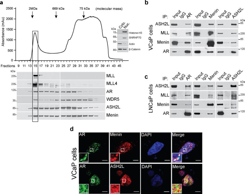
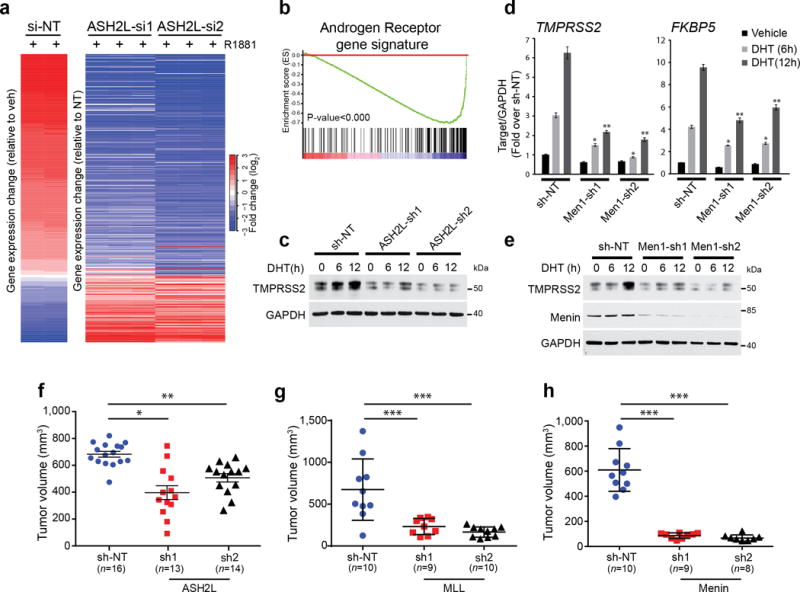
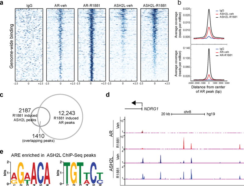
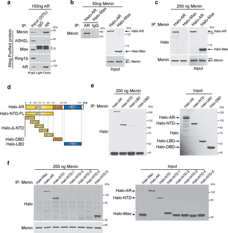
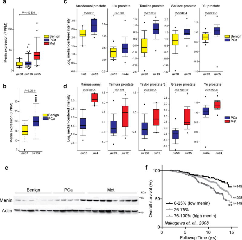
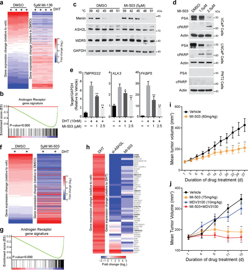
Resistance to androgen deprivation therapies and increased androgen receptor (AR) activity are major drivers of castration-resistant prostate cancer (CRPC). Although prior work has focused on targeting AR directly, co-activators of AR signaling, which may represent new therapeutic targets, are relatively underexplored. Here we demonstrate that the mixed-lineage leukemia protein (MLL) complex, a well-known driver of MLL fusion-positive leukemia, acts as a co-activator of AR signaling. AR directly interacts with the MLL complex via the menin-MLL subunit. Menin expression is higher in CRPC than in both hormone-naive prostate cancer and benign prostate tissue, and high menin expression correlates with poor overall survival of individuals diagnosed with prostate cancer. Treatment with a small-molecule inhibitor of menin-MLL interaction blocks AR signaling and inhibits the growth of castration-resistant tumors in vivo in mice. Taken together, this work identifies the MLL complex as a crucial co-activator of AR and a potential therapeutic target in advanced prostate cancer.


Conflict of interest statement

COMPETING FINANCIAL INTERESTS

The University of Michigan has filed a patent on the menin inhibitors described in this study and J.G. and T.C. are named as co-inventors.

Figures

Figure 1
Figure 1
Androgen receptor interacts with MLL complex proteins. (a) VCaP nuclear lysate fractions from Superose-6 gel filtration column were immunoblotted using indicated antibodies. Arrows indicate the approximate mass of the complexes eluted. Distribution of cytoplasmic or nuclear proteins demonstrates the efficiency of lysate fractionation (inset western blot). Shown are representative blots (n=3). (b–c) VCaP (b) and LNCaP (c) nuclear lysates were immunoprecipitated (IP) with AR, ASH2L and menin antibodies followed by immunoblotting (IB) using indicated antisera. Shown are representative blots from three independent experiments. (d) VCaP cells were fixed and immunostained using AR (green) and ASH2L (red) or menin (red) antibodies. Confocal microscopic images were analyzed by ImageJ software. Co-localization is represented by yellow puncta. Bar = 10μm. Inset bar = 2.5μm. Shown are representative images from three independent experiments.
Figure 2
Figure 2
MLL complex proteins are important for AR signaling and cell growth. (a) Microarray profiling of ASH2L knockdown (two independent siRNAs) and control non-target (NT) VCaP cells following 24 hr androgen (R1881) treatment. Heatmap displays the altered expression of the androgen-induced genes upon ASH2L knockdown. (b) Gene set enrichment analysis (GSEA) of microarray data shows enrichment of AR target gene signature. (c) Immunoblots for TMPRSS2 protein expression in VCaP cells stably expressing NT shRNA or one of two independent ASH2L shRNAs after DHT stimulation for indicated time points. Shown are representative blots (n=2). (d) Expression of FKBP5 and TMPRSS2 AR-target genes was measured using qPCR in LNCaP cells stably expressing non-targeting (NT) shRNA or one of two independent menin shRNAs, after DHT stimulation for indicated time points. * P < 0.01, **P < 0.001, compared with vehicle by one-way ANOVA. (n = 3, mean ± s.e.m) (e) Immunoblots for TMPRSS2 protein expression in VCaP cells stably expressing NT shRNA or one of two independent menin shRNAs after DHT stimulation for indicated time points. Shown are representative blots (n=3). (f,g) VCaP cells expressing NT shRNA or one of two independent ASH2L (f), MLL (g) or menin (h) shRNAs respectively were injected subcutaneously into both sides of mouse dorsal flank. Tumor volumes of xenografts were measured at 7 weeks post implantation. Each dot represents individual tumors. *, P<0.01; **, P<0.001, ***P<0.0001, compared with shNT by one-way ANOVA. All graphs are shown with mean ± s.e.m.
Figure 3
Figure 3
AR and ASH2L are recruited to the same genomic loci upon androgen stimulation. (a) A heat map representation of AR and ASH2L binding to promoter regions, 2.5kb flanking transcriptional start sites (TSS, indicated by 0) in vehicle and R1881 stimulated VCaP cells as assessed by ChIP-Sequencing. Gene promoters are rank-ordered by the level of AR enrichment at the TSS. (b) Average TSS-aligned profiles of AR and ASH2L occupancy for all annotated genes before and after vehicle or R1881 stimulation. (c) The overlap between R1881-induced AR and ASH2L peaks. (d) Representative gene (NDRG1) co-occupied by AR and ASH2L before and after AR stimulation. (e) De novo motif detection with MEME program identified enrichment of two half-androgen response elements (ARE) among ASH2L binding sites. (MEME E value 2.1e-025).
Figure 4
Figure 4
AR directly interacts with menin. (a) Purified untagged full-length (FL) AR was incubated with purified menin, ASH2L, Max or Ring1b proteins. Anti-AR antibody or control IgG immunoprecipitates (IP) were subjected to immunoblot (IB) analysis using indicated antibodies. (b) Halo-tagged FL-AR or FL-Max was incubated with purified menin. AR was immunoprecipitated using anti-AR and immunoprecipitates were subjected to immunoblot analysis with anti-menin. Immunoblots were stripped and re-probed using anti-Halo. (c) Purified menin was incubated with Halo-tagged FL-AR and FL-Max. Menin was immunoprecipitated using anti-menin and binding to AR was analyzed by immunoblotting with anti-Halo. (d) Schematic representation of Halo-tagged AR truncation mutants used in this study. (e,f) Halo-tagged FL and truncation mutants of AR were incubated with purified menin. Menin was immunoprecipitated and binding to either FL or truncation mutants of AR was analyzed by immunoblot analysis using anti-Halo. The amounts of Halo-tagged proteins used in the pull-down experiments were estimated by anti-Halo immunoblot analysis. All panels show representative blots of three independent experiments.
Figure 5
Figure 5
Menin is upregulated in both localized and metastatic castration resistant prostate cancer. (a,b) Menin expression in RNA-Sequencing (RNA-Seq) data from benign (n=38), localized (PCa) (n=118) and metastatic tumor (Met) (n=55) prostate tissues in Michigan Center for Translational Pathology (MCTP) cohort (a) and in benign (n=37) and localized tumor (n=137) prostate tissue samples in The Cancer Genome Atlas (TCGA) (b). The y-axis denotes Fragments Per Kilobase of transcript per Million mapped reads (FPKM). P values are calculated using one-way ANOVA (a) or t-test (b) (c,d) Menin transcript expression in multiple prostate cancer microarray studies from the Oncomine database. Datasets were analyzed for menin expression in benign vs localized prostate cancer (PCa) (c) and PCa vs metastatic castrate resistant prostate cancer (Met) (d). Study first author, statistical significance and number of samples are indicated. P values are calculated using two-sample, one-tailed Welch’s t-test. (e) Expression of menin protein in benign (n=6), PCa (n=5) and CRPC (n=8) tissues was assessed by immunoblotting. Loading control, β-actin (f) Menin mRNA expression correlates with poor overall survival by Kaplan-Meier analyses of prostate cancer outcome in the Nakagawa study. Samples were divided into quartiles based on menin expression. Expression of menin in the middle two quartiles was merged (25–75%). ** P<0.001, compared to low menin expressers based on log-rank (Mantel-Cox) test.
Figure 6
Figure 6
A Menin-MLL small molecule inhibitor impairs prostate cancer growth in mice. (a) Heat map representation of the impact of 5μM MI-136 treatment on DHT induced genes in VCaP cells as assessed by microarray. (b) GSEA was performed using an AR target gene signature (supplementary table S2). (c) VCaP cells were treated with 5μM MI-503 and incubated at indicated temperatures. Cells were lysed and soluble proteins were detected by immunoblotting. Shown are representative blots (n=2). (d) VCaP, LNCaP or PNT2 cells were treated with either DMSO or MI-503 for 48 hours and the effects on protein levels of PSA and cleaved PARP were determined by immunoblotting Shown are representative blots (n=3). (e) The effect on AR target gene expression (TMPRSS2, FKBP5, and KLK3) was quantified by qPCR in VCaP cells pre-treated with either DMSO or MI-503 and subsequently stimulated with 10nM DHT for 6 hours. *, P<0.01; **, P<0.001, compared with untreated by one-way ANOVA. (n = 3, mean ± s.e.m) (f) Heat map representation of the impact of 5μM MI-503 on DHT induced genes in VCaP cells was assessed by microarray. (g) GSEA was performed using an AR target gene signature (supplementary table S2). (h) Heat map representation demonstrates the effects on gene expression after ASH2L KD and MI-503 treatment. (i) Impact of MI-503 on growth of LNCaP-AR xenografts in castrated mice. LNCaP-AR xenografts were implanted in castrated mice. Once the tumors reach 80–100mm3, mice were treated daily with vehicle (n=20) or 60mg/kg MI-503 (n=18) intraperitoneally. (j) Impact of MI-503 on growth of castrate resistant VCaP xenografts. Castrate resistant VCaP tumors were generated as described in online methods. Once tumors reach 80–100mm3, mice were treated with vehicle (n=20), MI-503 (75mg/kg, n=16), MDV-3100 (10mg/kg, n=20) or combination (n=20). MI-503 was given intraperitoneally and MDV-3100 was given by oral route. Tumors are measured using caliper measurements taken bi-weekly. *, P<0.05; **, P<0.005. Compared to vehicle by a Student’s t test.

Comment in

  • Therapy: It's raining menin.
    Danovi S. Danovi S. Nat Rev Cancer. 2015 May;15(5):256-7. doi: 10.1038/nrc3951. Epub 2015 Apr 16. Nat Rev Cancer. 2015. PMID: 25877330 No abstract available.

References

    1. Damber JE, Aus G. Prostate cancer. Lancet. 2008;371:1710–1721. - PubMed
    1. Jemal A, et al. Global cancer statistics. CA Cancer J Clin. 2011;61:69–90. - PubMed
    1. de Bono JS, et al. Abiraterone and increased survival in metastatic prostate cancer. N Engl J Med. 2011;364:1995–2005. - PMC - PubMed
    1. Scher HI, et al. Increased survival with enzalutamide in prostate cancer after chemotherapy. N Engl J Med. 2012;367:1187–1197. - PubMed
    1. Tran C, et al. Development of a second-generation antiandrogen for treatment of advanced prostate cancer. Science. 2009;324:787–790. - PMC - PubMed

METHODS-ONLY REFERENCE

    1. Ritchie ME, et al. limma powers differential expression analyses for RNA-sequencing and microarray studies. Nucleic Acids Res. 2015;43 - PMC - PubMed
    1. Bailey TL, et al. MEME SUITE: tools for motif discovery and searching. Nucleic Acids Res. 2009;37:W202–208. - PMC - PubMed

Publication types

MeSH terms

Associated data