Comparative Study
. 2015 Aug;135(8):2068-2076.
doi: 10.1038/jid.2015.126. Epub 2015 Mar 30.

Narrow Band Ultraviolet B Treatment for Human Vitiligo Is Associated with Proliferation, Migration, and Differentiation of Melanocyte Precursors

Affiliations
Comparative Study

Narrow Band Ultraviolet B Treatment for Human Vitiligo Is Associated with Proliferation, Migration, and Differentiation of Melanocyte Precursors

Nathaniel B Goldstein et al. J Invest Dermatol. 2015 Aug.

Abstract

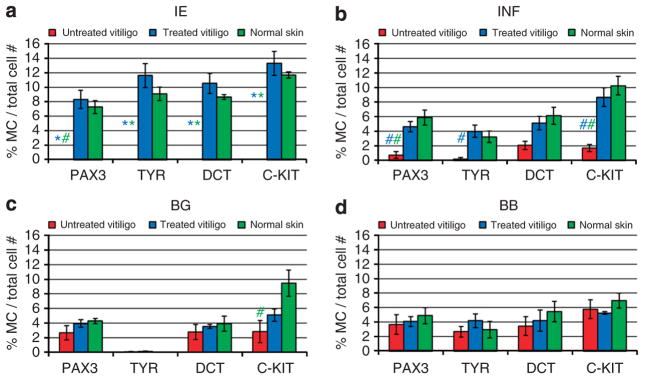
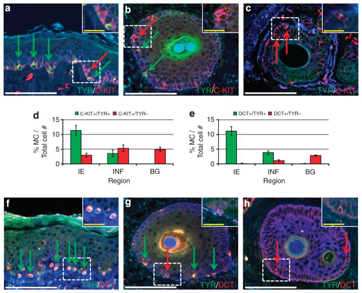
In vitiligo, the autoimmune destruction of epidermal melanocytes produces white spots that can be repigmented by melanocyte precursors from the hair follicles, following stimulation with UV light. We examined by immunofluorescence the distribution of melanocyte markers (C-KIT, DCT, PAX3, and TYR) coupled with markers of proliferation (KI-67) and migration (MCAM) in precursors and mature melanocytes from the hair follicle and the epidermis of untreated and narrow band UVB (NBUVB)-treated human vitiligo skin. NBUVB was associated with a significant increase in the number of melanocytes in the infundibulum and with restoration of the normal melanocyte population in the epidermis, which was lacking in the untreated vitiligo. We identified several precursor populations (melanocyte stem cells, melanoblasts, and other immature phenotypes), and progressively differentiating melanocytes, some with putative migratory and/or proliferative abilities. The primary melanocyte germ was present in the untreated and treated hair follicle bulge, whereas a possible secondary melanocyte germ composed of C-KIT+ melanocytes was found in the infundibulum and interfollicular epidermis of UV-treated vitiligo. This is an exceptional model for studying the mobilization of melanocyte stem cells in human skin. Improved understanding of this process is essential for designing better treatments for vitiligo, ultimately based on melanocyte stem cell activation and mobilization.


Conflict of interest statement

CONFLICT OF INTEREST

The authors state no conflict of interest.

Figures

Figure 1
Figure 1. Mapping the anatomic regions of the hair follicle (HF)
Anti-human K15 (green; labels epithelial stem cells) was paired with anti-human desmin (red; labels the arrector pili muscle (APM)); anti-human CD200 (green; labels epithelial stem cells) was immunostained by itself. HF infundibulum (INF) (a) was the region K15−/CD200− located between interfollicular epidermis (IE) and insertion of sebaceous gland (SG). Bulge (BG) (b and c) was the region K15+/CD200+ delineated by the SG duct superiorly and by the insertion of the APM inferiorly. The sub-BG (SBG) was located proximal to the bulge and distal to the bulb (BB). The bulb (d) was the region K15−/CD200− located at the base of HF, in which the dermal papilla (DP; delineated with blue line) was visible internally.
Figure 2
Figure 2. Graphical representation of melanocyte markers expression (PAX3, tyrosinase (TYR), dopachrome-tautomerase (DCT), and C-KIT)
Marker expression was tested in interfollicular epidermis (IE) (a), hair follicle infundibulum (INF) (b), hair follicle bulge (BG) (c), and hair follicle bulb (BB) (d) of untreated vitiligo skin (red bars), narrow band UVB (NBUVB)-treated vitiligo skin (blue bars), and normal control skin (green bars). Significant differences in expression were observed for all markers in the epidermis (a) and some markers in the INF (b) of untreated vitiligo skin, as compared with NBUVB-treated or with normal skin. No significant differences were observed in the BG (c) and BB (d) of untreated vitiligo skin, as compared with NBUVB-treated vitiligo skin or with normal skin, except for C-KIT expression in the untreated BG compared with normal skin. (P<1.0E-03 (*); 1.0E-03 ≤ P ≤ 5.0E-02 (#); all other comparisons showed P>5.0E-02 (not significant)). One-way analysis of variance with Tukey’s multiple comparisons tests were used for statistical comparison. Each bar represents mean ± SEM (n =4, untreated vitiligo patients; n = 8, NBUVB-treated vitiligo patients).
Figure 3
Figure 3. Distribution of C-KIT/tyrosinase (TYR) and dopachrome-tautomerase (DCT)/TYR populations in the interfollicular epidermis (IE) and hair follicles (HFs)
Narrow band UVB (NBUVB)-treated skin sections, showing expression of melanocyte markers: anti-TYR (green) combined with either anti-C-KIT (red) or anti-DCT (red). Anti-K14 (blue) labeled basal keratinocytes. The precursor phenotypes—(C-KIT+/TYR−) and (DCT+/TYR−)—are shown by red arrows and the differentiated phenotypes—(C-KIT+/TYR+) and (DCT+/TYR+)—by green arrows in the IE (a and f), infundibulum (INF) (b and g), and bulge (BG) (c and h). Graphical representation in the regions tested of percent (C-KIT+/TYR−) precursors (red bars) and (C-KIT+/TYR+) differentiated cells (green bars) (d); percent (DCT+/TYR−) precursors (red bars) and (DCT+/TYR+) differentiated cells (green bars) (e). Each bar represents mean ± SEM (n = 8, NBUVB-treated vitiligo patients). White bars =100 μm, yellow bars =25 μm.
Figure 4
Figure 4. Distribution of melanocyte stem cell and melanoblast phenotypes and their proliferative capacity
(a) Untreated vitiligo: graphical representation of percent of melanoblast phenotype (C-KIT+/DCT+) (ai), C-KIT+/DCT− phenotype (aii), and melanocyte stem cell phenotype (C-KIT−/DCT+) (aiii) per total melanocytes (MC) in interfollicular epidermis (IE), infundibulum (INF), and bulge (BG). The most prevalent in INF and BG were non-proliferative melanoblasts (DCT+/C-KIT+/KI-67−; yellow bars, ai) and non-proliferative stem cells (DCT+/C-KIT−/KI-67−; green bars, aiii). Small subpopulation of proliferating melanoblasts DCT+/C-KIT+/KI-67+ are shown in the BG (white bar, ai). Small proliferative population of melanocyte stem cells (DCT+/C-KIT−/KI-67+) shown in the BG and IFN (light-green bars, aiii). (b) Narrow band UVB (NBUVB)-treated vitiligo: representative images of immunostaining in the IE and hair follicle, showing expression of anti-KI-67 (blue) combined with anti-C-KIT (red) and anti-DCT (green). Red arrows in the INF (bii) and red bars (cii) indicate cells that expressed only C-KIT (non-proliferating MC). The blue arrow shows cells that expressed only the KI-67 marker (likely keratinocytes) (bi and bii). Yellow Bar =25 μm. (c) NBUVB-treated vitiligo: graphical representation of percent melanoblasts (C-KIT+/DCT+) (ci), C-KIT+/DCT− phenotype (cii), and the stem cells (DCT+/C-KIT−) (ciii) per total MC in IE, INF, and BG. The most prevalent phenotype observed in all three regions tested was non-proliferative (DCT+/C-KIT +/KI-67−) (bi, yellow arrow, and ci, yellow bars). A small population of proliferating MC DCT+/C-KIT+/KI-67+ decreased from the epidermal surface with the depth of the INF (ci, white bars, bi, white arrows). Each bar represents mean ± SEM (n = 5, untreated vitiligo patients; n =5, NBUVB-treated vitiligo patients).
Figure 5
Figure 5. Assessment of putatively migratory (melanoma cell adhesion molecule (MCAM+)), differentiating (tyrosinase (TYR)+), and proliferative (KI-67+) phenotypes
(a) Representative immunostaining pictures of narrow band UVB (NBUVB)-treated vitiligo showing expression of markers in the interfollicular epidermis (IE): KI-67 (blue image, blue arrows) combined with MCAM (red image, red arrows) and dopachrome-tautomerase (DCT) (green image, green arrows). Triple-positive cells (DCT+/MCAM+/KI-67+) are presented in the merged image (gray arrows), as well as one cell DCT+ only (green arrow). Yellow bar = 25 μm. (b) Graphical representation of the percent presumably migrating (MCAM+) and non-migrating (MCAM−) melanocytes co-expressing DCT and KI-67 (bi) or TYR and KI-67 (bii) per total melanocytes in IE, infundibulum (INF), and bulge (BG) of NBUVB-treated vitiligo skin. bi shows a large population of putatively non-migratory and non-proliferative DCT+/MCAM−/KI-67− cells in all three regions (green bars), an increasing population of presumably migrating but non-proliferating melanocytes (DCT+/MCAM+/KI-67−) from the BG to the IE (yellow bars), and a small population of putatively migrating proliferative melanocytes (DCT+/MCAM+/Ki-67+) highest in the IE (gray bars). bii shows similar trends for TYR. The BG was not analyzed for TYR as the melanocytes were TYR−. Each bar represents mean ± SEM. (c): Representative immunostaining images of NBUVB-treated vitiligo showing expression of markers in the INF: anti-KI-67 (blue image, blue arrows) combined with anti-MCAM (red image, red arrows) and anti-TYR (green image, green arrows). A single triple-positive cell is seen in the merged image (white arrow), as well as two TYR+ cells (green arrows) and a KI-67+ keratinocyte (blue arrow). Each bar represents mean ± SEM (n = 5, NBUVB-treated vitiligo patients). Yellow bar =25 μm.
Figure 6
Figure 6. Diagram of melanocyte precursors and more differentiated phenotypes in treated and untreated vitiligo
The precursors and more differentiated phenotypes are represented with different colors in the bulge (BG), infundibulum (INF), and interfollicular epidermis (IE). The relative diameter of each circle in each study region represents the estimated percent of melanocytes exhibiting particular phenotypes in each study region, normalized to the average number of melanocytes in each region (melanocyte percentages presented in the Figure 2 and Supplementary Table S1 online). Percentages of each phenotype and circle diameters are summarized in the Supplementary Tables S3 and S4 online.

References

    1. Birlea SA, Spritz RA, Norris DA. Vitiligo. In: Wolff K, Goldsmith LA, Katz SI, Gilchrest BA, Paller AS, Leffell DJ, editors. Fitzpatrick’s Dermatology in General Medicine. 8. McGraw-Hill; New York: 2012. pp. 792–803.
    1. Birlea SA, Serota M, Norris DA. Non-Bullous Skin Diseases: Alopecia Areata, Vitiligo, Psoriasis and Urticaria. In: Mackay, Rose, editors. Autoimmune Diseases. 5. Oxford, Elsevier Academic Press; London, UK: 2013. pp. 971–89.
    1. Box GE, Hunter JS, Hunter WG. Wiley, editor. Statistics for Experimenters: Design, Innovation, and Discovery. 2. John Wiley&Sons, Inc; Hoboken, New Jersey, USA: 2005.
    1. Chou WC, Takeo M, Rabbani P, et al. Direct migration of follicular melanocyte stem cells to the epidermis after wounding or UVB irradiation is dependent on Mc1r signaling. Nat Med. 2013;19:924–9. - PMC - PubMed
    1. Collins LM, Dziak JJ, Li R. Design of experiments with multiple independent variables: a resource management perspective on complete and reduced factorial designs. Psychol Methods. 2009;14:202–24. - PMC - PubMed

Publication types

MeSH terms

Substances