. 2015 Mar 30;10(3):e0120195.
doi: 10.1371/journal.pone.0120195. eCollection 2015.

Human gastroenteropancreatic expression of melatonin and its receptors MT1 and MT2

Affiliations

Human gastroenteropancreatic expression of melatonin and its receptors MT1 and MT2

Fanny Söderquist et al. PLoS One. .

Abstract

Background and aim: The largest source of melatonin, according to animal studies, is the gastrointestinal (GI) tract but this is not yet thoroughly characterized in humans. This study aims to map the expression of melatonin and its two receptors in human GI tract and pancreas using microarray analysis and immunohistochemistry.

Method: Gene expression data from normal intestine and pancreas and inflamed colon tissue due to ulcerative colitis were analyzed for expression of enzymes relevant for serotonin and melatonin production and their receptors. Sections from paraffin-embedded normal tissue from 42 individuals, representing the different parts of the GI tract (n=39) and pancreas (n=3) were studied with immunohistochemistry using antibodies with specificity for melatonin, MT1 and MT2 receptors and serotonin.

Results: Enzymes needed for production of melatonin are expressed in both GI tract and pancreas tissue. Strong melatonin immunoreactivity (IR) was seen in enterochromaffin (EC) cells partially co-localized with serotonin IR. Melatonin IR was also seen in pancreas islets. MT1 and MT2 IR were both found in the intestinal epithelium, in the submucosal and myenteric plexus, and in vessels in the GI tract as well as in pancreatic islets. MT1 and MT2 IR was strongest in the epithelium of the large intestine. In the other cell types, both MT2 gene expression and IR were generally elevated compared to MT1. Strong MT2, IR was noted in EC cells but not MT1 IR. Changes in gene expression that may result in reduced levels of melatonin were seen in relation to inflammation.

Conclusion: Widespread gastroenteropancreatic expression of melatonin and its receptors in the GI tract and pancreas is in agreement with the multiple roles ascribed to melatonin, which include regulation of gastrointestinal motility, epithelial permeability as well as enteropancreatic cross-talk with plausible impact on metabolic control.


Conflict of interest statement

Competing Interests: The authors have declared that no competing interests exist.

Figures

Fig 1
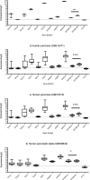
Fig 1. Normalized gastropancreatic gene expression data (log2) for key enzymes in tryptophan serotonin and melatonin metabolism and receptors for melatonin and serotonin.
a) Human small intestinal epithelium (GSE9576) b) Human pancreas (GSE15471) c) Human pancreas (GSE16515) d) Human pancreatic islets (GSE38642). MT2 expression levels exceed MT1 levels in small intestine, whole pancreas and islets. (* p<0.05, *** p<0.001, **** p<0.0001).
Fig 2
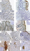
Fig 2. Immunohistochemical staining of gastrointestinal tract and pancreas tissue with antibodies against melatonin and receptors MT1 and MT2.
A) Strong MT1 receptor immunoreactivity (IR) in epithelium in pyloric mucosa. B) Strong MT2 receptor IR in epithelium and endocrine cell (arrow) in pyloric mucosa. C) Weak MT1 receptor IR in epithelium in ileal mucosa. D) MT2 receptor IR is negative in epithelial cells but strong in endocrine cells (arrow) in duodenal mucosa E) Strong MT1 receptor IR in epithelium of colon mucosa. Insert shows neutralization test for MT1. F) Strong MT2 receptor IR in epithelium and endocrine cells (arrow) in colon mucosa. Insert shows neutralization test for MT2. G) Strong melatonin IR in endocrine cells in pyloric mucosa. H) Strong melatonin IR in endocrine cells in ileal mucosa. I) Strong melatonin IR in epithelial cells and endocrine cells in colon mucosa. Insert shows neutralization test for melatonin. A-I: Magnification 100X. J) Strong melatonin IR in endocrine cells in pancreatic islets. Inset shows negative serotonin IR. K) Strong MT1 receptor IR in endocrine cells in pancreatic islets. L) Strong MT2 receptor IR in endocrine cells in pancreatic islets and pancreatic ducts. (J-K: Magnification 200X).
Fig 3
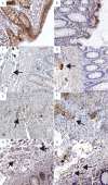
Fig 3. Immunohistochemical staining of melatonin receptors in specific cell types.
A) Positive melatonin receptor 1 (MT1) immunoreactivity (IR) in epithelial cells. B) Melatonin receptor 2 (MT2) IR in epithelial cells. C) Arrow indicates negative MT1 IR in the submucosal plexus. D) Arrow indicates positive MT2 IR in the submucosal plexus. E) Arrow indicates a cell showing positive MT1 IR in the myenteric plexus; muscle cells are negative. F) Large arrow indicates positive MT2 IR in the myenteric plexus, small arrow indicates positive IR in muscle tissue. G) Large arrow indicates weak MT1 IR in the endothelium of arterioles and venules. H) Large arrow indicates MT2 IR in the endothelium and smooth muscle of arterioles and venules. Magnification 200X.
Fig 4
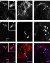
Fig 4. Confocal images of double immunofluorescence staining of crypts of Lieberkühn in ileum mucosa.
A: Melatonin immunoreactive (IR) cells. B: Serotonin IR cells. C: Merge A-C; Inset: magnification of cell in circle. Arrow indicates structures positive for serotonin but not melatonin. Square indicates cell where melatonin IR is strong compared to serotonin IR. D: Melatonin receptor MT1 IR in crypt epithelium. E: Serotonin IR cell. F: Merge D-F; Arrow indicates serotonin IR cell negative for melatonin receptor MT1. G: Melatonin receptor MT2 IR. H: Serotonin IR cells. I: Merge GI: Arrow indicates serotonin IR cell positive for melatonin receptor MT2. White bar indicates 20 μm.

References

    1. Slominski RM, Reiter RJ, Schlabritz-Loutsevitch N, Ostrom RS, Slominski AT. Melatonin membrane receptors in peripheral tissues: distribution and functions. Mol Cell Endocrinol, 2012. 351(2): p. 152–66. 10.1016/j.mce.2012.01.004 - DOI - PMC - PubMed
    1. Ramracheya RD, Muller DS, Squires PE, Brereton H, Sugden D, Huang GC, et al. Function and expression of melatonin receptors on human pancreatic islets. J Pineal Res, 2008. 44(3): p. 273–9. 10.1111/j.1600-079X.2007.00523.x - DOI - PubMed
    1. Lyssenko V, Nagorny CL, Erdos MR, Wierup N, Jonsson A, Spegel P, et al. Common variant in MTNR1B associated with increased risk of type 2 diabetes and impaired early insulin secretion. Nat Genet, 2009. 41(1): p. 82–8. 10.1038/ng.288 - DOI - PMC - PubMed
    1. Bahr I, Muhlbauer E, Schucht H, Peschke E. Melatonin stimulates glucagon secretion in vitro and in vivo. J Pineal Res, 2011. 50(3): p. 336–44. 10.1111/j.1600-079X.2010.00848.x - DOI - PubMed
    1. Calvo JR, Gonzalez-Yanes C, Maldonado MD. The role of melatonin in the cells of the innate immunity: a review. J Pineal Res, 2013. 55(2): p. 103–20. 10.1111/jpi.12075 - DOI - PubMed

Publication types

MeSH terms

Associated data

LinkOut - more resources