. 2015 Jun 18;522(7556):345-348.
doi: 10.1038/nature14282. Epub 2015 Mar 30.

IL-17-producing γδ T cells and neutrophils conspire to promote breast cancer metastasis

Affiliations

IL-17-producing γδ T cells and neutrophils conspire to promote breast cancer metastasis

Seth B Coffelt et al. Nature. .

Abstract

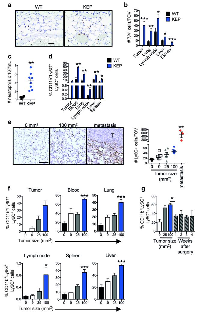
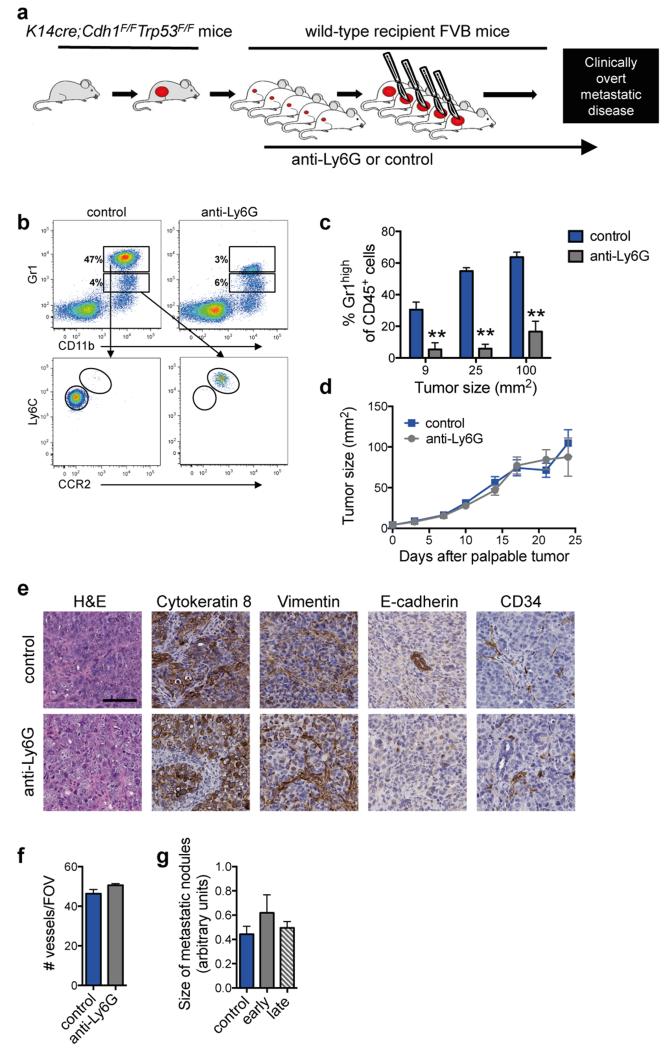
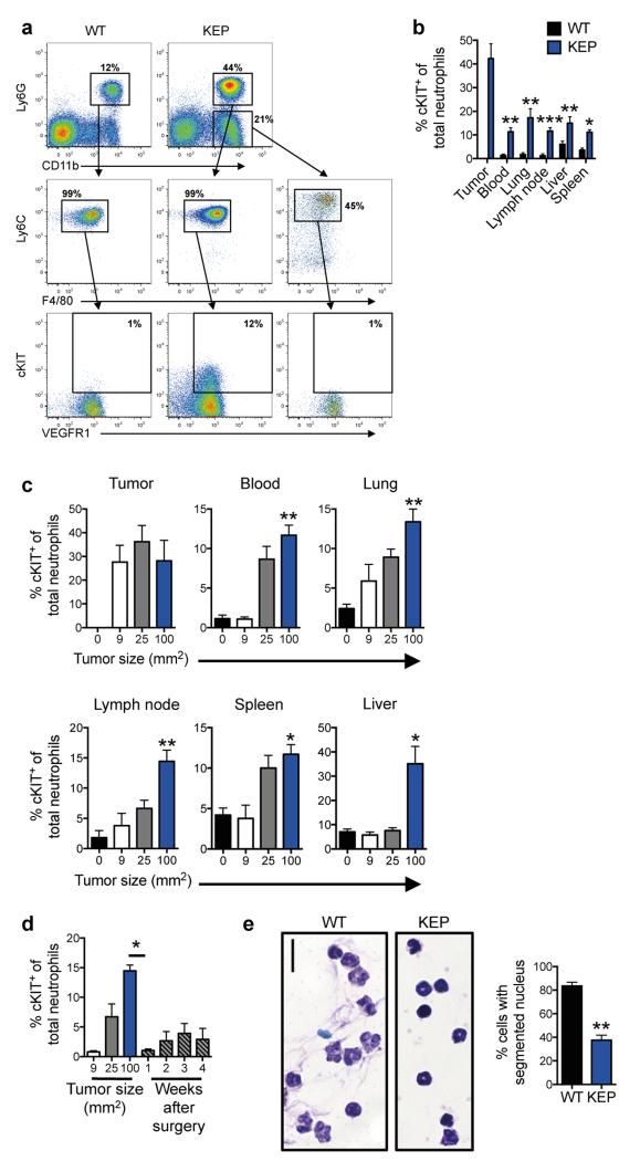
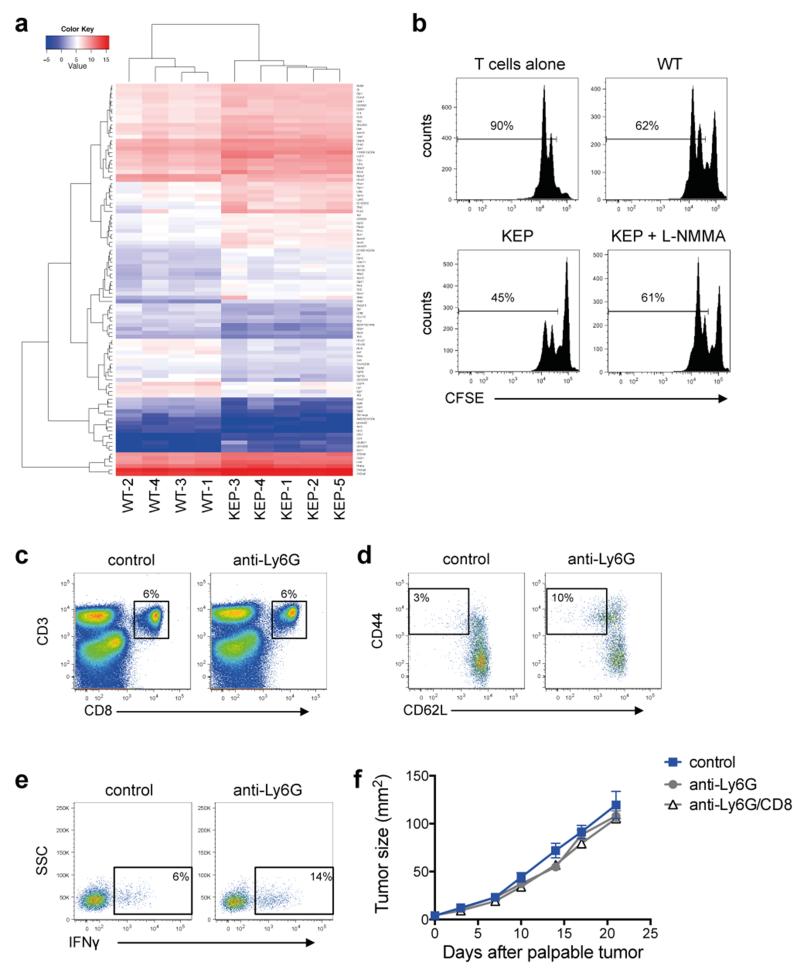
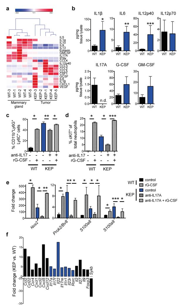
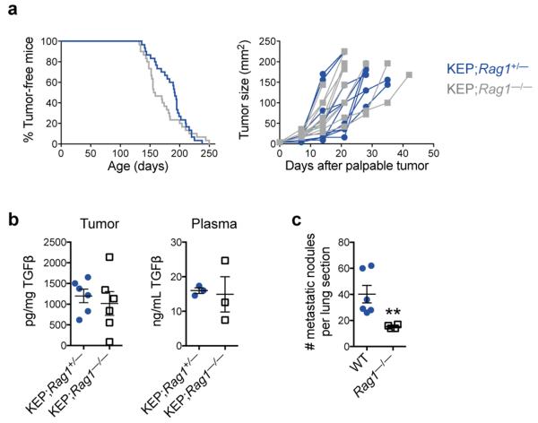
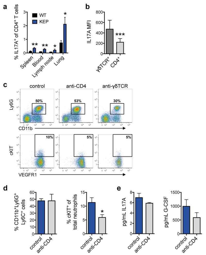
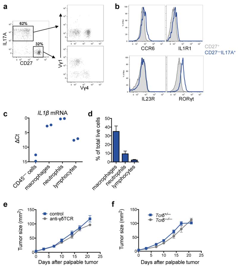
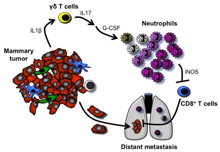
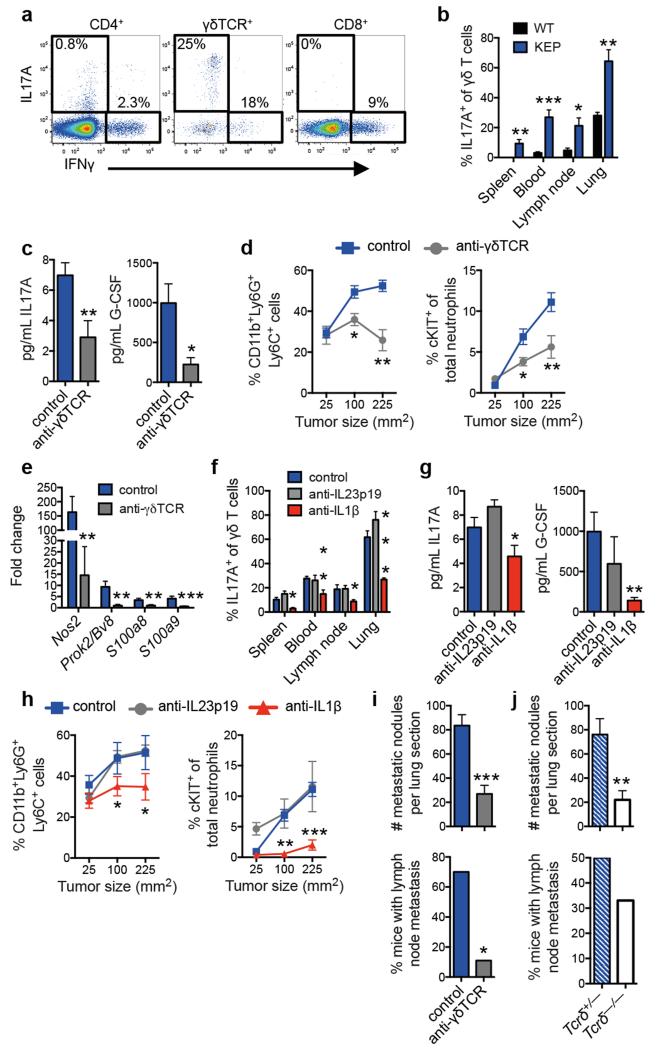
Metastatic disease remains the primary cause of death for patients with breast cancer. The different steps of the metastatic cascade rely on reciprocal interactions between cancer cells and their microenvironment. Within this local microenvironment and in distant organs, immune cells and their mediators are known to facilitate metastasis formation. However, the precise contribution of tumour-induced systemic inflammation to metastasis and the mechanisms regulating systemic inflammation are poorly understood. Here we show that tumours maximize their chance of metastasizing by evoking a systemic inflammatory cascade in mouse models of spontaneous breast cancer metastasis. We mechanistically demonstrate that interleukin (IL)-1β elicits IL-17 expression from gamma delta (γδ) T cells, resulting in systemic, granulocyte colony-stimulating factor (G-CSF)-dependent expansion and polarization of neutrophils in mice bearing mammary tumours. Tumour-induced neutrophils acquire the ability to suppress cytotoxic T lymphocytes carrying the CD8 antigen, which limit the establishment of metastases. Neutralization of IL-17 or G-CSF and absence of γδ T cells prevents neutrophil accumulation and downregulates the T-cell-suppressive phenotype of neutrophils. Moreover, the absence of γδ T cells or neutrophils profoundly reduces pulmonary and lymph node metastases without influencing primary tumour progression. Our data indicate that targeting this novel cancer-cell-initiated domino effect within the immune system--the γδ T cell/IL-17/neutrophil axis--represents a new strategy to inhibit metastatic disease.


Figures

Extended Data Figure 1
Extended Data Figure 1. Systemic neutrophil expansion and accumulation in mammary tumor-bearing K14cre;Cdh1F/F;Trp53F/F (KEP) mice and the metastasis model
a, Representative images of neutrophils identified by the 7/4 antibody in lung sections in wild-type (WT) or KEP mice. Scale bar = 50 μm. b, Quantification of neutrophil accumulation per field of view (FOV) in various organs by immunohistochemistry using the 7/4 antibody (n = 6 WT, 9 KEP mice). c, Absolute neutrophil counts in blood of WT and tumor-bearing KEP mice (n = 4 WT, 8 KEP). d, Quantification of neutrophil accumulation in various organs determined by flow cytometry and gated on CD45+ cells. Neutrophils were not detectable in WT mammary glands (n = 5 WT, 7 KEP mice). e, Representative images of Ly6G-stained lung sections and quantification of neutrophil accumulation in the metastasis model. Data were generated from mock-transplanted, non-tumor-bearing mice (0 mm2), or tumor-transplanted recipient mice sacrificed when tumors reached the tumor size shown or when mice exhibited signs of respiratory distress due to pulmonary metastasis. For quantification in lungs with metastases, neutrophils residing inside metastases were excluded. T = pulmonary metastatic lesion. Scale bar = 100 μm (n = 3, 5, 6, 6 and 3 mice for 0, 9, 25, 100 mm2 and metastasis respectively). f, Kinetics of neutrophil accumulation in various organs of the metastasis model by flow cytometry after gating on CD45+ cells. Recipient mice transplanted with KEP tumor pieces were sacrificed at the tumor size shown (n = 6, 5, 6, and 7 mice for 0, 9, 25, 100 mm2 respectively). g, Kinetics of neutrophil proportions in blood (gated on CD45+ cells), before and after surgical removal of their primary tumor (n = 5). All data are mean + s.e.m. *p<0.05, **p<0.01, ***p<0.001 as determined by Mann-Whitney U test or Kruskal-Wallis test followed by Dunn’s post test.
Extended Data Figure 2
Extended Data Figure 2. Neutrophil depletion does not affect primary tumor or metastatic nodule growth
a, Schematic illustration of the neutrophil depletion experiment in the spontaneous metastasis model. b, Representative dot plots of neutrophils gated on CD45+ cells in blood of control and anti-Ly6G-treated recipient mice. The Gr1 antibody was used here to avoid false negative results since the anti-Ly6G depleting antibody may mask the Ly6G epitope. CD11b+Gr1high cells were Ly6C+CCR2, indicating that these cells were neutrophils. CD11b+Gr1low cells that were Ly6C+ and CCR2+ represented the monocytic fraction. c, Quantification of neutrophil depletion in blood of control and anti-Ly6G-treated recipient mice at the tumor size indicated (n = 8 control, 5 anti-Ly6G; **p<0.01 as determined by Mann-Whitney U test). d, Primary tumor growth kinetics of mice treated as indicated (n = 12 control, 14 anti-Ly6G). e, Representative images of primary tumors in the metastasis model treated as shown and stained with H&E, cytokeratin 8, vimentin, E-cadherin and CD34. Scale bar = 100 μm. f, Quantification of blood vessels per field of view (FOV) in control and neutrophil-depleted mice by anti-CD34 immunohistochemistry (n = 10). g, Quantification of pulmonary metastatic nodule size in control and neutrophil-depleted mice (n = 9 control, 11 early phase, 14 late phase mice). All data are mean + s.e.m.
Extended Data Figure 3
Extended Data Figure 3. Subpopulations of neutrophils in mammary tumor-bearing mice are immature
a, Gating strategy for identification of neutrophils (CD45+CD11b+Ly6G+Ly6C+F4/80 cells), cKIT+ neutrophils and monocytes (CD45+CD11b+Ly6GLy6C+F4/80 cells) by flow cytometry. Blood cells from WT and tumor-bearing KEP mice are shown here. b, Quantification of cKIT+ neutrophil accumulation in various organs determined by flow cytometry after gating on CD45+CD11b+Ly6G+Ly6C+F4/80 cells. cKIT+ neutrophils were not detectable in WT mammary glands (n = 5 WT, 7 KEP; Mann-Whitney U test). c, cKIT+ neutrophil proportions in various organs of the metastasis model as determined by flow cytometry after gating on CD45+CD11b+Ly6G+Ly6C+F4/80 cells. Mice were sacrificed at the tumor size shown (n = 5, 5, 5, and 8 mice for 0, 9, 25, 100 mm2 respectively; Kruskal–Wallis test followed by Dunn’s post test). d, Kinetics of cKIT+ neutrophil proportions in blood (gated on CD45+CD11b+Ly6G+Ly6C+F4/80 cells), before and after surgical removal of their primary tumor (n = 5/group; Mann-Whitney U test). e, Representative images and quantification of neutrophil nuclear morphology. Ly6G+ cells were isolated from blood of WT and tumor-bearing KEP mice then assessed by Giemsa stain. Hyper-segmented cells were considered mature, whereas all other cells were considered immature. Scale bar = 10 μm. (n = 6 WT, 5 KEP mice; Mann-Whitney U test). All data are mean + s.e.m. *p<0.05, **p<0.01, ***p<0.001.
Extended Data Figure 4
Extended Data Figure 4. Neutrophils influence the function and phenotype of CD8+ T cells
a, Unsupervised hierarchical clustering of RNA-Seq analysis depicting 100 differentially expressed genes between circulating neutrophils from WT and tumor-bearing KEP mice. P value (0.05) was used as cutoff (n = 4 WT, 5 KEP mice). See also Extended Data Table 1 for top 50 genes ranked by fold change. b, Circulating neutrophils from either WT or tumor-bearing KEP mice were incubated with CFSE-labeled splenic CD8+ T cells from WT mice and CD3/CD28 stimulation beads. The iNOS inhibitor, L-NMMA, was added where indicated. After 48 hours, CD8+ T cell proliferation was measured by flow cytometry. c, Dot plots depicting live cell-gated CD8+ T cell proportions in lungs of mice in control and neutrophil-depleted mice, sacrificed when transplanted tumors reached 100 mm2. d, Dot plots of effector CD8+ T cell (CD62LCD44+) proportions in lungs of transplanted mammary tumor-bearing mice that were sacrificed when tumors reached 100 mm2. e, IFNγ expression by CD8+ T cells in lungs of transplanted mammary tumor-bearing mice that were sacrificed when tumors reached 100 mm2. f, Tumor growth kinetics in neutrophil-depleted or combined neutrophil- and CD8+ T cell-depleted, mammary tumor-transplanted recipient mice, as compared with control (n = 13 control, 21 anti-Ly6G, 14 anti-Ly6G/CD8). Data are mean + s.e.m.
Extended Data Figure 5
Extended Data Figure 5. Cytokine expression levels in tumors and T cells, and their effects on neutrophils
a, Unsupervised clustering of cytokine expression analysis in WT mammary glands and KEP tumors. Protein lysates were prepared as previously described from whole tissue and analyzed for expression of various cytokines by Luminex-based assay (n = 5/group). b, Protein levels of indicated cytokines in WT mammary glands and KEP tumors, determined by Luminex-based cytokine profiling. n.d. = not detectable (n = 10/group; Mann-Whitney U test). c, d, Quantification of neutrophil and cKIT-expressing neutrophil accumulation in blood as determined by flow cytometry and gated on CD45+ cells. WT (n = 4) or tumor-bearing KEP mice (n = 9) were treated with anti-IL17 (n = 8) and/or recombinant G-CSF (rG-CSF; n = 4) where indicated (Mann-Whitney U test or Kruskal–Wallis test followed by Dunn’s post test). e, Gene expression in circulating neutrophils from WT control (n = 5), rG-CSF-treated WT mice (n = 4), KEP control (n = 10), anti-IL17-treated (n = 6), anti-IL17 + rG-CSF-treated KEP mice (n = 4; Mann-Whitney U test or Kruskal-Wallis test followed by Dunn’s post test). f, Spleens of three WT mice and three KEP mice were pooled and CD3+ T cells were isolated. These cells were analyzed by a real-time PCR array containing 86 different genes. Gene expression changes of greater than three-fold are shown. Members of the IL17 signaling pathway are depicted in blue. *p<0.05, **p<0.01, ***p<0.001. All data are mean + s.e.m.
Extended Data Figure 6
Extended Data Figure 6. Absence of the adaptive immune system reduces metastasis
a, Graphic representation of mammary tumor latency (left panel) and tumor growth (right panel) in lymphocyte-proficient KEP;Rag1+/− and lymphocyte-deficient KEP;Rag1−/− mice (n = 30/group). b, Levels of TGFβ1 in mammary tumors and the plasma of tumor-bearing mice (n = 6 tumor, 3 plasma). c, Quantification of metastatic burden in lungs of recipient WT or Rag1−/− mice that were transplanted with KEP mammary tumor fragments and underwent surgical removal of the primary tumor (n = 6 WT, 4 Rag1−/− mice; **p<0.01, Mann-Whitney U test). Data are mean + s.e.m.
Extended Data Figure 7
Extended Data Figure 7. Depletion of CD4+ T cells does not affect systemic cytokine levels or neutrophil expansion
a, The proportion of IL17A+ cells among CD4+ T cells in organs of wild-type (WT) and tumor-bearing K14cre;Cdh1F/F;Trp53F/F (KEP) mice (n = 6/group; Mann-Whitney U test). b, Median fluorescence intensity (MFI) of IL17A expression in circulating γδ and CD4+ T cells from tumor-bearing KEP mice, as determined by flow cytometry (n = 11/group; Wilcoxon matched-pairs test). c, Representative dot plots depicting total neutrophil and cKIT+ proportions in blood of control, anti-CD4- and anti-γδTCR-treated tumor-bearing KEP mice. d, Quantification of total neutrophil and cKIT+ neutrophil proportions in blood of control and anti-CD4-treated tumor-bearing KEP mice (n = 7/group; Mann-Whitney U test). e, Serum levels of IL17A and G-CSF in control and anti-CD4-treated tumor-bearing KEP mice (n = 10 control, 6 anti-CD4; Mann-Whitney U test). *p<0.05, **p<0.01. All data are mean + s.e.m.
Extended Data Figure 8
Extended Data Figure 8. γδ T cell phenotype in KEP mice and their lack of influence on tumor growth in the metastasis model
a, γδ T cells from lungs of tumor-bearing KEP mice were analyzed by flow cytometry for IL17, CD27, Vγ1, and Vγ4 expression. Two major populations of γδ T cells were observed including IL17+CD27 and IL17CD27+ cells. b, Representative histograms of CCR6, IL1R1, IL23R and RORγt expression in IL17+CD27 and IL17CD27+ γδ T cell populations shown in a. c, Il1β gene expression in various cell populations isolated from transplanted KEP tumors. Tumors from three mice were pooled to form one group. CD45 cells (which includes cancer cells, endothelial cells and fibroblasts), CD45+CD11b+F4/80+ macrophages, CD45+CD11b+Ly6G+ neutrophils and CD45+CD11b lymphocytes were sorted from two pooled groups. Real time-PCR was performed on individual cell populations for Il1β expression. Relative expression among different cells is shown. d, Graphic representation of immune cell proportions in KEP tumors (n = 4). e, Primary tumor growth kinetics of control and γδ T cell-depleted tumor transplant recipient mice (n = 13/group). f, Growth kinetics of primary tumors transplanted into Tcrδ+/− (n = 10) and Tcrδ−/− mice (n = 6). All data are mean + s.e.m.
Extended Data Figure 9
Extended Data Figure 9. The γδ T cell-IL17-neutrophil axis promotes metastatic breast cancer
Mammary tumors evoke a systemic inflammatory cascade that is initiated by IL1β production. Tumor-derived IL1β activates γδ T cells to produce IL17. Increased systemic IL17 levels lead to upregulation of G-CSF, which subsequently causes neutrophil expansion and alteration of neutrophil phenotype. These phenotypically altered neutrophils produce iNOS that suppresses the activity of anti-tumor CD8+ T cells. As a result of this systemic inflammatory cascade, the chance that disseminated cancer cells can establish metastases in distant organs is maximized.
Figure 1
Figure 1. Neutrophils promote breast cancer metastasis
a, Spontaneous metastasis model. Tumor fragments from KEP mice are orthotopically transplanted into WT recipient mice (designated by #1), allowed to proliferate (#2), then surgically resected (#3). Metastases develop in 100% of recipient mice. Antibody-mediated depletion experiments were performed in three ways: from palpable tumors to metastasis-related sacrifice (continuous treatment), during primary tumor growth (early phase), or after surgery until metastasis-related sacrifice (late phase). b, Neutrophil proportions in lungs at the indicated tumor size (n = 6, 5, 6 and 8 mice for 0, 9, 25 and 100 mm2, respectively; Kruskal-Wallis test followed by Dunn’s post test). c, d, Images of cytokeratin 8-stained lung sections, quantification of lung metastases and incidence of metastasis in lymph nodes. Neutrophils were depleted continuously until metastasis-related sacrifice in c (n = 11 mice/group; Mann-Whitney U test and Fisher’s exact test) or depleted during the early or late phases in d (n = 9 control, 11 early phase, 14 late phase; Kruskal-Wallis test followed by Dunn’s post test and Fisher’s exact test). Data in d are representative of two independent experiments. All data are mean + s.e.m. *p<0.05, **p<0.01, ***p<0.001. Scale bars = 6 mm.
Figure 2
Figure 2. Neutrophils suppress CD8+ T cell activation to facilitate metastasis
a, Gene expression in circulating neutrophils (n = 5 WT, 10 KEP mice). b, Circulating neutrophils from either WT (n = 7) or tumor-bearing KEP mice (n = 8) were incubated with CFSE-labeled splenic CD8+ T cells from WT mice and CD3/CD28 stimulation beads. The iNOS inhibitor, L-NMMA, was added where indicated (n = 8). After 48 hours, CD8+ T cell proliferation was measured. c, CD8+ T cell activation status in lungs of transplanted tumor-bearing control and neutrophil-depleted mice (n = 6/group). d, Quantification of lung metastases and incidence of lymph node metastasis following neutrophil and CD8+ T cell depletion (n = 11 control, 16 anti-Ly6G, 8 anti-Ly6G/CD8; Kruskal-Wallis test followed by Dunn’s post test and Fisher’s exact test). All data are mean + s.e.m. *p<0.05, **p<0.01, ***p<0.001.
Figure 3
Figure 3. Lymphocyte-derived IL17 is required for G-CSF-induced neutrophil expansion and phenotype
a, Cytokine levels in serum of WT (n = 5), tumor-bearing KEP mice (n = 9), and anti-IL17- (n = 7) or anti-G-CSF-treated KEP mice (n = 6). b, Proportions of circulating neutrophils and cKIT-expressing neutrophils in KEP mice during primary tumor growth (n = 9 control, 8 anti-IL17, 6 anti-G-CSF). c, Gene expression in circulating neutrophils from tumor-bearing KEP control mice (n = 9), anti-IL17- (n = 6) or anti-G-CSF-treated KEP mice (n = 6). d, Cytokine levels in serum of tumor-bearing KEP;Rag1+/− (n = 9) and KEP;Rag1−/− mice (n = 7). e, Absolute blood neutrophil counts in tumor-bearing KEP;Rag1+/− mice (n = 8) or KEP;Rag1−/− mice (n = 5). f, Gene expression in circulating neutrophils from KEP;Rag1+/− (n = 10) and KEP;Rag1−/− mice (n = 8). g, Percentage of tumor-bearing mice with lung or lymph node metastasis (n = 50 KEP;Rag1+/−, 32 KEP;Rag1−/− mice). All data are mean + s.e.m. *p<0.05, **p<0.01, ***p<0.001 as determined by Mann-Whitney U test or Fisher’s exact test.
Figure 4
Figure 4. IL1β-activated, IL17-producing γδ T cells regulate neutrophil expansion, neutrophil phenotype and metastasis
a, Intracellular staining within circulating T cells of tumor-bearing KEP mice. b, Proportion of IL17A-producing γδ T cells (WT, n = 5; KEP, n = 6). c, Cytokine levels in serum of control (n = 10) and anti-γδTCR-treated (n = 7) KEP mice. d, Proportions of circulating neutrophils and cKIT-expressing neutrophils in KEP mice during primary tumor growth (n = 8/group). e, Gene expression in circulating neutrophils from tumor-bearing KEP control mice (n = 10) and anti-γδTCR-treated KEP mice (n = 6). f, Proportion of IL17A-producing γδ T cells in tumor-bearing mice (n = 6 KEP control, 5 anti-IL23p19, 5 anti-IL1β). g, Cytokine levels in serum (n = 9 KEP control, 5 anti-IL23p19, 6 anti-IL1β). h, Proportions of circulating neutrophils and cKIT-expressing neutrophils in KEP mice during primary tumor growth (n = 9 control, 5 anti-IL23p19, 5 anti-IL1β). i, j, Quantification of lung metastases and incidence of lymph node metastasis in the metastasis model (n = 10 control, 9 anti-γδTCR-treated mice; n = 9/group Tcrδ+/− and Tcrδ−/− mice,). All data are mean + s.e.m. *p<0.05, **p<0.01, ***p<0.001 as determined by Mann-Whitney U test or Fisher’s exact test.

Comment in

Similar articles

Cited by

References

    1. Quail DF, Joyce JA. Microenvironmental regulation of tumor progression and metastasis. Nat Med. 2013;19:1423–1437. - PMC - PubMed
    1. McAllister SS, Weinberg RA. The tumour-induced systemic environment as a critical regulator of cancer progression and metastasis. Nat Cell Biol. 2014;16:717–727. - PMC - PubMed
    1. Noh H, Eomm M, Han A. Usefulness of pretreatment neutrophil to lymphocyte ratio in predicting disease-specific survival in breast cancer patients. J Breast Cancer. 2013;16:55–59. - PMC - PubMed
    1. Azab B, et al. Usefulness of the neutrophil-to-lymphocyte ratio in predicting short- and long-term mortality in breast cancer patients. Ann Surg Oncol. 2012;19:217–224. - PubMed
    1. Granot Z, et al. Tumor entrained neutrophils inhibit seeding in the premetastatic lung. Cancer Cell. 2011;20:300–314. - PMC - PubMed

ADDITIONAL REFERENCES

    1. Ciampricotti M, et al. Development of metastatic HER2(+) breast cancer is independent of the adaptive immune system. J Pathol. 2011;224:56–66. - PubMed
    1. de Visser KE, Korets LV, Coussens LM. De novo carcinogenesis promoted by chronic inflammation is B lymphocyte dependent. Cancer Cell. 2005;7:411–423. - PubMed
    1. Ciampricotti M, Hau CS, Doornebal CW, Jonkers J, de Visser KE. Chemotherapy response of spontaneous mammary tumors is independent of the adaptive immune system. Nat Med. 2012;18:344–346. author reply 346. - PubMed
    1. Girardi M, et al. Regulation of cutaneous malignancy by gammadelta T cells. Science. 2001;294:605–609. - PubMed

Publication types

MeSH terms

Substances

Associated data