

Multi-layer electrospun membrane mimicking tendon sheath for prevention of tendon adhesions
- PMID: 25822877
- PMCID: PMC4424997
- DOI: 10.3390/ijms16046932
Multi-layer electrospun membrane mimicking tendon sheath for prevention of tendon adhesions
Abstract
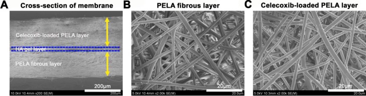
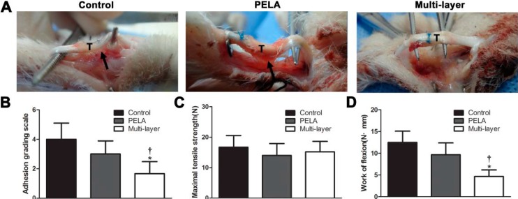
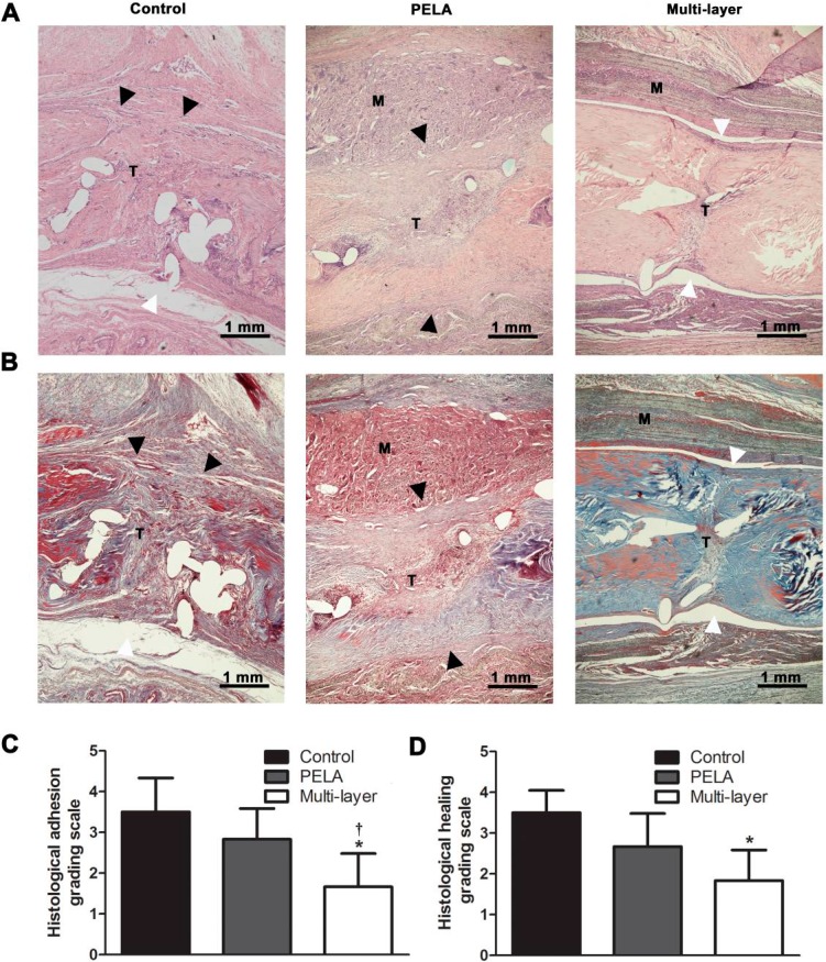
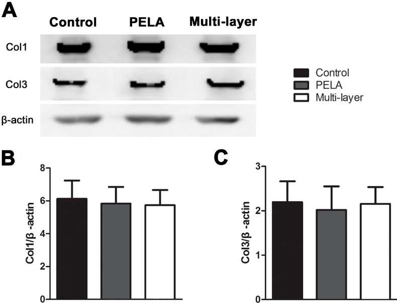
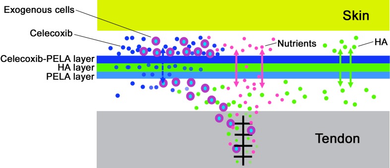
Defect of the tendon sheath after tendon injury is a main reason for tendon adhesions, but it is a daunting challenge for the biomimetic substitute of the tendon sheath after injury due to its multi-layer membrane-like structure and complex biologic functions. In this study, a multi-layer membrane with celecoxib-loaded poly(l-lactic acid)-polyethylene glycol (PELA) electrospun fibrous membrane as the outer layer, hyaluronic acid (HA) gel as middle layer, and PELA electrospun fibrous membrane as the inner layer was designed. The anti-adhesion efficacy of this multi-layer membrane was compared with a single-layer use in rabbit flexor digitorum profundus tendon model. The surface morphology showed that both PELA fibers and celecoxib-loaded PELA fibers in multi-layer membrane were uniform in size, randomly arrayed, very porous, and smooth without beads. Multi-layer membrane group had fewer peritendinous adhesions and better gliding than the PELA membrane group and control group in gross and histological observation. The similar mechanical characteristic and collagen expression of tendon repair site in the three groups indicated that the multi-layer membrane did not impair tendon healing. Taken together, our results demonstrated that such a biomimetic multi-layer sheath could be used as a potential strategy in clinics for promoting tendon gliding and preventing adhesion without poor tendon healing.
Figures
Similar articles
-
Release of celecoxib from a bi-layer biomimetic tendon sheath to prevent tissue adhesion.Mater Sci Eng C Mater Biol Appl. 2016 Apr 1;61:220-6. doi: 10.1016/j.msec.2015.12.028. Epub 2015 Dec 17. Mater Sci Eng C Mater Biol Appl. 2016. PMID: 26838844
-
Biomimetic sheath membrane via electrospinning for antiadhesion of repaired tendon.Biomacromolecules. 2012 Nov 12;13(11):3611-9. doi: 10.1021/bm301022p. Epub 2012 Oct 17. Biomacromolecules. 2012. PMID: 23025492
-
Coaxial Electrospun Nanofibrous Membranes as Dual-Functional Biomimetic Tendon Sheath for Tendon Repair and Anti-Peritendinous Adhesion.Adv Healthc Mater. 2025 Jan;14(1):e2402074. doi: 10.1002/adhm.202402074. Epub 2024 Nov 26. Adv Healthc Mater. 2025. PMID: 39600050
-
[Research progress of mechanism and prevention of peritendinous adhesions].Zhongguo Xiu Fu Chong Jian Wai Ke Za Zhi. 2013 May;27(5):633-6. Zhongguo Xiu Fu Chong Jian Wai Ke Za Zhi. 2013. PMID: 23879107 Review. Chinese.
-
Electrospun Nanofibrous Membranes for Preventing Tendon Adhesion.ACS Biomater Sci Eng. 2020 Aug 10;6(8):4356-4376. doi: 10.1021/acsbiomaterials.0c00201. Epub 2020 Jul 17. ACS Biomater Sci Eng. 2020. PMID: 33455173 Review.
Cited by
-
Nanofiber Graft Therapy to Prevent Shoulder Stiffness and Adhesions after Rotator Cuff Tendon Repair: A Comprehensive Review.Biomedicines. 2024 Jul 19;12(7):1613. doi: 10.3390/biomedicines12071613. Biomedicines. 2024. PMID: 39062186 Free PMC article. Review.
-
Polymer-Based Constructs for Flexor Tendon Repair: A Review.Polymers (Basel). 2022 Feb 23;14(5):867. doi: 10.3390/polym14050867. Polymers (Basel). 2022. PMID: 35267690 Free PMC article. Review.
-
Advances in the application of hydrogel-based scaffolds for tendon repair.Genes Dis. 2023 Jul 7;11(4):101019. doi: 10.1016/j.gendis.2023.04.039. eCollection 2024 Jul. Genes Dis. 2023. PMID: 38560496 Free PMC article. Review.
-
Bletilla striata Polysaccharide-Containing Carboxymethyl Cellulose Bilayer Structure Membrane for Prevention of Postoperative Adhesion and Achilles Tendon Repair.Biomacromolecules. 2024 Sep 9;25(9):5786-5797. doi: 10.1021/acs.biomac.4c00463. Epub 2024 Jun 27. Biomacromolecules. 2024. PMID: 38935055 Free PMC article.
-
Functional Hyaluronic Acid-Polylactic Acid/Silver Nanoparticles Core-Sheath Nanofiber Membranes for Prevention of Post-Operative Tendon Adhesion.Int J Mol Sci. 2021 Aug 16;22(16):8781. doi: 10.3390/ijms22168781. Int J Mol Sci. 2021. PMID: 34445516 Free PMC article.
References
-
- Sharma P., Maffulli N. Biology of tendon injury: Healing, modeling and remodeling. J. Musculoskelet. Neuronal Interact. 2006;6:181–190. - PubMed
Publication types
MeSH terms
Substances
LinkOut - more resources
Full Text Sources
Other Literature Sources
Medical
