. 2015 Mar 30;10(3):e0120674.
doi: 10.1371/journal.pone.0120674. eCollection 2015.

Deficiency of angulin-2/ILDR1, a tricellular tight junction-associated membrane protein, causes deafness with cochlear hair cell degeneration in mice

Affiliations

Deficiency of angulin-2/ILDR1, a tricellular tight junction-associated membrane protein, causes deafness with cochlear hair cell degeneration in mice

Tomohito Higashi et al. PLoS One. .

Abstract

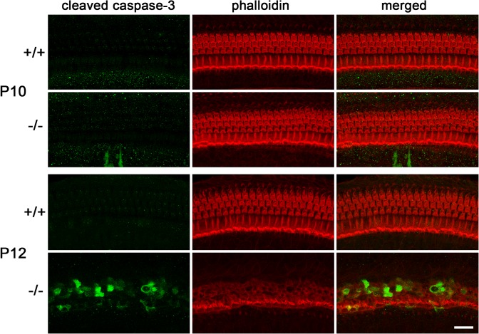
Tricellular tight junctions seal the extracellular spaces of tricellular contacts, where the vertices of three epithelial cells meet, and are required for the establishment of a strong barrier function of the epithelial cellular sheet. Angulins and tricellulin are known as specific protein components of tricellular tight junctions, where angulins recruit tricellulin. Mutations in the genes encoding angulin-2/ILDR1 and tricellulin have been reported to cause human hereditary deafness DFNB42 and DFNB49, respectively. To investigate the pathogenesis of DFNB42, we analyzed mice with a targeted disruption of Ildr1, which encodes angulin-2/ILDR1. Ildr1 null mice exhibited profound deafness. Hair cells in the cochlea of Ildr1 null mice develop normally, but begin to degenerate by two weeks after birth. Tricellulin localization at tricellular contacts of the organ of Corti in the cochlea was retained in Ildr1 null mice, but its distribution along the depth of tricellular contacts was affected. Interestingly, compensatory tricellular contact localization of angulin-1/LSR was observed in the organ of Corti in Ildr1 null mice although it was hardly detected in the organ of Corti in wild-type mice. The onset of hair cell degeneration in Ildr1 null mice was earlier than that in the reported Tric mutant mice, which mimic one of the tricellulin mutations in DFNB49 deafness. These results indicate that the angulin-2/ILDR1 deficiency causes the postnatal degenerative loss of hair cells in the cochlea, leading to human deafness DFNB42. Our data also suggest that angulin family proteins have distinct functions in addition to their common roles of tricellulin recruitment and that the function of angulin-2/ILDR1 for hearing cannot be substituted by angulin-1/LSR.


Conflict of interest statement

Competing Interests: The authors have declared that no competing interests exist.

Figures

Fig 1
Fig 1. Confirmation of ILDR1/angulin-2 gene targeting in Ildr1 k-/- mice.
A. Restriction maps for wild-type (WT) and targeted (KO) allele. Exons 3, 4 and 5 are replaced with a splicing acceptor-IRES-LacZ (SA-IRES-LacZ) and neomycin-resistance gene (neo) cassette in the KO allele. The position of the probe for Southern blotting and primer pairs for genotyping PCR are indicated as a bar and arrows, respectively. B, BamH I. B. Genotype analyses by Southern blotting of BamH I-digested genomic DNA from wild-type (+/+), heterozygous (+/-) and homozygous (-/-) mice for the mutant ILDR1 gene allele. Southern blotting with the probe indicated in A yielded a 9.2- and 4.7-kbp band from WT and KO allele, respectively. C. Genotype analyses by PCR amplification of genomic DNA. Primer pairs indicated in (A) yielded a 680- and 388-bp band from WT and KO allele, respectively. D. Loss of the angulin-2/ILDR1 protein in the organ of Corti in Ildr1 k-/- mice at P3 examined by immunofluorescence microscopy. The tissue was double-stained with anti-angulin-2/ILDR1 pAb (green) and anti-occludin mAb (red). In the wild-type tissue, angulin-2/ILDR1 was concentrated at TCs of hair cells and supporting cells, whereas in the Ildr1 k-/- tissue these signals became undetectable. Bar, 10 μm.
Fig 2
Fig 2. Hearing loss in Ildr1 k-/- mice.
A. Hearing thresholds at sound frequencies of 10, 20 and 40 kHz of wild-type (+/+) (n = 3) and homozygous (-/-) mice (n = 4) at P35. Ildr1 k-/- mice showed increased thresholds (85- dB sound pressure level (SPL)) compared with wild-type mice (5–40 dB SPL). All average data on the graphs are shown as mean ± SEM. In all instances, P values were less than 0.01 and considered to be significant. B. ABRs to stimuli of 5–90 dB SPL at 20 kHz in P35 wild-type and Ildr1 k-/- mice. Typical data for a wild-type mouse and an Ildr1 k-/- mouse are shown. In wild-type mice, typical ABR waveform can be observed by small sound stimuli until 30dB. In contrast, Ildr1 k-/- mice did not respond to large sound stimuli over 90dB.
Fig 3
Fig 3. Morphological analyses of cochlea from Ildr1 k-/- mice.
A. Toluidine-blue-stained Epon semi-thin sections of the middle turn of cochlea in wild-type (+/+) and Ildr1 k-/- (-/-) mice at P35. Higher magnification of organ of Corti is shown in the bottom panels. The structure of the organ of Corti in Ildr1 k-/- mice was remarkably disrupted in contrast to wild-type. Bars, 50 μm. B. Scanning electron microscopic images of the surface of the organ of Corti in wild-type (+/+) and Ildr1 k-/- (-/-) mice at various time points. The images were obtained from the middle turn. The surface structures were indistinguishable between wild-type and Ildr1 k-/- mice at P3 and P10 while the OHCs of Ildr1 k-/- mice were partially (P12) or completely (P15 and P35) lost and the IHCs were also affected at P35. Bar, 10 μm. Insets in P35 show close-up views of IHCs. Bar, 2 μm.
Fig 4
Fig 4. Apoptotic degenerative death of hair cells in Ildr1 k-/- mice.
Immunofluorescence microscopic images of the organ of Corti in the middle turn in wild-type (+/+) and Ildr1 k-/- (-/-) mice at P10 and P12 using anti-cleaved caspase-3 antibody (green) and phalloidin (red). At P10, cleaved caspase-3 signal, which indicates apoptotic cells, was detected in the organ of Corti neither in wild-type nor in Ildr1 k-/- mice. At P12, cleaved caspase-3 signal was hardly detected in wild-type tissue whereas prominent signals were detected in the OHCs and IHCs in Ildr1 k-/- mice. Bar, 20 μm.
Fig 5
Fig 5. Mechanotransduction activity of neonatal Ildr1 k-/- mice.
Fluorescence microscopic images of the P3 mouse cochlea (Ildr1 k-/-, n = 3; wild-type, n = 5) exposed to 5 μM FM1-43 for 10 sec. Both wild-type (+/+) and Ildr1 k-/- (-/-) mouse IHCs (#) and OHCs (*) showed robust uptake of FM1-43 with green signals, whereas both wild-type and Ildr1 k-/- mouse hair cells pretreated with 5 mM BAPTA did not take up FM1-43. No remarkable differences were observed between wild-type and Ildr1 k-/- mice. The lower panels contain lower magnification images of the upper panels. Bars: 200 μm, upper panels; 20 μm, lower panels.
Fig 6
Fig 6. Localization of tricellulin in the organ of Corti in Ildr1 k-/- mice.
Immunofluorescence microscopic images of the organ of Corti in the middle turn in wild-type (+/+) and Ildr1 k-/- (-/-) mice at various time points using anti-tricellulin mAb (green) and anti-occludin pAb (red). Tricellulin was concentrated at TCs in the organ of Corti in both wild-type and Ildr1 k-/- mice at every time point, although its staining patterns from P12-35 in Ildr1 k-/- mice are different from those in wild-type mice probably because of the disorganization of the organ of Corti by the hair cell loss. At least two wild-type mice and Ildr1 k-/- mice were analyzed for each time point and the consistent results were obtained. Bars, 10 μm.
Fig 7
Fig 7. Change in the distribution of tricellulin along the length of TCs in the organ of Corti in Ildr1 k-/- mice.
A–B, Immunofluorescence microscopic images of the organ of Corti in the middle turn in wild-type (A) and Ildr1 k/- (B) mice at P10 using anti-tricellulin (green) and anti-occludin (red) antibodies. A z-view of the area indicated by the yellow rectangle is also shown (lower left panels). Centroids of the fluorescence signals of tricellulin and occludin at TCs (blue rectangles) were determined using ImageJ software and the relative depths of the tricellulin signals were calculated by dividing by the depths of the occludin signals. Bars, 2 μm. C. Depths of tricellulin signals relative to occludin signals at 100 TCs were determined from two mice. Error bars indicate s.d. *p<0.005 (Student's t-test). Note that tricellulin was distributed more basolaterally within TCs in Ildr1 k/- mice (-/-) compared with wild-type mice (+/+).
Fig 8
Fig 8. Compensatory accumulation of angulin-1/LSR in the organ of Corti in Ildr1 k-/- mice.
A. Immunofluorescence microscopic images of the organ of Corti in the middle turn in wild-type (+/+) and Ildr1 k-/- (-/-) mice at various time points using anti-angulin-1/LSR mAb (green) and anti-occludin pAb (red). Note that the angulin-1/LSR signals are evident at TCs in the organ of Corti in Ildr1 k-/- mice, but not in wild-type mice. B. The fluorescence intensities of angulin-1/LSR and occludin at TCs in the organ of Corti in wild-type mice and Ildr1 k/- mice at P10 were quantified. Error bars indicate s.d. *p<0.005 (Student’s t-test). Note that the intensity of angulin-1/LSR was significantly up-regulated at TCs in Ildr1 k/- mice, whereas that of occludin was unchanged. At least two wild-type mice and Ildr1 k-/- mice were analyzed for each time point and consistent results were obtained. Bars, 10 μm.

Similar articles

Cited by

References

    1. Anderson JM, Van Itallie CM (2009) Physiology and function of the tight junction. Cold Spring Harb Perspect Biol 1: a002584 10.1101/cshperspect.a002584 - DOI - PMC - PubMed
    1. Staehelin LA (1973) Further observations on the fine structure of freeze-cleaved tight junctions. J Cell Sci 13: 763–786. - PubMed
    1. Furuse M, Tsukita S (2006) Claudins in occluding junctions of humans and flies. Trends Cell Biol 16: 181–188. - PubMed
    1. Angelow S, Ahlstrom R, Yu AS (2008) Biology of claudins. Am J Physiol Renal Physiol 295: F867–876. 10.1152/ajprenal.90264.2008 - DOI - PMC - PubMed
    1. Gunzel D, Fromm M (2012) Claudins and other tight junction proteins. Compr Physiol 2: 1819–1852. 10.1002/cphy.c110045 - DOI - PubMed

Publication types