. 2016 Jan 21;35(3):333-43.
doi: 10.1038/onc.2015.86. Epub 2015 Mar 30.

Mitochondrial ATAD3A combines with GRP78 to regulate the WASF3 metastasis-promoting protein

Affiliations

Mitochondrial ATAD3A combines with GRP78 to regulate the WASF3 metastasis-promoting protein

Y Teng et al. Oncogene. .

Abstract

AAA domain containing 3A (ATAD3A) is an integral mitochondrial membrane protein with unknown function, although we now show that high-level expression is associated with poor survival in breast cancer patients. Using a mass spectrometry approach we have demonstrated that ATAD3A interacts with the WASF3 metastasis-promoting protein. Knockdown of ATAD3A leads to decreased WASF3 protein levels in breast and colon cancer cells. Silencing ATAD3A also results in loss of both cell anchorage-independent growth and invasion and suppression of tumor growth and metastasis in vivo using immuno-compromised mice. HSP70 is responsible for stabilizing WASF3 in the cytoplasm, but inactivation of HSP70 does not lead to the loss of WASF3 stability at the mitochondrial membrane, where presumably it is protected through its interaction with ATAD3A. In response to endoplasmic reticulum (ER) stress, increases in the GRP78 protein level leads to increased WASF3 protein levels. We also show that ATAD3A was present in a WASF3-GRP78 complex, and suppression of GRP78 led to destabilization of WASF3 at the mitochondrial membrane, which was ATAD3A dependent. Furthermore, ATAD3A-mediated suppression of CDH1/E-cadherin occurs through its regulation of GRP78-mediated WASF3 stability. Proteolysis experiments using isolated mitochondria demonstrates the presence of the N-terminal end of WASF3 within the mitochondria, which is the interaction site with the N-terminal end of ATAD3A. It appears, therefore, that stabilization of WASF3 function occurs through its interaction with ATAD3A and GRP78, which may provide a bridge between the ER and mitochondria, allowing communication between the two organelles. These findings also suggest that pharmacologic inhibition of ATAD3A could be an effective therapeutic strategy to treat human cancer.


Conflict of interest statement

Conflict of interest: The authors declare no conflicts of interest related to this work.

Figures

Figure 1
Figure 1. ATAD3A is a novel WASF3-interacting protein
(A) IP from MDA-MB-231 and SW620 cells using a WASF3 antibody identified ATAD3A in the immunocomplex (arrow). Smaller bands in the lower panel represent the IgG heavy chain. (B) Mitochondria from MDA-MB-231 cells expressing an HA-tagged ATAD3A gene were isolated. IP with HA shows that WASF3 was present in the immunocomplex of the whole cell lysate as expected as well as in isolated mitochondria. Purity of the mitochondrial fraction was noted by the COXIV mitochondrial specific protein and absence of the cytoplasmic GAPDH protein. (C) MDA-MB-231 cells were transfected with HA-tagged full length ATAD3A (HA-3A), N-terminal ATAD3A (amino acids 1–250, HA-3A-Nter) or C-terminal ATAD3A (amino acids 320–586, HA-3A-Cter). IP with HA shows that WASF3 is only present in the immunocomplexes where the N-terminal part of ATAD3A is present. (D) WASF3 is associated with isolated mitochondria where the purity of the fragment is demonstrated by the presence of the Cytochrome C mitochondrial-specific protein and absence of cytosolic GAPDH. Treatment of the isolated mitochondria with trypsin shows progressive reduction in overall WASF3 levels over 30 minutes but a resistant subpopulation of a smaller 40 kD WASF3 protein. (E) Following treatment with HSP90 inhibitors AUY922 or 17-AAG, IP of WASF3 from MDA-MB-231 cells shows decreased levels of phosphoactivated WASF3, which significantly reduces the specific binding to ATAD3A. In (A) and (B), preimmune IgG was used as a negative control.
Figure 2
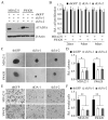
Figure 2. ATAD3A knockdown inhibits cancer cell anchorage-independent growth and invasion in vitro
(A) Western blot analysis shows that ATAD3A protein levels in MDA-MB-231 and SW620 cells are much lower in independently derived ATAD3A knockdown cells (sh3A-1, sh3A-2) compared with that in the knockdown control cells (shGFP). (B) Knockdown of ATAD3A does not affect cell proliferation over 6 days in both MDA-MB-231 and SW620 cells. Silencing ATAD3A inhibits the cell anchorage-independent growth (C) and invasion (E) in both MDA-MB-231 and SW620 cells. Quantification of the relative colony number and relatively invading cell number from the three independent experiments is shown in (D) and (F), respectively. * p<0.05, ** p<0.01.
Figure 3
Figure 3. ATAD3A knockdown inhibits tumor growth and metastasis in vivo
(A) Kaplan-Meier survival curves demonstrate that high ATAD3A expression is associated with low overall survival rate of breast cancer patients. (B) In NSG mice, knockdown of ATAD3A (sh3A) remarkably inhibits tumor growth, showing a smaller tumor size (upper panel) and reduced tumor weight (lower panel) compared with tumors derived from the knockdown control cells (shGFP). (C-D) In NSG mice, knockdown of ATAD3A inhibits tumor induced angiogenesis compared with tumors derived from the control cells. The representative images are shown in (C) and the CD31-positive microvessels are marked (arrows). Quantification of the CD31-positive microvessels is shown in (D). (E-F) Knockdown of ATAD3A significantly decreases lung weight (E) and the number of metastatic nodules on the lung surface (F). (G) Histological analysis shows that only a few, small-sized, metastatic tumors are observed in the lungs from the mice that were injected with ATAD3A-knockdown cells (arrow) compared with extensive infiltration seen in the knockdown control animals (arrow). Images on the right represent higher magnification of the boxed area in the left hand image. Error bars represent SD (n = 10), * p<0.05 and ** p<0.01.
Figure 4
Figure 4. ATAD3A regulates WASF3 protein stability associated with mitochondria
(A) Western blot analysis shows that suppressing ATAD3A expression effectively reduces WASF3 and GRP78 protein levels in MDA-MB-231 and SW620 cells. No concomitant decrease in WASF3 mRNA levels, however, was detected in ATAD3A knockdown cells (below). Levels of other chaperone proteins are relatively unaffected. Quantification of the WASF3 protein levels from the three independent experiments is shown in (B), ** p<0.01. (C) Western blot analysis shows significantly reduced WASF3 protein levels in the tumors from the primary mouse xenografts from ATAD3A knockdown MDA-MB-231 cells. (D) Immunofluorescence assays show that the general distribution of the WASF3 protein in the cytoplasm is generally lost with the treatment of HSP70 inhibitor PES, except at a perinuclear location. (E) Western blot analysis shows a significant reduction of WASF3 in the cytoplasmic (Cyto) fraction but not in the isolated mitochondria (Mito) when treated with PES. ATAD3A protein levels, however, were unaffected in the mitochondrial fraction following PES treatment. GAPDH and COXIV serve as specific markers for the cytoplasm and mitochondria respectively, demonstrating the purity of the fractions. (F) In the presence of PES, Western blot analysis shows dramatic loss of WASF3 protein in ATAD3A knockdown cells compared with the knockdown control cells. (G) Immunofluorescence analysis shows a depletion of WASF3 in mitochondria in ATAD3A knockdown MDA-MB-231 cells. When the ATAD3A knockdown cells are treated with PES, WASF3 is undetectable in both mitochondria and cytoplasmic fraction. Mitochondria are labeled with MitoTracker dyes (red).
Figure 5
Figure 5. GRP78 is required for ATAD3A-mediated WASF3 protein stability
(A) Western blot analysis shows that overexpression of GRP78 rescues WASF3 protein levels in ATAD3A knockdown MDA-MB-231 and SW620 cells. (B) Inhibition of protein synthesis by CHX followed by Western blot at the indicated times shows that the half-life of WASF3 is increased in the MDA-MB-231 cells when co-transfected with pCDH-HA-WASF3 (W3) and pCMV-Flag-GRP78 (GRP78) vectors, compared with that in the cells which co-transfected with pCDH-HA-WASF3 (W3) and pCMV-Flag (EV) vectors. The same results were observed from SW620 cells. (C) WASF3 protein levels are normalized to β-Actin and the WASF3 half-life from three independent experiments was quantified (lower panel). * p<0.05 and ** p<0.01. (D) Western blot shows that knockdown of GRP78 (sh78-1, sh78-2) in MDA-MB-231 or SW620 cells leads to a decrease in WASF3 protein levels compared with the knockdown control cells (shGFP). RT-PCR shows that mRNA expression levels of WASF3 were not affected as a result of GRP78 knockdown. (E) Western blot analysis shows increased protein levels of both GRP78 and WASF3, but not HSP70, when MDA-MB-231 cells were treated with the TG ER stress inducer. (F) IP analysis shows more GRP78 protein in the WASF3 immunocomplex following the treatment with TG. (G) Western blot analysis shows a more dramatic reduction in WASF3 protein levels in GRP78 knockdown MDA-MB-231 cells when compared with the knockdown control cells. (H) IP analysis shows that ATAD3A binds to GRP78, but not to HSP70 in MDA-MB-231 cells. (I) Western blot shows that the loss of WASF3 in ATAD3A knockdown cells cannot be rescued by overexpression of HSP70. (J) IP analysis shows that binding of WASF3 to HSP70 is impaired, but does not affect WASF3 binding to GRP78 following PES treatment.
Figure 6
Figure 6. WASF3 is involved in ATAD3A-mediated alteration of CDH1 expression
(A) Heatmap analysis of microarray data shows differential gene expression in MDA-MB-231 as a result of ATAD3A knockdown. Red square: up-regulated genes (fold change more 2.0); blue square: down-regulated genes (fold change less than 0.5). (B) GO analysis for the biological functions of differentially expressed genes shows most of the dysregulated genes in the ATAD3A knockdown cells are involved in the cancer cell motility and metastasis phenotypes. (C) QRT-PCR analysis of selected gene expression shows their altered expression levels in ATAD3A knockdown cells, which is consistent with those seen in the microarray experiments. Relative expression levels are presented as a percentage of control expression levels. Error bars represent SD (n = 3). (D) Western blot shows increased CDH1 protein levels in ATAD3A knockdown MDA-MB-231 cells. 1, 2 and 3 indicate three independent repeats. (E) Western blot analysis shows that overexpressing GRP78 in the ATAD3A knockdown MDA-MD-231 cells leads to a suppression of CDH1 upregulation mediated by loss of ATAD3A. However, CDH1 levels did not change in WASF3 knockdown cells whether or not GRP78 was overexpressed. (F) Matrigel invasion assays show a significant increase in invasion potential when GRP78 was overexpressed in ATAD3A knockdown cells, but there was no change in invasion potential when GRP78 was overexpressed in WASF3 knockdown cells. Error bars represent SD (n = 3), * p<0.05.

Similar articles

Cited by

References

    1. Sossey-Alaoui K, Su G, Malaj E, Roe B, Cowell JK. WAVE3, an actin-polymerization gene, is truncated and inactivated as a result of a constitutional t(1;13)(q21;q12) chromosome translocation in a patient with ganglioneuroblastoma. Oncogene. 2002;21:5967–5974. - PubMed
    1. Rotty JD, Wu C, Bear JE. New insights into the regulation and cellular functions of the ARP2/3 complex. Nat Rev Mol Cell Biol. 2013;14:7–12. - PubMed
    1. Sossey-Alaoui K, Head K, Nowak N, Cowell JK. Genomic organization and expression profile of the human and mouse WAVE gene family. Mamm Genome. 2003;14:314–322. - PubMed
    1. Bisi S, Disanza A, Malinverno C, Frittoli E, Palamidessi A, Scita G. Membrane and actin dynamics interplay at lamellipodia leading edge. Curr Opin Cell Biol. 2013;25:565–573. - PubMed
    1. Sossey-Alaoui K, Li X, Ranalli TA, Cowell JK. WAVE3-mediated cell migration and lamellipodia formation are regulated downstream of phosphatidylinositol 3-kinase. J Biol Chem. 2005;280:21748–21755. - PubMed

Publication types

MeSH terms

Substances