. 2015 Mar;32(3):769-78.
doi: 10.1007/s11095-013-1219-9.

Silencing the double-stranded RNA binding protein DGCR8 inhibits ovarian cancer cell proliferation, migration, and invasion

Silencing the double-stranded RNA binding protein DGCR8 inhibits ovarian cancer cell proliferation, migration, and invasion

Yuqi Guo et al. Pharm Res. 2015 Mar.

Abstract

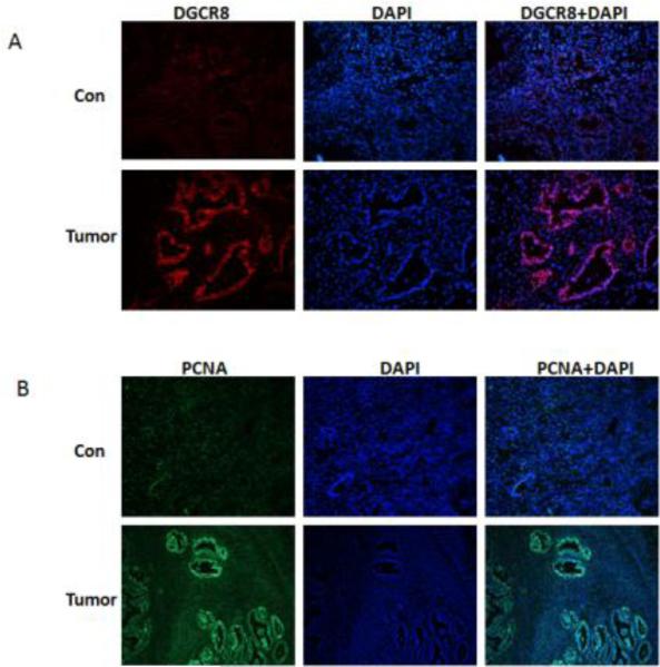
Purpose: To evaluate the role of DiGeorge Critical Region 8 (DGCR8), a key component of miRNA biogenesis pathway in ovarian cancer.

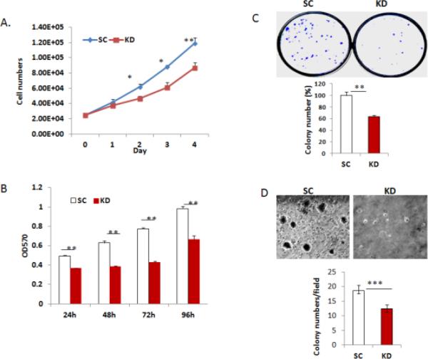
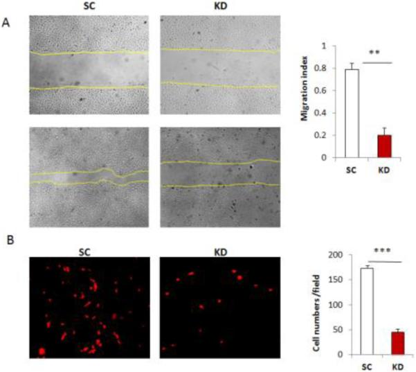
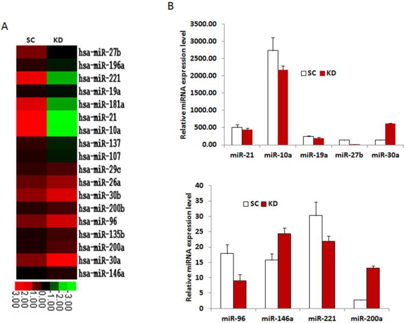
Methods: The expression of DGCR8 in ovarian cancer was detected by immunostaining and DGCR8 knockdown in ovarian cancer cells was achieved using lentiviral shRNA. Differential expression of miRNAs was determined using Nanostring miRNA arrays and validated by real-time RT-PCR.

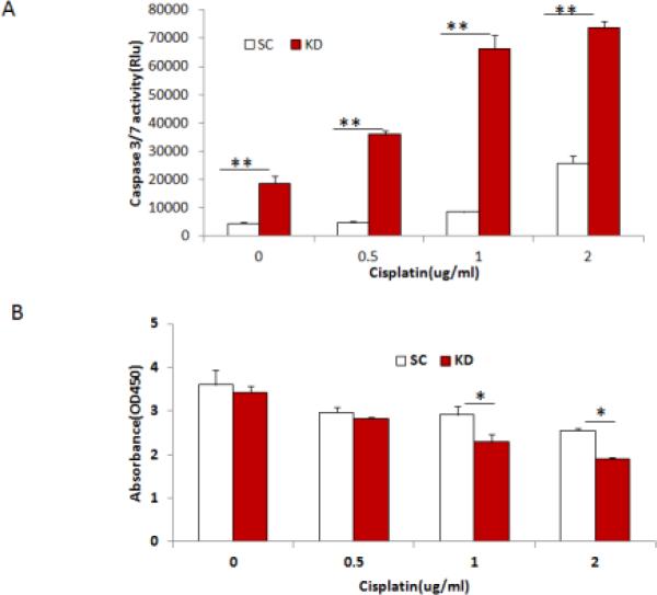
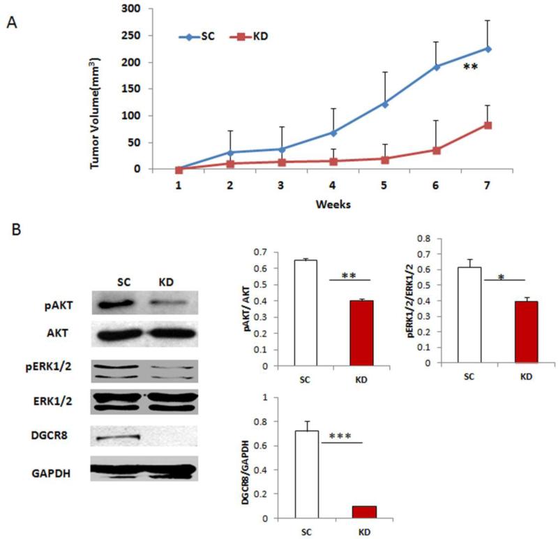
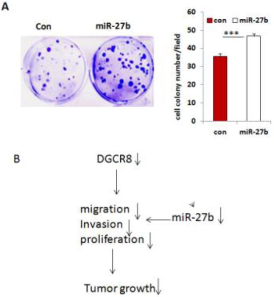
Results: DGCR8 was highly expressed in ovarian cancer. Knockdown of DGCR8 expression inhibits cell proliferation, migration, and invasion, as well as sensitizes cells to apoptosis induced by the chemotherapeutic drug cisplatin. Cellular survival pathways including ERK1/2 mitogen-activated protein kinase and phosphatidylinositol 3-kinase/AKT were attenuated in DGCR8 knockdown cells. DGCR8 knockdown results in dysregulated miRNA gene expression. miR-27b was identified as the most highly down-regulated miRNA in DGCR8 knockdown cells and promoted cell proliferation in ovarian cancer cells.

Conclusions: DGCR8 functions as an oncogene in ovarian cancer, which is in part mediated by miR-27b.


Figures

Figure 1
Figure 1. DGCR8 is highly expressed in ovarian cancer
A. The expression of DGCR8 in immunostained sections of ovarian cancer and normal ovary tissue B. Cell proliferations in sections of ovarian cancer and normal ovary were stained for the proliferation marker PCNA.
Figure 2
Figure 2. Silencing DGCR8 in ovarian cancer cells reduces cell proliferation
A. Cell growth curves in DGCR8KD and control cells were determined by cell counting (* p<0.05; ** p<0.01). B. Cell proliferation was examined by using MTT assay in DGCR8KD and control cells(**p<0.01). C. Colony formation by DGCR8KD and control cells (**p<0.01). D. Soft agar colony formation assay was performed in a 6-well plate in triplicates. Colonies were photographed and counted after 3 weeks (***p<0.001).
Figure 3
Figure 3. Silencing DGCR8 inhibits cell migration and invasion in ovarian cancer cells
A. Wound healing assays were performed to examine the migration rate of SKOV3 cells transduced with DGCR8 shRNA and scramble control. Photographs were taken at 0 and 24 h following initial scratch. The migration rates were quantified by measuring the injured area from triplicates. Three separate experiments were performed ( **p<0.01). B. Cell invasion assay was performed with Matrigel transwell plates and cells migrating through Matrigel after 24h were stained with PI, photographed after fluorescent microscopy and counted. Data were collected from three separate experiments and analyzed using student T-tests (***p<0.001).
Figure 4
Figure 4. Knockdown of DGCR8 sensitizes cells to cisplatin induced cell apoptosis
A. DGCR8KD and control SKOV3 cells were treated with different doses of cisplatin and apoptosis was examined at 24h following the treatment by measuring caspase3/7 activity. Data were presented from one representative experiment of three experiments (**p<0.01). B. Cell viabilities were examined in DGCR8 KD and control SKOV3 cells following treatment with various doses of cisplatin . Three separate experiments were repeated (*p<0.05).
Figure 5
Figure 5. Knockdown of DGCR8 attenuates cellular survival pathways
A. Tumors were continuously grown in mice injected with control cells whereas tumor significantly were reduced in mice injected with DGCR8KD cells(n=4,**p<0.01). B. DGCR8, phospho and total AKT, phospho and totalERK1/2 and GAPDH expression in DGCR8KD and control SKOV3 cells were detected by Western blot. Protein bands were quantified by densitometry (**p<0.01, ***p<0.001).
Figure 6
Figure 6. Knockdown of DGCR8 in SKOV3 cells leads to dysregulation of miRNA expression
A. miRNA expressions inDGCR8KD and SC SKOV3 cells were detected in duplicate using Nano-string miRNA array, and the averaged signaling intensities were displayed in heat-maps for the highest up and down-regulated miRNAs (>2 fold) in DGCR8KD compared to SC control cells. B. Selected miRNA expressions were verified by polyA tailing real-time RT-PCR.
Figure 7
Figure 7. miR-27b promotes cell proliferation in ovarian cancer cells
A. Colony formations were significantly increased in SKOV3 cells transduced with lentiviral miR-27b overexpression vector compared to controls. B. Molecular model for the oncogenic properties of DGCR8.

Similar articles

Cited by

References

    1. Gregory RI, Yan KP, Amuthan G, Chendrimada T, Doratotaj B, Cooch N, Shiekhattar R. Nature. 2004;432:235–240. - PubMed
    1. Han J, Lee Y, Yeom KH, Kim YK, Jin H, Kim VN. Genes Dev. 2004;18:3016–3027. - PMC - PubMed
    1. Landthaler M, Yalcin A, Tuschl T. Curr Biol. 2004;14:2162–2167. - PubMed
    1. Han L, Zhang A, Zhou X, Xu P, Wang GX, Pu PY, Kang CS. Int J Oncol. 2010;37:299–305. - PubMed
    1. Sand M, Gambichler T, Skrygan M, Sand D, Scola N, Altmeyer P, Bechara FG. Cancer Invest. 2010;28:649–653. - PubMed

Publication types

MeSH terms