. 2015;37(4-5):321-37.
doi: 10.1159/000375368. Epub 2015 Mar 24.

Does Caspase-6 Have a Role in Perinatal Brain Injury?

Affiliations

Does Caspase-6 Have a Role in Perinatal Brain Injury?

Ana A Baburamani et al. Dev Neurosci. 2015.

Abstract

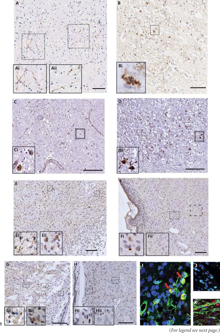
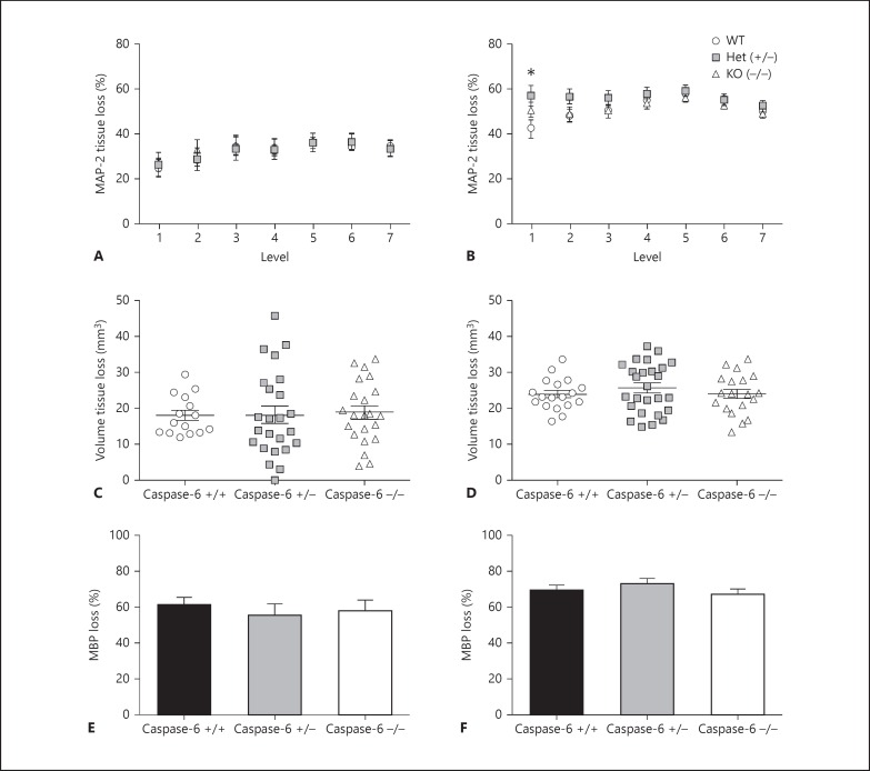
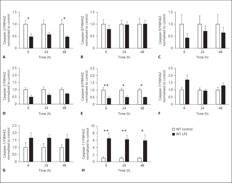
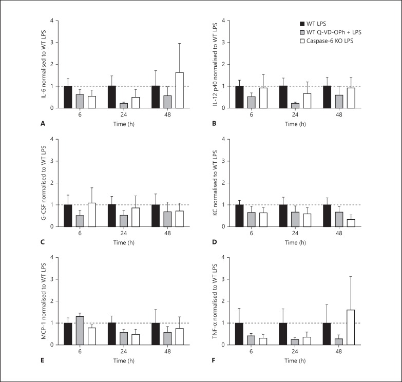
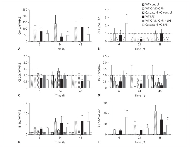
Apoptotic mechanisms are centre stage for the development of injury in the immature brain, and caspases have been shown to play a pivotal role during brain development and in response to injury. The inhibition of caspases using broad-spectrum agents such as Q-VD-OPh is neuroprotective in the immature brain. Caspase-6, an effector caspase, has been widely researched in neurodevelopmental disorders and found to be important following adult stroke, but its function in the neonatal brain has yet to be detailed. Furthermore, caspases may be important in microglial activation; microglia are required for optimal brain development and following injury, and their close involvement during neuronal cell death suggests that apoptotic cues such as caspase activation may be important in microglial activation. Therefore, in this study we aimed to investigate the possible apoptotic and non-apoptotic functions caspase-6 may have in the immature brain in response to hypoxia-ischaemia. We examined whether caspases are involved in microglial activation. We assessed cleaved caspase-6 expression following hypoxia-ischaemia and conducted primary microglial cultures to assess whether the broad-spectrum inhibitor Q-VD-OPh or caspase-6 gene deletion affected lipopolysaccharide (LPS)-mediated microglial activation and phenotype. We observed cleaved caspase-6 expression to be low but present in the cell body and cell processes in both a human case of white matter injury and 72 h following hypoxia-ischaemia in the rat. Gene deletion of caspase-6 did not affect the outcome of brain injury following mild (50 min) or severe (60 min) hypoxia-ischaemia. Interestingly, we did note that cleaved caspase-6 was co-localised with microglia that were not of apoptotic morphology. We observed that mRNA of a number of caspases was modulated by low-dose LPS stimulation of primary microglia. Q-VD-OPh treatment and caspase-6 gene deletion did not affect microglial activation but modified slightly the M2b phenotype response by changing the time course of SOCS3 expression after LPS administration. Our results suggest that the impact of active caspase-6 in the developing brain is subtle, and we believe there are predominantly other caspases (caspase-2, -3, -8, -9) that are essential for the cell death processes in the immature brain.


Figures

Fig. 1
Fig. 1
Cleaved caspases in the human preterm brain and in a neonatal hypoxic-ischaemic rat model. Immunoreactivity of cl-C6 (A, B) and cl-C3 (C, D) was expressed in a human preterm white matter injury case in the periventricular white matter (A, C) and anterior limb of the internal capsule (B, D). cl-C6 staining appeared fibrous (Ai, Aii) and perinuclear (Bi). Sporadic and nuclear cl-C3 was seen in the periventricular white matter region (C, Ci). However, the internal capsule the cl-C3 immunoreactivity was both nuclear and cytoplasmic (D). Following neonatal hypoxia-ischaemia in rats, cl-C6 (E, F) was observed in both the ipsilateral (E) and contralateral (F) hemispheres. Consistent with the observations made in the human tissue, cl-C6 again appeared to be fibrous in the cortex (Ei) and perinuclear in white matter (Eii) in the ipsilateral hemisphere, whereas in the contralateral side, there was perinuclear staining seen in the white matter (Fi) but not in the cortex (Fii). cl-C6 immunoreactivity was co-localised mainly in the microglia (tomato lectin; G) but not with oligodendroglia (Olig-2; H) or astroglia (GFAP; I). cl-C3 was also observed in rats following neonatal hypoxia-ischaemia, (J, K). Nuclear and punctate staining of cl-C3 was abundant in the ipsilateral side (J, Ji, Jii), but the contralateral side had primarily nuclear staining (K, Ki, Kii). Red arrows indicate co-labelled cells. Scale bar = 100 µm (A-F, J, K) and 10 µm (G-I).
Fig. 2
Fig. 2
Neuropathology assessment of caspase-6 WT, Het and KO mice 72 h following 50 or 60 min of hypoxia-ischaemia at P9. Tissue loss (A, B) was assessed using MAP-2 staining at 7 levels (posterior to anterior) of the brain in caspase-6 +/+ (WT; open black circles), caspase-6 +/- (Het; grey squares) and caspase-6 -/- (KO; open black triangles). Volume tissue loss assessment (C, D) and subcortical white matter loss at the level of the hippocampus (E, F) showed no significant difference between genotypes following 50 min (A, C, E) or 60 min (B, D, F) of hypoxia-ischaemia (H-I). Mean ± SEM. * p < 0.05
Fig. 3
Fig. 3
Caspase mRNA in primary microglia following LPS (10 ng/ml). Gene expression for the following caspases was assessed in WT primary microglia: initiator - caspase-2 (A), caspase-8 (B) and caspase-9 (C); effector - caspase-3 (D), caspase-6 (E) and caspase-7 (F), and inflammatory - caspase-1 (G) and caspase-11 (H). LPS-treated microglia (black bars) were normalised and compared with control (white bars) cells for each time point. Data were analysed with a two-way ANOVA (for time and treatment) and, when significant, a Sidak post hoc analysis was conducted. Data are mean ± SEM (n = 5 independent experiments). * p < 0.05, ** p < 0.01.
Fig. 4
Fig. 4
Cytokine expression from microglial cell supernatants. IL-6 (A), IL-12 p40 (B), G-CSF (C), KC (D), MCP-1 (E) and TNF-α (F) measured from microglial cell supernatants at 6, 24 and 48 h. Data are expressed as a fold change from WT LPS treatment for each time point. Data were analysed with a two-ANOVA for time and treatment. Data are mean ± SEM (n = 5-6 independent experiments). Dotted line indicates 1.
Fig. 5
Fig. 5
Gene expression of microglial phenotype markers in WT primary microglia following LPS (10 ng/ml), Q-VD-OPh (20 µm), Q-VD-OPh + LPS, and caspase-6 KO control and LPS. Expression of genes indicative of M1: Cox-2 (A) and iNOS (B); M2a: CD206 (C) and IGF-1 (D), and M2b: IL-1ra (E) and SOCS3 (F), was normalised to WT control (black checked bars) for WT LPS (black bars), WT Q-VD-OPh (dark grey checked bars), WT Q-VD-OPh + LPS (dark grey bars) and caspase-6 KO LPS (white bars) was normalised to caspase-6 KO control (checked bars). For iNOS (B) gene expression, only LPS-treated cells were detected, so data were normalised to WT LPS. Data were analysed with a two-way ANOVA (for time and treatment) and, when significant, a Dunnett's post hoc analysis was conducted comparing with WT LPS treatment. ND = Not detected. Data are mean ± SEM (n = 4-5 independent experiments; iNOS: n = 3-4 independent experiments). Dotted line indicates 1. * p < 0.05.

References

    1. Soane L, Siegel ZT, Schuh RA, Fiskum G. Postnatal developmental regulation of Bcl-2 family proteins in brain mitochondria. J Neurosci Res. 2008;86:1267–1276. - PMC - PubMed
    1. Hagberg H, Mallard C, Rousset CI, Thornton C. Mitochondria: hub of injury responses in the developing brain. Lancet Neurol. 2014;13:217–232. - PubMed
    1. Blomgren K, Hagberg H. Free radicals, mitochondria, and hypoxia-ischemia in the developing brain. Free Rad Biol Med. 2006;40:388–397. - PubMed
    1. Taylor DL, Edwards AD, Mehmet H. Oxidative metabolism, apoptosis and perinatal brain injury. Brain Pathol. 1999;9:93–117. - PMC - PubMed
    1. Troy CM, Akpan N, Jean YY. Regulation of caspases in the nervous system implications for functions in health and disease. Prog Mol Biol Transl Sci. 2011;99:265–305. - PMC - PubMed

Publication types

LinkOut - more resources