The brain timewise: how timing shapes and supports brain function
- PMID: 25823867
- PMCID: PMC4387511
- DOI: 10.1098/rstb.2014.0170
The brain timewise: how timing shapes and supports brain function
Abstract
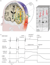
We discuss the importance of timing in brain function: how temporal dynamics of the world has left its traces in the brain during evolution and how we can monitor the dynamics of the human brain with non-invasive measurements. Accurate timing is important for the interplay of neurons, neuronal circuitries, brain areas and human individuals. In the human brain, multiple temporal integration windows are hierarchically organized, with temporal scales ranging from microseconds to tens and hundreds of milliseconds for perceptual, motor and cognitive functions, and up to minutes, hours and even months for hormonal and mood changes. Accurate timing is impaired in several brain diseases. From the current repertoire of non-invasive brain imaging methods, only magnetoencephalography (MEG) and scalp electroencephalography (EEG) provide millisecond time-resolution; our focus in this paper is on MEG. Since the introduction of high-density whole-scalp MEG/EEG coverage in the 1990s, the instrumentation has not changed drastically; yet, novel data analyses are advancing the field rapidly by shifting the focus from the mere pinpointing of activity hotspots to seeking stimulus- or task-specific information and to characterizing functional networks. During the next decades, we can expect increased spatial resolution and accuracy of the time-resolved brain imaging and better understanding of brain function, especially its temporal constraints, with the development of novel instrumentation and finer-grained, physiologically inspired generative models of local and network activity. Merging both spatial and temporal information with increasing accuracy and carrying out recordings in naturalistic conditions, including social interaction, will bring much new information about human brain function.
Keywords: brain imaging; hierarchical order; magnetoencephalography; networks; prediction; time.
Figures

References
Publication types
MeSH terms
Grants and funding
LinkOut - more resources
Full Text Sources
Other Literature Sources
