. 2015 May;4(5):513-22.
doi: 10.5966/sctm.2014-0267. Epub 2015 Mar 30.

Human umbilical cord mesenchymal stem cell exosomes enhance angiogenesis through the Wnt4/β-catenin pathway

Affiliations

Human umbilical cord mesenchymal stem cell exosomes enhance angiogenesis through the Wnt4/β-catenin pathway

Bin Zhang et al. Stem Cells Transl Med. 2015 May.

Abstract

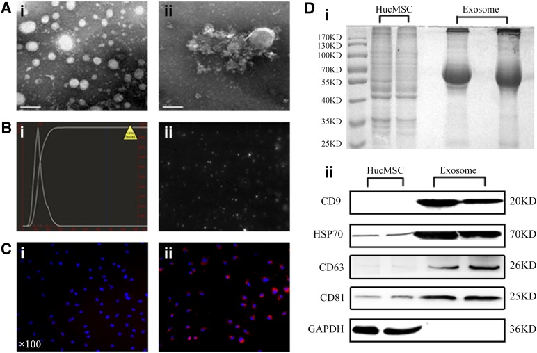
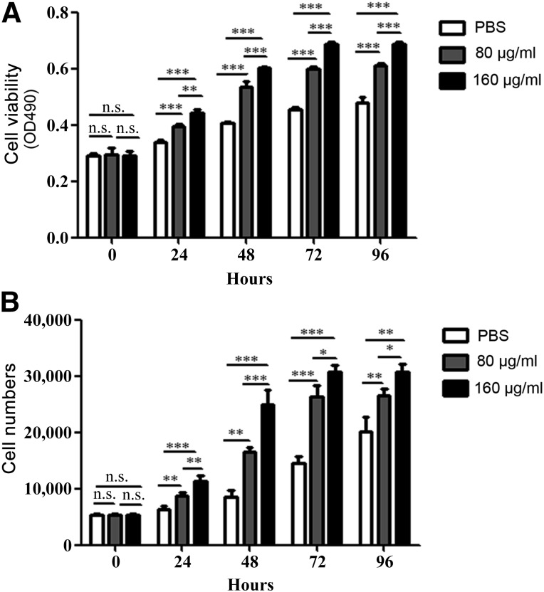
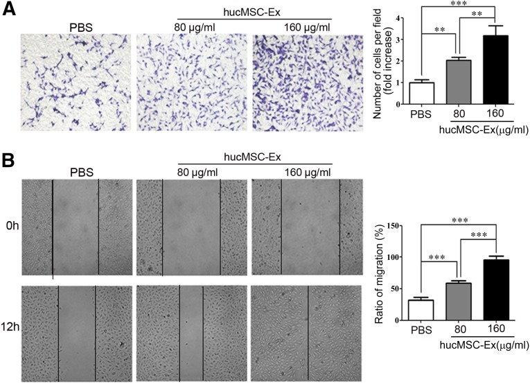
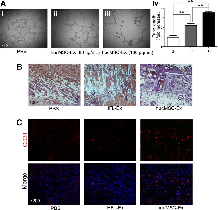
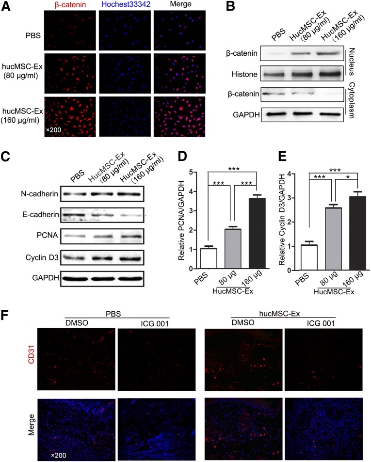
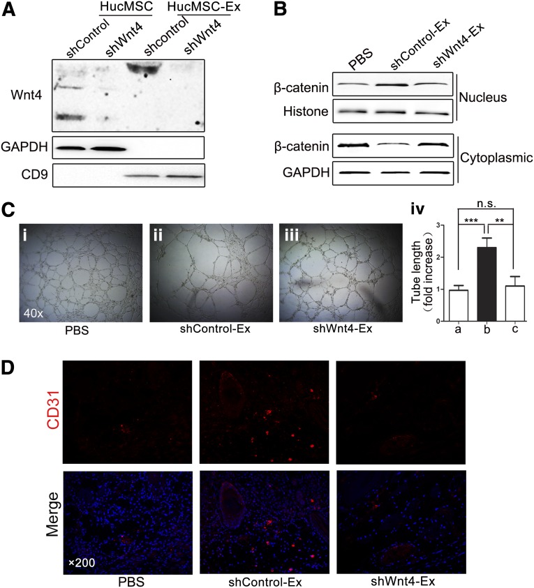
Human umbilical cord mesenchymal stem cells (hucMSCs) and their exosomes have been considered as potential therapeutic tools for tissue regeneration; however, the underlying mechanisms are still not well understood. In this study, we isolated and characterized the exosomes from hucMSCs (hucMSC-Ex) and demonstrated that hucMSC-Ex promoted the proliferation, migration, and tube formation of endothelial cells in a dose-dependent manner. Furthermore, we demonstrated that hucMSC-Ex promoted wound healing and angiogenesis in vivo by using a rat skin burn model. We discovered that hucMSC-Ex promoted β-catenin nuclear translocation and induced the increased expression of proliferating cell nuclear antigen, cyclin D3, N-cadherin, and β-catenin and the decreased expression of E-cadherin. The activation of Wnt/β-catenin is critical in the induction of angiogenesis by hucMSC-Ex, which could be reversed by β-catenin inhibitor ICG-001. Wnt4 was delivered by hucMSC-Ex, and the knockdown of Wnt4 in hucMSC-Ex abrogated β-catenin nuclear translocation in endothelial cells. The in vivo proangiogenic effects were also inhibited by interference of Wnt4 expression in hucMSC-Ex. Taken together, these results suggest that hucMSC-Ex-mediated Wnt4 induces β-catenin activation in endothelial cells and exerts proangiogenic effects, which could be an important mechanism for cutaneous wound healing.

Keywords: Angiogenesis; Exosomes; Regenerative medicine; Wnt4; β-Catenin.


Figures

Figure 1.
Figure 1.
Exosome characterization and internalization. (Ai, Aii): Morphologic analysis of hucMSC-derived exosomes (hucMSC-Ex) by transmission electron microscopy (scale bar = 100 nm). (B): Size distribution measurements under flow conditions by nanoparticle tracking (Bi) with the corresponding video frame (Bii). (C): Phosphate-buffered saline was used as a control (×100) (Ci), and CM-Dil-labeled exosomes were taken up by EA.hy926 cells (×100) (Cii). (Di): SDS-polyacrylamide gel electrophoresis analysis of the protein profiles of cell lysates and exosomes. (Dii): Detection of exosomal marker expression in hucMSC-Ex by Western blot. Abbreviation: hucMSC, human umbilical cord mesenchymal stem cells.
Figure 2.
Figure 2.
Exosomes from human umbilical cord mesenchymal stem cells (HucMSC-Ex) prompted the proliferation of endothelial cells. (A): MTT assay for the proliferating ability of EA.hy926 cells with or without hucMSC-Ex treatment. (B): Cell-counting assay for the proliferating ability of hucMSC-Ex-treated EA.hy926 cells. ∗, p < .05; ∗∗, p < .01; ∗∗∗, p < .001. Abbreviations: n.s., not significant; PBS, phosphate-buffered saline.
Figure 3.
Figure 3.
HucMSC-Ex prompted the migration of endothelial cells. (A): EA.hy926 cells were treated with different concentrations of exosomes (80, 160 μg/ml) or PBS for 12 hours. Transwell migration assay was performed to analyze the migratory ability of the cells (×100). Cells that migrated to the bottom were counted. ∗∗, p < .01; ∗∗∗, p < .001. (B): EA.hy926 cells treated with different concentrations of exosomes (80, 160 μg/ml) were subjected to a wound-healing assay for 12 hours (×100), and the percentage of closure of the wounded areas was measured. ∗∗, p < .01; ∗∗∗, p < .001. Abbreviations: hucMSC-Ex, exosomes from human umbilical cord mesenchymal stem cells; PBS, phosphate-buffered saline.
Figure 4.
Figure 4.
HucMSC-Ex improved the tube-formation ability of endothelial cells and vascular density in cutaneous burn model. (Ai–Aiii): Exosomes increased the tube length in a dose-dependent manner (×40). (Aiv): Measurement of the tube length by ImageJ software. ∗∗, p < .01. (B): Representative micrographs of wound histological images (hematoxylin and eosin staining) at 1 week after treatment with hucMSC-Ex, HFL1-Ex, or PBS (×100). (C): Representative immunofluorescence images of CD31 expression in the wound area after treatment with hucMSC-Ex, HFL1-Ex, or PBS (×200). Abbreviations: HFL-Ex, exosomes from human lung fibroblast; hucMSC-Ex, exosomes from human umbilical cord mesenchymal stem cells; PBS, phosphate-buffered saline.
Figure 5.
Figure 5.
HucMSC-Ex prompted angiogenesis by activating Wnt/β-catenin signaling. (A): β-Catenin expression in EA.hy926 cells after exosome treatment (80 or 160 μg/ml) treatment was detected by immunofluorescence staining (×200). ∗∗, p < .01. (B): Cytoplasm and nuclear fractions were prepared from EA.hy926 cells treated with indicated concentrations of hucMSC-Ex or PBS. β-Catenin protein levels were determined using Western blot. (C): EA.hy926 cells were treated with different concentrations of exosomes (80 or 160 μg/ml). The expression of N-cadherin, E-cadherin, PCNA, and cyclin D3 proteins was detected using Western blot. (D): Densitometric analysis of the PCNA Western blot bands (∗∗∗, p < .001) compared with the PBS group (n = 3). (E): Densitometric analysis of the cyclin D3 Western blot bands (∗, p < .05 and ∗∗∗, p < .001) compared with the PBS group (n = 3). (F): The cutaneous-burn rat models were injected with hucMSC-Ex with or without ICG-001 (1 mg per rat). PBS and DMSO were used as controls for hucMSCs and ICG-001, respectively. Representative immunofluorescence images of CD31 expression in the wound area are shown (×200). Abbreviations: DMSO, dimethyl sulfoxide; GAPDH, glyceraldehyde-3-phosphate dehydrogenase; hucMSC-Ex, exosomes from human umbilical cord mesenchymal stem cells; PBS, phosphate-buffered saline; PCNA, proliferating cell nuclear antigen.
Figure 6.
Figure 6.
HucMSC-Ex-delivered Wnt4 was critical for angiogenesis in vitro and in vivo. (A): HucMSCs were transfected with lentivirus Wnt4-shRNA or control-shRNA. The expression of Wnt4 in hucMSCs and hucMSC-Ex was determined using Western blot. (B): Cytoplasm and nuclear fractions were prepared from EA.hy926 cells treated with exosomes from Wnt4-shRNA or control-shRNA-transfected hucMSCs. β-catenin protein levels were determined using Western blot. (Ci–Ciii): The tube-formation ability of EA.hy926 cells treated with PBS, shWnt4-Ex, or shControl-Ex was determined (×40). (Civ): Measurement of the tube length by Image J software. ∗∗, p < .01; ∗∗∗, p < .001. (D): The rat wound models were treated with PBS, shControl-Ex, or shWnt4-Ex for 1 week. The expression of CD31 in the wound area was determined using immunofluorescence staining (×200). Abbreviations: GAPDH, glyceraldehyde-3-phosphate dehydrogenase; hucMSCs, human umbilical cord mesenchymal stem cells; hucMSC-Ex, exosomes from human umbilical cord mesenchymal stem cells; n.s., not significant; PBS, phosphate-buffered saline; PCNA, proliferating cell nuclear antigen; shControl, control-short hairpin RNA lentiviral expression vector; shControl-Ex, exosomes from control-short hairpin RNA-transfected hucMSCs; shWnt4, Wnt4-short hairpin RNA lentiviral expression vector; shWnt4-Ex, exosomes from Wnt4-short hairpin RNA-transfected hucMSCs.

References

    1. Batsali AK, Kastrinaki MC, Papadaki HA, et al. Mesenchymal stem cells derived from Wharton’s jelly of the umbilical cord: Biological properties and emerging clinical applications. Curr Stem Cell Res Ther. 2013;8:144–155. - PubMed
    1. Yoo HS, Yi T, Cho YK, et al. Mesenchymal stem cell lines isolated by different isolation methods show variations in the regulation of graft-versus-host disease. Immune Netw. 2013;13:133–140. - PMC - PubMed
    1. Sun L, Wang D, Liang J, et al. Umbilical cord mesenchymal stem cell transplantation in severe and refractory systemic lupus erythematosus. Arthritis Rheum. 2010;62:2467–2475. - PubMed
    1. Wang X, Wang Y, Gou W, et al. Role of mesenchymal stem cells in bone regeneration and fracture repair: A review. Int Orthop. 2013;37:2491–2498. - PMC - PubMed
    1. Laroni A, Novi G, Kerlero de Rosbo N, et al. Towards clinical application of mesenchymal stem cells for treatment of neurological diseases of the central nervous system. J Neuroimmune Pharmacol. 2013;8:1062–1076. - PubMed

Publication types

MeSH terms