Review
. 2015 May 7;125(19):2898-907.
doi: 10.1182/blood-2015-02-355974. Epub 2015 Mar 30.

Activated protein C: biased for translation

Affiliations
Review

Activated protein C: biased for translation

John H Griffin et al. Blood. .

Abstract

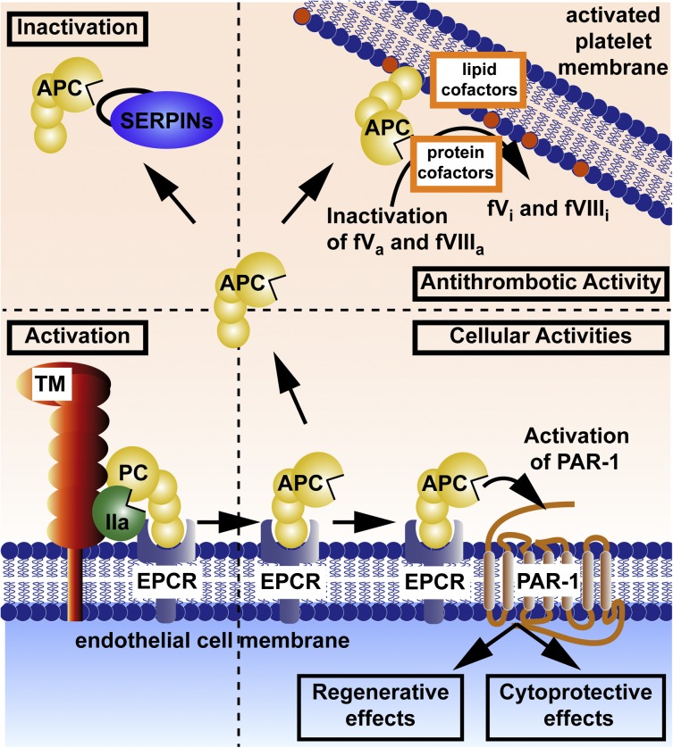
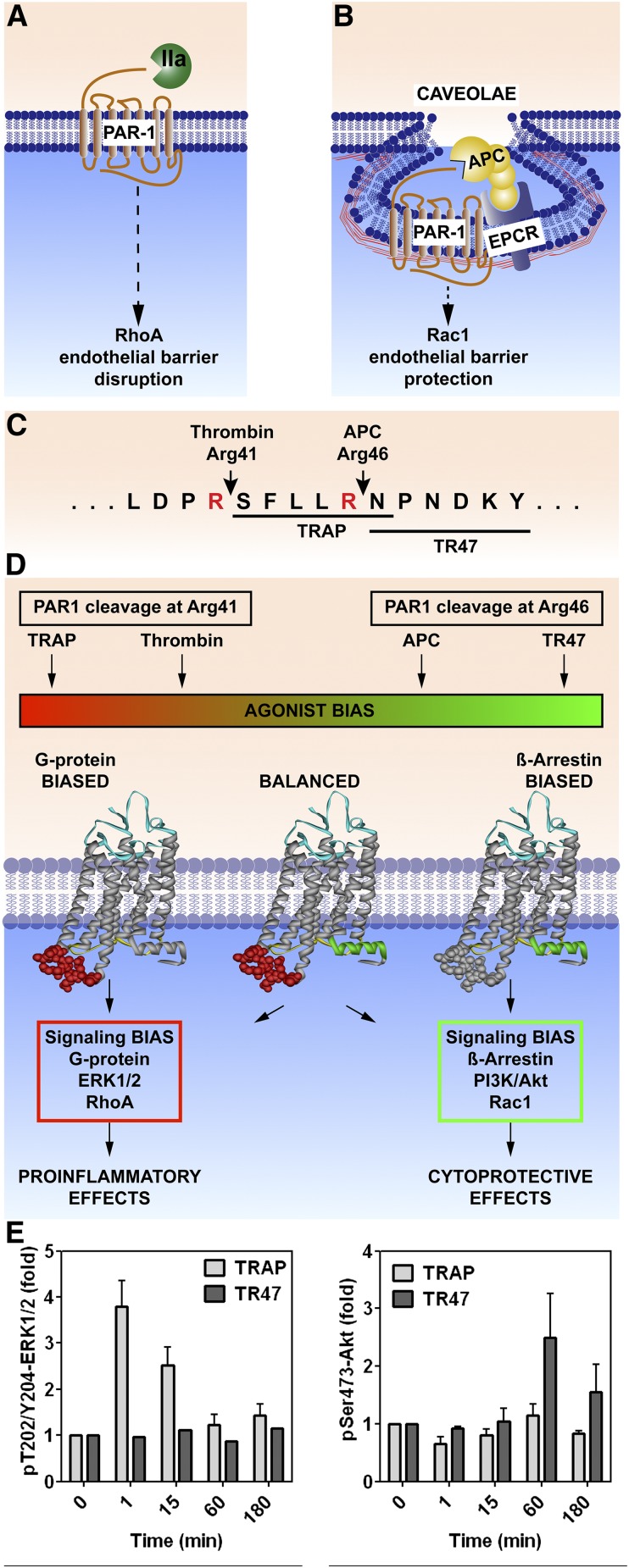
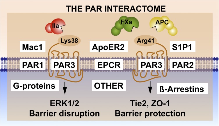
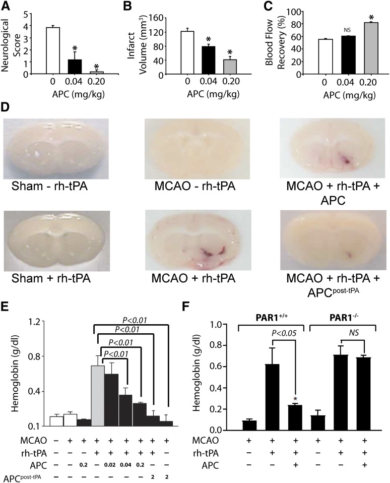
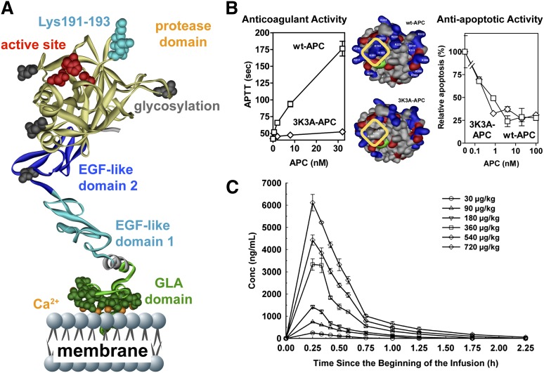
The homeostatic blood protease, activated protein C (APC), can function as (1) an antithrombotic on the basis of inactivation of clotting factors Va and VIIIa; (2) a cytoprotective on the basis of endothelial barrier stabilization and anti-inflammatory and antiapoptotic actions; and (3) a regenerative on the basis of stimulation of neurogenesis, angiogenesis, and wound healing. Pharmacologic therapies using recombinant human and murine APCs indicate that APC provides effective acute or chronic therapies for a strikingly diverse range of preclinical injury models. APC reduces the damage caused by the following: ischemia/reperfusion in brain, heart, and kidney; pulmonary, kidney, and gastrointestinal inflammation; sepsis; Ebola virus; diabetes; and total lethal body radiation. For these beneficial effects, APC alters cell signaling networks and gene expression profiles by activating protease-activated receptors 1 and 3. APC's activation of these G protein-coupled receptors differs completely from thrombin's activation mechanism due to biased signaling via either G proteins or β-arrestin-2. To reduce APC-associated bleeding risk, APC variants were engineered to lack >90% anticoagulant activity but retain normal cell signaling. Such a neuroprotective variant, 3K3A-APC (Lys191-193Ala), has advanced to clinical trials for ischemic stroke. A rich data set of preclinical knowledge provides a solid foundation for potential translation of APC variants to future novel therapies.


Figures

Figure 1
Figure 1
Protein C activation and expression of APC’s multiple activities. Activation of the EPCR-bound protein C (PC) zymogen (bottom left) is accomplished by thrombomodulin (TM)-bound thrombin (IIa). Anticoagulant activity (upper right) is based on limited proteolysis, causing irreversible inactivation (i) of the activated clotting factors (f)Va and fVIIIa for which various lipids and protein cofactors play essential roles, as shown for this reaction on platelet membranes. Cytoprotective actions of APC (bottom right) include its antiapoptotic and anti-inflammatory activities, its ability to stabilize endothelial barriers to prevent vascular leakage, and its ability to alter gene expression profiles for many genes. APC’s various cytoprotective activities and regenerative effects generally require EPCR and PAR1. Not depicted here is the fact that APC’s cytoprotective or regenerative actions sometimes require PAR3 and/or other receptors, depending on the biological context, cell type, and organ. Inactivation of circulating APC by plasma serine protease inhibitors (SERPINs; upper left) is a major mechanism for clearance of APC. Coloring of molecules is as follows: protein C zymogen and active protease, APC (yellow); IIa (green); TM (red); EPCR (blue); and SERPINs (purple).
Figure 2
Figure 2
PAR1-dependent biased signaling initiated by thrombin or APC. PAR1 subpopulations are localized either in membrane sections that lack caveolin-1 (A) or in caveolae that contain caveolin-1 and EPCR (B). (C) PAR1 cleavage by thrombin at Arg41 generates the N-terminal tethered-peptide agonist that begins with residue 42, whereas APC cleavage at Arg46 generates a different N-terminal tethered agonist that begins with residue 47. (D) The former cleavage results in G protein–dependent signaling, whereas the latter cleavage results in β-arrestin-2–dependent signaling. Synthetic peptides known as TRAPs that begin with amino acid 42 cause thrombinlike effects on cells, whereas a 20-mer synthetic peptide that begins with amino acid 47 (TR47) causes APC-like effects on cells .,, (E) Such effects are illustrated by the differences in phosphorylations of ERK1/2 compared to Akt, because TRAP induces phosphorylation of ERK1/2 but not Akt, whereas TR47 induces phosphorylation of Akt but not ERK1/2. IIa, thrombin; TRAP, thrombin receptor–activating peptide.
Figure 3
Figure 3
Noncanonical PAR3 activation and signaling selection by the PAR3 interactome. Activation of PAR3 can occur by thrombin (IIa)-mediated cleavage at Lys38 (canonical cleavage) or by APC and factor (F)Xa cleavage at noncanonical Arg41., The latter cleavage results in selective activation of Tie2 and ZO-1 that promotes stabilization of the tight junctions, whereas the former cleavage enhances PAR1-induced ERK1/2 activation that results in barrier-disruptive effects. Because PAR3 is considered a nonsignaling receptor, other PAR3 effectors appear to be required for signaling induction, diversification, and regulation. Collectively referred to as the PAR interactome, these components may include EPCR, which binds extracellular proteases and membrane proteins; PAR1 and/or PAR2, which can form heterodimers; other receptors such as Mac1 (CD11b/CD18), ApoER2, and Tie2, which initiate signaling; and intracellular signaling system components such as G proteins or β-arrestins.
Figure 4
Figure 4
APC provides extensive PAR1-dependent neuroprotective effects after murine ischemia-induced stroke in the absence or presence of tPA. When APC was given 10 minutes after onset of murine middle cerebral artery occlusion (MCAO), it had beneficial effects on motor neurologic score (A), total brain infarct volume (B), and postischemic cerebral blood flow (C). (D) In MCAO studies, recombinant human (rh)-tPA was given, and tPA-induced brain hemorrhage was visualized as hemoglobin leakage at 24 hours (tPA, 0.2 mg/kg). (E) For other studies, mice received either 0.2 mg/kg APC with tPA or 2.0 mg/kg APC at 3 hours post-tPA, and bleeding in the ischemic hemisphere was quantified. Results showed that APC decreased tPA-induced bleeding. (F) Similar studies using PAR1 null mice (PAR1−/−) showed that PAR1 was required for the ability of APC to prevent tPA-induced bleeding. Panels A-C reprinted from Cheng et al with permission; panels D-F reprinted from Cheng et al with permission.
Figure 5
Figure 5
3K3A-APC with reduced anticoagulant activity but normal cytoprotective activity is safe in humans when given as a high-dose bolus regimen. (A) The polypeptide ribbon model of APC depicts the N-terminal Gla domain at the bottom, which binds EPCR and phospholipid membranes; the EGF-1 and EGF-2 domains in the middle; and the protease domain at the top, with the “active site” triad residues (His211, Asp257, Ser360) labeled in red. Light blue coloring highlights 3 Lys residues (KKK191-193) on top of the protease domain, which form a positively charged exosite that recognizes factor Va. (B) Mutation of these Lys residues to Ala residues in the human APC variant 3K3A-APC (area outlined in yellow, middle image) deletes a factor Va binding site and consequently reduces anticoagulant activity by >90% (left graph) but does not affect 3K3A-APC’s antiapoptotic activity when tested in endothelial cell apoptosis assays (right graph). This 3K3A-APC variant is designated to be “signaling-selective” because it lacks most anticoagulant activity but retains normal cell-signaling actions. (C) In a phase 1 clinical study of 3K3A-APC, high-dose boluses were safe when administered to healthy adults and formed the basis for US Food and Drug Administration approval of a phase 2 study of 3K3A-APC in ischemic stroke patients (http://www.neuronext.org/neuronext-pleased-announce-funding-our-fourth-approved-trial-safety-evaluation-3k3a-apc-ischemic). Panel C reprinted from Lyden et al with permission.

References

    1. Mosnier LO, Zlokovic BV, Griffin JH. The cytoprotective protein C pathway. Blood. 2007;109(8):3161–3172. - PubMed
    1. Dömötör E, Benzakour O, Griffin JH, Yule D, Fukudome K, Zlokovic BV. Activated protein C alters cytosolic calcium flux in human brain endothelium via binding to endothelial protein C receptor and activation of protease activated receptor-1. Blood. 2003;101(12):4797–4801. - PubMed
    1. Griffin JH, Zlokovic B, Fernández JA. Activated protein C: potential therapy for severe sepsis, thrombosis, and stroke. Semin Hematol. 2002;39(3):197–205. - PubMed
    1. Gupta A, Williams MD, Macias WL, Molitoris BA, Grinnell BW. Activated protein C and acute kidney injury: Selective targeting of PAR-1. Curr Drug Targets. 2009;10(12):1212–1226. - PubMed
    1. Danese S, Vetrano S, Zhang L, Poplis VA, Castellino FJ. The protein C pathway in tissue inflammation and injury: pathogenic role and therapeutic implications. Blood. 2010;115(6):1121–1130. - PMC - PubMed

Publication types