

Kif18a is specifically required for mitotic progression during germ line development
- PMID: 25824710
- PMCID: PMC4450139
- DOI: 10.1016/j.ydbio.2015.03.011
Kif18a is specifically required for mitotic progression during germ line development
Abstract
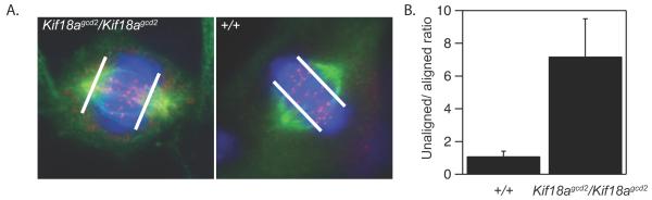
Genome integrity in the developing germ line is strictly required for fecundity. In proliferating somatic cells and in germ cells, there are mitotic checkpoint mechanisms that ensure accurate chromosome segregation and euploidy. There is growing evidence of mitotic cell cycle components that are uniquely required in the germ line to ensure genome integrity. We previously showed that the primary phenotype of germ cell deficient 2 (gcd2) mutant mice is infertility due to germ cell depletion during embryogenesis. Here we show that the underlying mutation is a mis-sense mutation, R308K, in the motor domain of the kinesin-8 family member, KIF18A, a protein that is expressed in a variety of proliferative tissues and is a key regulator of chromosome alignment during mitosis. Despite the conservative nature of the mutation, we show that its functional consequences are equivalent to KIF18A deficiency in HeLa cells. We also show that somatic cells progress through mitosis, despite having chromosome alignment defects, while germ cells with similar chromosome alignment defects undergo mitotic arrest and apoptosis. Our data provide evidence for differential requirements for chromosome alignment in germ and somatic cells and show that Kif18a is one of a growing number of genes that are specifically required for cell cycle progression in proliferating germ cells.
Keywords: Cell cycle; Gametogenesis; Germ cells; Kinesin; Laboratory mouse; Mitosis; Mitotic spindle.
Copyright © 2015 Elsevier Inc. All rights reserved.
Figures


Similar articles
-
vasa and piwi are required for mitotic integrity in early embryogenesis in the spider Parasteatoda tepidariorum.Dev Biol. 2015 Jun 15;402(2):276-90. doi: 10.1016/j.ydbio.2014.08.032. Epub 2014 Sep 23. Dev Biol. 2015. PMID: 25257304
-
Defects in chromosome congression and mitotic progression in KIF18A-deficient cells are partly mediated through impaired functions of CENP-E.Cell Cycle. 2009 Aug 15;8(16):2643-9. doi: 10.4161/cc.8.16.9366. Epub 2009 Aug 29. Cell Cycle. 2009. PMID: 19625775 Free PMC article.
-
Kif18A is involved in human breast carcinogenesis.Carcinogenesis. 2010 Sep;31(9):1676-84. doi: 10.1093/carcin/bgq134. Epub 2010 Jul 1. Carcinogenesis. 2010. PMID: 20595236
-
Chromokinesins: multitalented players in mitosis.Trends Cell Biol. 2005 Jul;15(7):349-55. doi: 10.1016/j.tcb.2005.05.006. Trends Cell Biol. 2005. PMID: 15946846 Review.
-
The control of cell cycle in mouse primordial germ cells: old and new players.Curr Pharm Des. 2012;18(3):233-44. doi: 10.2174/138161212799040448. Curr Pharm Des. 2012. PMID: 22229562 Review.
Cited by
-
KIFC1 is essential for normal spermatogenesis and its depletion results in early germ cell apoptosis in the Kuruma shrimp, Penaeus (Marsupenaeus) japonicus.Aging (Albany NY). 2019 Dec 29;11(24):12773-12792. doi: 10.18632/aging.102601. Epub 2019 Dec 29. Aging (Albany NY). 2019. PMID: 31895691 Free PMC article.
-
Modification of the neck-linker of KIF18A alters Microtubule subpopulation preference.Mol Biol Cell. 2024 Jan 1;35(1):ar3. doi: 10.1091/mbc.E23-05-0167. Epub 2023 Oct 30. Mol Biol Cell. 2024. PMID: 37903223 Free PMC article.
-
Whole-genome doubling in tissues and tumors.Trends Genet. 2023 Dec;39(12):954-967. doi: 10.1016/j.tig.2023.08.004. Epub 2023 Sep 14. Trends Genet. 2023. PMID: 37714734 Free PMC article. Review.
-
Weakened APC/C activity at mitotic exit drives cancer vulnerability to KIF18A inhibition.EMBO J. 2024 Mar;43(5):666-694. doi: 10.1038/s44318-024-00031-6. Epub 2024 Jan 26. EMBO J. 2024. PMID: 38279026 Free PMC article.
-
Mitotic chromosome alignment ensures mitotic fidelity by promoting interchromosomal compaction during anaphase.J Cell Biol. 2019 Apr 1;218(4):1148-1163. doi: 10.1083/jcb.201807228. Epub 2019 Feb 7. J Cell Biol. 2019. PMID: 30733233 Free PMC article.
References
-
- Wilkinson DG. In Situ Hybridization: A Practical Approach. Vol. 224. Oxford University Press; USA: 1992.
-
- Tam PP, Snow MH. Proliferation and migration of primordial germ cells during compensatory growth in mouse embryos. Journal of embryology and experimental morphology. 1981;64:133–47. - PubMed
Publication types
MeSH terms
Substances
Grants and funding
LinkOut - more resources
Full Text Sources
Other Literature Sources
Molecular Biology Databases
