. 2015 Apr 30;43(8):4284-95.
doi: 10.1093/nar/gkv189. Epub 2015 Mar 30.

The hexameric structure of the human mitochondrial replicative helicase Twinkle

Affiliations

The hexameric structure of the human mitochondrial replicative helicase Twinkle

Pablo Fernández-Millán et al. Nucleic Acids Res. .

Abstract

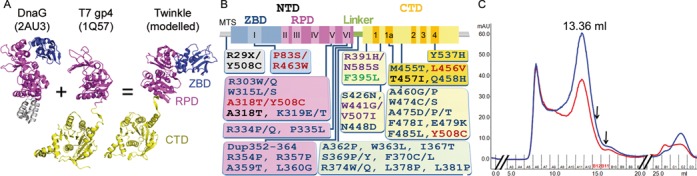
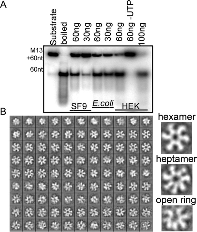
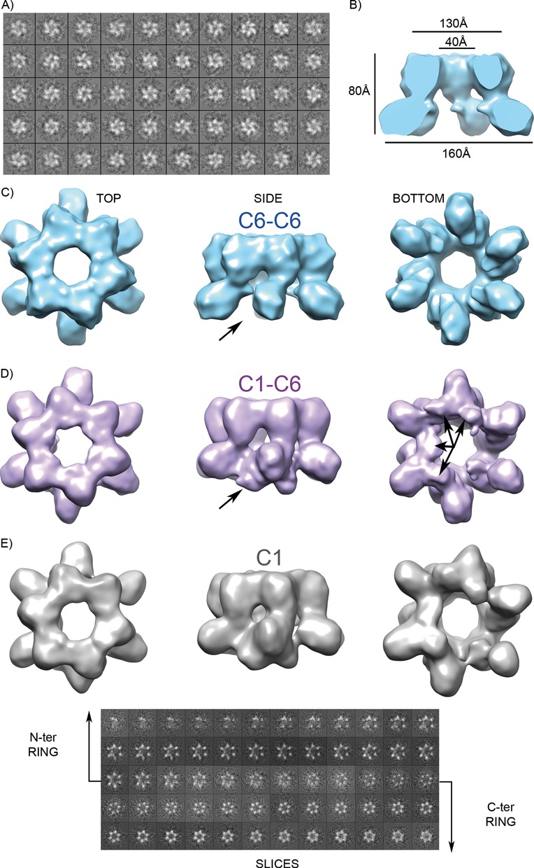
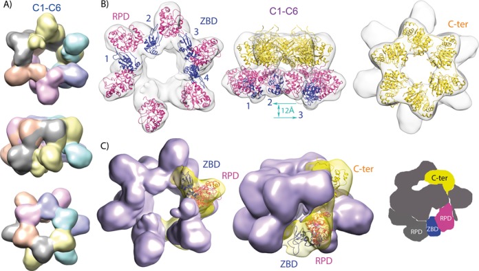
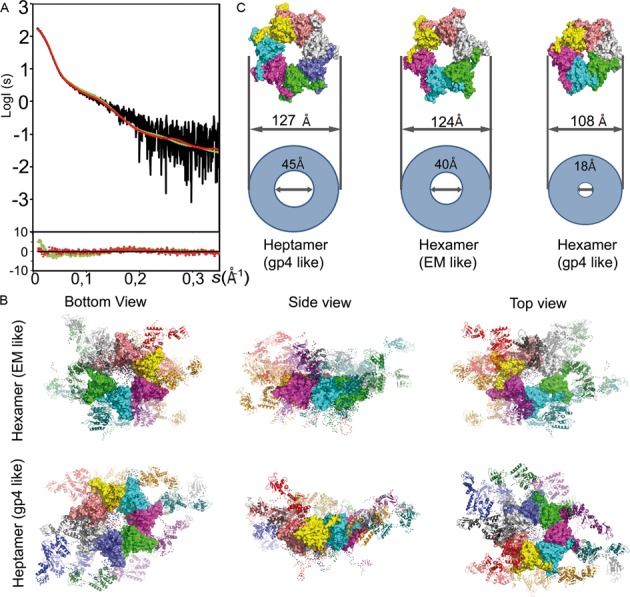
The mitochondrial replicative helicase Twinkle is involved in strand separation at the replication fork of mitochondrial DNA (mtDNA). Twinkle malfunction is associated with rare diseases that include late onset mitochondrial myopathies, neuromuscular disorders and fatal infantile mtDNA depletion syndrome. We examined its 3D structure by electron microscopy (EM) and small angle X-ray scattering (SAXS) and built the corresponding atomic models, which gave insight into the first molecular architecture of a full-length SF4 helicase that includes an N-terminal zinc-binding domain (ZBD), an intermediate RNA polymerase domain (RPD) and a RecA-like hexamerization C-terminal domain (CTD). The EM model of Twinkle reveals a hexameric two-layered ring comprising the ZBDs and RPDs in one layer and the CTDs in another. In the hexamer, contacts in trans with adjacent subunits occur between ZBDs and RPDs, and between RPDs and CTDs. The ZBDs show important structural heterogeneity. In solution, the scattering data are compatible with a mixture of extended hexa- and heptameric models in variable conformations. Overall, our structural data show a complex network of dynamic interactions that reconciles with the structural flexibility required for helicase activity.


Figures

Figure 1.
Figure 1.
Model, mutants and purification of Twinkle. (A) Ribbon representation of DnaG (PDB code 2AU3), the natural short form of T7 gp4 (PDB 1Q57) and the modeled Twinkle monomer. ZBD is shown in blue, RPD in magenta and CTD in gold. The DnaB interaction domain of DnaG is in gray. (B) Representation of disease-causing mutations of human c10orf2 gene on a scheme showing the Twinkle domains (IOSCA, red; adPEO, blue; MDS, black; Perrault Syndrome, purple; Renal Tubulopathy, green) (references in Supplementary Table S1). Domains are represented with same colors as in panel A. MTS stands for mitochondrial targeting sequence; motifs I to VI of the primase domain and motifs 1 to 4 in the helicase domain are indicated. (C) Size exclusion chromatography profile of Twinkle produced in SF9 insect cells (see experimental procedures). The blue and red curves correspond to the absorption at 280 and 260 nm, respectively. The peak of Twinkle is labeled with the elution volume. The fractions used for GraFix analysis are indicated by arrows.
Figure 2.
Figure 2.
Activity and oligomers of Twinkle. (A) DNA unwinding activity of the three forms of Twinkle, expressed in SF9 insect cells (SF9, lanes 3 and 4), in E. coli (lanes 5 and 6) and in HEK mammalian cells (HEK, lanes 7 to 9, produced as previously reported) (25), was analyzed by monitoring the unwound ssDNA radiolabeled product of 60 bases from the M13(+) ssDNA plasmid. Lane 1 contains substrate; lane 2, boiled substrate; and the remaining lanes contain the indicated amounts of Twinkle (from 30ng to 100ng). Helicase activity of Twinkle was tested in the presence of UTP, except lane 8 for the HEK cells (lane 60ng -UTP). (B) Samples of Twinkle produced in insect cells purified by size exclusion chromatography were analyzed by negative staining EM. The two-dimensional class averages obtained by classification show the presence of hexamers, heptamers and open rings.
Figure 3.
Figure 3.
Mitochondrial helicase 3D reconstruction. (A) Cryo-EM 2D classes of a crosslinked GraFix fraction (see Results) showed hexamers. (B) The 3D reconstruction applying C6 symmetry to the whole particle (C6-C6 map) shows two stacked rings, one with continuous density and the other with no density between lobules. (C) Three views of the electron density map after applying 6-fold symmetry to the whole particle. (D) Relaxation of the symmetry at the discontinuous ring (C1-C6 map) resulted in extra density between lobules, indicated by arrows. (E) Rendering of the 3D map without imposed symmetry (C1) is displayed together with slices of the reconstructed volume. The sequence of slices reveals 6-fold symmetry in the C-terminal ring but open asymmetric rings at the N-terminal region.
Figure 4.
Figure 4.
Model fitting. (A) Segmentation of the C1-C6 map into monomers upon structural fitting based on the T7 gp4 short form structural arrangement (12), in which each RPD contacts the C-terminal domain of the next subunit. (B) Organization of the molecular model upon flexible fitting into the C1-C6 3D map. The ZBD domains are numbered and represented in blue, RPDs in magenta and CTDs in gold. The C- (or N)-terminal domains are not depicted in the left (or right) volume, for clarity. The vertical blue arrow symbolizes the level difference between ZBD2 and ZBD3 (12 Å). (C) Left and central volumes are segmented around one subunit with the homology model fitted in, with same colors as in B. Right, scheme showing a ZBD contacting the RPD from its own subunit and from its neighbor simultaneously.
Figure 5.
Figure 5.
Analysis of Twinkle in solution. (A) Left panel: the experimental scattering-intensity curve (in black) is represented on a logarithmic scale as a function of the momentum transfer, s = 4πsin(θ) λ−1 (2θ, scattering angle; λ = 0.9919 Å, X-ray wavelength). The fitted EOM (see Experimental Procedures) curve for the mixture of flexible hexamers and heptamers (in red, χ2 = 0.71) and for the flexible gp4-like hexamer (in green, χ2 = 1.80) is shown. Below the panel, residuals show respective quality of fit. (B) Top and lateral molecular representation of the sub-ensemble of two models of T7 gp4 structure-based (1Q57, gp4 like) heptamers (right column) and three of EM-based hexamers (EM like, left) that collectively fit the data, superimposed by their CTD. ZBDs and RPDs are represented in ribbons while CTD rings are represented as a surface; each subunit has a different color. Red dots represent the position of Cα atoms of the flexible linkers and N- and C-terminal tails. (C) Surface representations of Twinkle CTD rings based on the T7 gp4 heptamer (PDB 1Q57) and hexamer (1E0J) and the cryo EM structure described here. Relative internal and external diameters values are represented by circles.

Similar articles

Cited by

References

    1. Spelbrink J.N., Li F.Y., Tiranti V., Nikali K., Yuan Q.P., Tariq M., Wanrooij S., Garrido N., Comi G., Morandi L., et al. Human mitochondrial DNA deletions associated with mutations in the gene encoding Twinkle, a phage T7 gene 4-like protein localized in mitochondria. Nat. Genet. 2001;28:223–231. - PubMed
    1. Leipe D.D., Aravind L., Grishin N.V., Koonin E.V. The bacterial replicative helicase DnaB evolved from a RecA duplication. Genome Res. 2000;10:5–16. - PubMed
    1. Shutt T.E., Gray M.W. Twinkle, the mitochondrial replicative DNA helicase, is widespread in the eukaryotic radiation and may also be the mitochondrial DNA primase in most eukaryotes. J. Mol. Evol. 2006;62:588–599. - PubMed
    1. Singleton M.R., Dillingham M.S., Wigley D.B. Structure and mechanism of helicases and nucleic acid translocases. Annu. Rev. Biochem. 2007;76:23–50. - PubMed
    1. Ziebarth T.D., Farr C.L., Kaguni L.S. Modular architecture of the hexameric human mitochondrial DNA helicase. J. Mol. Biol. 2007;367:1382–1391. - PMC - PubMed

Publication types