. 2015 Jun;282(12):2379-93.
doi: 10.1111/febs.13286. Epub 2015 Apr 16.

Novel familial dilated cardiomyopathy mutation in MYL2 affects the structure and function of myosin regulatory light chain

Affiliations

Novel familial dilated cardiomyopathy mutation in MYL2 affects the structure and function of myosin regulatory light chain

Wenrui Huang et al. FEBS J. 2015 Jun.

Abstract

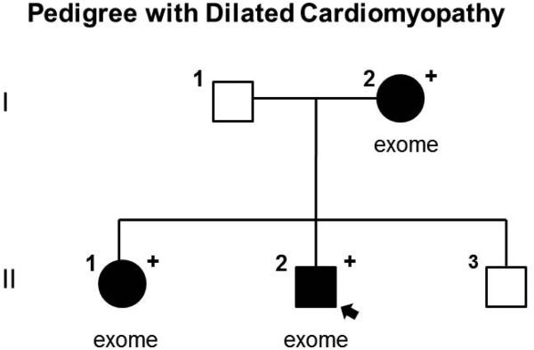
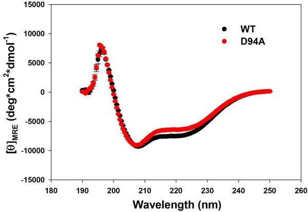
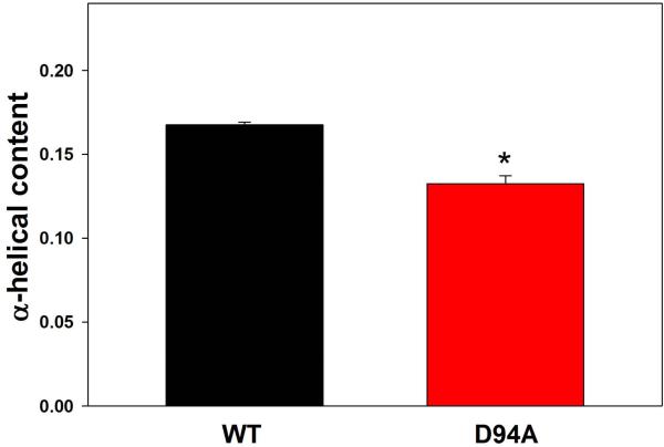
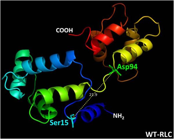
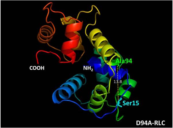
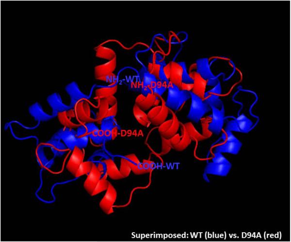
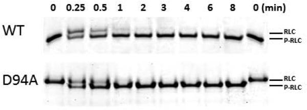
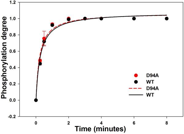
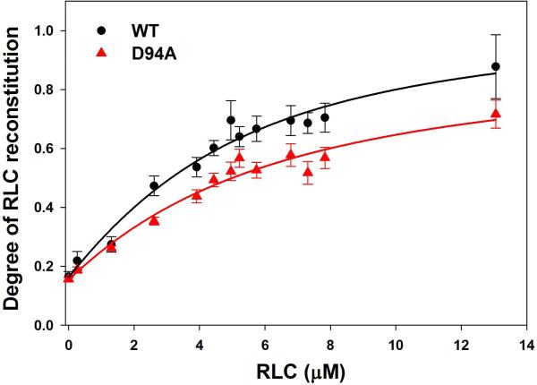
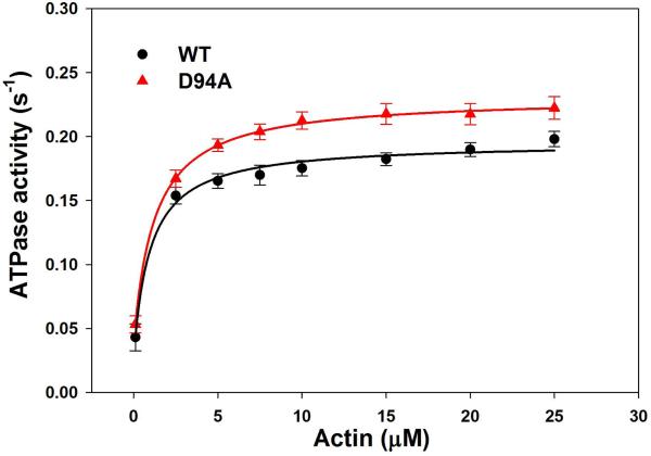
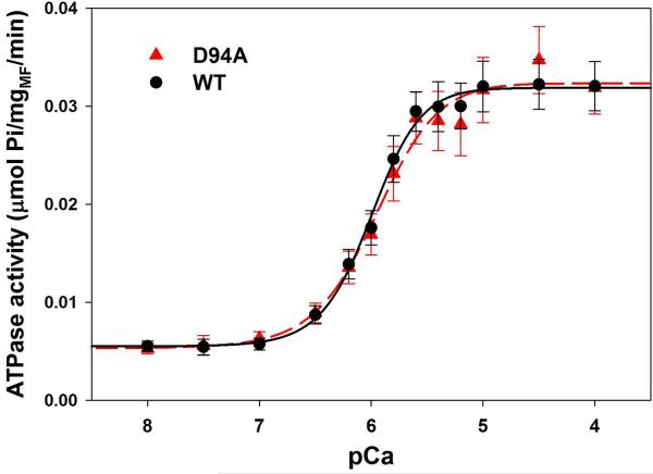
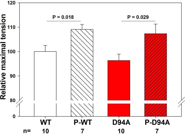
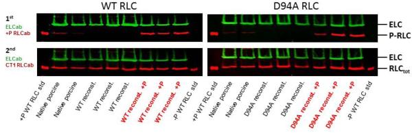
Dilated cardiomyopathy (DCM) is a disease of the myocardium characterized by left ventricular dilatation and diminished contractile function. Here we describe a novel DCM mutation in the myosin regulatory light chain (RLC), in which aspartic acid at position 94 is replaced by alanine (D94A). The mutation was identified by exome sequencing of three adult first-degree relatives who met formal criteria for idiopathic DCM. To obtain insight into the functional significance of this pathogenic MYL2 variant, we cloned and purified the human ventricular RLC wild-type (WT) and D94A mutant proteins, and performed in vitro experiments using RLC-mutant or WT-reconstituted porcine cardiac preparations. The mutation induced a reduction in the α-helical content of the RLC, and imposed intra-molecular rearrangements. The phosphorylation of RLC by Ca²⁺/calmodulin-activated myosin light chain kinase was not affected by D94A. The mutation was seen to impair binding of RLC to the myosin heavy chain, and its incorporation into RLC-depleted porcine myosin. The actin-activated ATPase activity of mutant-reconstituted porcine cardiac myosin was significantly higher compared with ATPase of wild-type. No changes in the myofibrillar ATPase-pCa relationship were observed in wild-type- or D94A-reconstituted preparations. Measurements of contractile force showed a slightly reduced maximal tension per cross-section of muscle, with no change in the calcium sensitivity of force in D94A-reconstituted skinned porcine papillary muscle strips compared with wild-type. Our data indicate that subtle structural rearrangements in the RLC molecule, followed by its impaired interaction with the myosin heavy chain, may trigger functional abnormalities contributing to the DCM phenotype.

Keywords: ATPase activity; RLC-reconstituted β-myosin; muscle contraction; phosphorylation; secondary structure.


Figures

Figure 1
Figure 1
Pedigree with DCM, solid symbols denote DCM, open symbols denote unaffected, “+” denotes heterozygous for D94A mutation, arrow points to the proband. Individuals who underwent exome sequencing are denoted “exome”.
Figure 2
Figure 2
A. Effect of the D94A RLC mutation on the CD spectra of RLC. Far-UV CD was performed utilizing a 1-mm path quartz cell in a Jasco J-720 spectropolarimeter. Spectra were recorded at 190–250 nm with a bandwidth of 1 nm. B. The [θ]222nm value was used to calculate the α-helical content (fH) using the following equation: [θ]222 = −30,300 fH − 2,340. Note significant reduction in α-helical content in D94A mutant compared to WT.
Figure 2
Figure 2
A. Effect of the D94A RLC mutation on the CD spectra of RLC. Far-UV CD was performed utilizing a 1-mm path quartz cell in a Jasco J-720 spectropolarimeter. Spectra were recorded at 190–250 nm with a bandwidth of 1 nm. B. The [θ]222nm value was used to calculate the α-helical content (fH) using the following equation: [θ]222 = −30,300 fH − 2,340. Note significant reduction in α-helical content in D94A mutant compared to WT.
Figure 3
Figure 3
Modeled structure of human ventricular RLCs, WT (A), D94A (B) and superimposed structures of WT (blue) and D94A (red) (C), using I-TASSER. Note that the Ala94 site is positioned closer to Ser15 than Asp94. The distance between the C-α of Ser15 and C-α of Asp94 is 21.9 Å while that of Ala94 is 13.8 Å. The predicted structures of RLCs were based on 3jvtB, 1prwA, 4ik1A, 2mysA, 4i2yA and 2w4aB PDB structures.
Figure 3
Figure 3
Modeled structure of human ventricular RLCs, WT (A), D94A (B) and superimposed structures of WT (blue) and D94A (red) (C), using I-TASSER. Note that the Ala94 site is positioned closer to Ser15 than Asp94. The distance between the C-α of Ser15 and C-α of Asp94 is 21.9 Å while that of Ala94 is 13.8 Å. The predicted structures of RLCs were based on 3jvtB, 1prwA, 4ik1A, 2mysA, 4i2yA and 2w4aB PDB structures.
Figure 4
Figure 4
A. Representative Urea-gel picture of the time course of phosphorylation of WT and D94A RLCs. Note a charge based separation of the phosphorylated (P-RLC) and non-phosphorylated RLC bands. B. Time course of MLCK-dependent phosphorylation of WT and D94A RLCs (0 to 8 minutes). Data points were fitted to Eq. 3 to generate phosphorylation kinetics rates (k). Note that both WT and D94A were phosphorylated with the rate of 1.1 min−1 (n=3 independent experiments) and reached the same maximal level of RLC phosphorylation.
Figure 4
Figure 4
A. Representative Urea-gel picture of the time course of phosphorylation of WT and D94A RLCs. Note a charge based separation of the phosphorylated (P-RLC) and non-phosphorylated RLC bands. B. Time course of MLCK-dependent phosphorylation of WT and D94A RLCs (0 to 8 minutes). Data points were fitted to Eq. 3 to generate phosphorylation kinetics rates (k). Note that both WT and D94A were phosphorylated with the rate of 1.1 min−1 (n=3 independent experiments) and reached the same maximal level of RLC phosphorylation.
Figure 5
Figure 5
The interaction of WT and D94A with the myosin heavy chain. A. Representative 15% SDS-PAGE gel picture of titration experiments of RLC-depleted porcine cardiac myosin with increasing concentrations of WT or D94A RLCs. ELC that remains intact during the depletion-reconstitution procedures was used as a loading control. B. Binding isotherms of WT or D94A to RLC-depleted porcine myosin. The data points were fitted into the nonlinear binding model (Eq. 4 and 5) yielding the apparent dissociation constant (Kd) and stoichiometry (n). Compared to WT, the D94A mutation significantly decreased the maximal level of RLC reconstitution.
Figure 5
Figure 5
The interaction of WT and D94A with the myosin heavy chain. A. Representative 15% SDS-PAGE gel picture of titration experiments of RLC-depleted porcine cardiac myosin with increasing concentrations of WT or D94A RLCs. ELC that remains intact during the depletion-reconstitution procedures was used as a loading control. B. Binding isotherms of WT or D94A to RLC-depleted porcine myosin. The data points were fitted into the nonlinear binding model (Eq. 4 and 5) yielding the apparent dissociation constant (Kd) and stoichiometry (n). Compared to WT, the D94A mutation significantly decreased the maximal level of RLC reconstitution.
Figure 6
Figure 6
ATPase activity of WT and D94A reconstituted porcine cardiac myosin and myofibrils. A. Actin activated myosin ATPase activity. Data points were fitted to the Michaelis-Menten equation to generate Vmax and Km. Note that the D94A mutation significantly increased the maximal level of ATPase activity with no change in Km compared with WT. B. Myofibrillar ATPase activity assay and the ATPase-pCa relationship in WT and D94A reconstituted porcine cardiac myofibrils. Small, but not significant, decreases in the Ca2+ sensitivity and nH were observed for the mutant compared with WT reconstituted myofibrils.
Figure 6
Figure 6
ATPase activity of WT and D94A reconstituted porcine cardiac myosin and myofibrils. A. Actin activated myosin ATPase activity. Data points were fitted to the Michaelis-Menten equation to generate Vmax and Km. Note that the D94A mutation significantly increased the maximal level of ATPase activity with no change in Km compared with WT. B. Myofibrillar ATPase activity assay and the ATPase-pCa relationship in WT and D94A reconstituted porcine cardiac myofibrils. Small, but not significant, decreases in the Ca2+ sensitivity and nH were observed for the mutant compared with WT reconstituted myofibrils.
Figure 7
Figure 7
Maximal force generation (A) and the force-pCa relationship (B) in skinned papillary muscle strips reconstituted with WT and D94A RLCs (C). A. The difference in maximal tension per cross section of muscle strip was ~3 kN/m2 (P>0.05). B. The force-pCa curve of D94A was overlapping that of WT, indicating no changes in the calcium sensitivity or Hill coefficient. C. Representative force-pCa traces in WT-reconstituted porcine papillary muscle strips.
Figure 7
Figure 7
Maximal force generation (A) and the force-pCa relationship (B) in skinned papillary muscle strips reconstituted with WT and D94A RLCs (C). A. The difference in maximal tension per cross section of muscle strip was ~3 kN/m2 (P>0.05). B. The force-pCa curve of D94A was overlapping that of WT, indicating no changes in the calcium sensitivity or Hill coefficient. C. Representative force-pCa traces in WT-reconstituted porcine papillary muscle strips.
Figure 8
Figure 8
The effect of RLC phosphorylation on maximal steady state force in WT- and D94A-reconstituted porcine papillary muscle strips. A. RLC phosphorylation increased maximal force generation by ~1.1-fold in both WT- and D94A-reconstituted fibers. B. Representative Western blots of WT- and D94A-reconstituted fibers before and after the treatment with MLCK.
Figure 8
Figure 8
The effect of RLC phosphorylation on maximal steady state force in WT- and D94A-reconstituted porcine papillary muscle strips. A. RLC phosphorylation increased maximal force generation by ~1.1-fold in both WT- and D94A-reconstituted fibers. B. Representative Western blots of WT- and D94A-reconstituted fibers before and after the treatment with MLCK.

References

    1. Schonberger J, Seidman CE. Many roads lead to a broken heart: the genetics of dilated cardiomyopathy. Am J Hum Genet. 2001;69:249–60. - PMC - PubMed
    1. Hershberger RE, Hedges DJ, Morales A. Dilated cardiomyopathy: the complexity of a diverse genetic architecture. Nature reviews Cardiology. 2013;10:531–47. - PubMed
    1. Millat G, Bouvagnet P, Chevalier P, Sebbag L, Dulac A, Dauphin C, Jouk PS, Delrue MA, Thambo JB, Le Metayer P, Seronde MF, Faivre L, Eicher JC, Rousson R. Clinical and mutational spectrum in a cohort of 105 unrelated patients with dilated cardiomyopathy. Eur J Med Genet. 2011;54:e570–5. - PubMed
    1. Szczesna-Cordary D, Morimoto S, Gomes AV, Moore JR. Cardiomyopathies: classification, clinical characterization, and functional phenotypes. Biochem Res Int. 2012;2012:870942. - PMC - PubMed
    1. Parvari R, Levitas A. The mutations associated with dilated cardiomyopathy. Biochem Res Int. 2012;2012:639250. - PMC - PubMed

Publication types

MeSH terms

Supplementary concepts