. 2015 Mar 31;131(13):1171-80.
doi: 10.1161/CIRCULATIONAHA.114.010482. Epub 2015 Feb 17.

Targeting pathogenic postischemic self-recognition by natural IgM to protect against posttransplantation cardiac reperfusion injury

Affiliations

Targeting pathogenic postischemic self-recognition by natural IgM to protect against posttransplantation cardiac reperfusion injury

Carl Atkinson et al. Circulation. .

Abstract

Background: Natural IgM antibodies represent a class of innate pattern recognition receptors that recognize danger-associated molecular patterns expressed on stressed or dying cells. They play important roles in tissue homeostasis by disposing of prenecrotic cells and suppressing inflammation. However, ischemic insult leads to a pathogenic level of IgM binding and complement activation, resulting in inflammation and injury. We investigate the role of self-reactive IgM in the unique setting of transplantation where the donor organ undergoes both cold and warm ischemia and global ischemic insult.

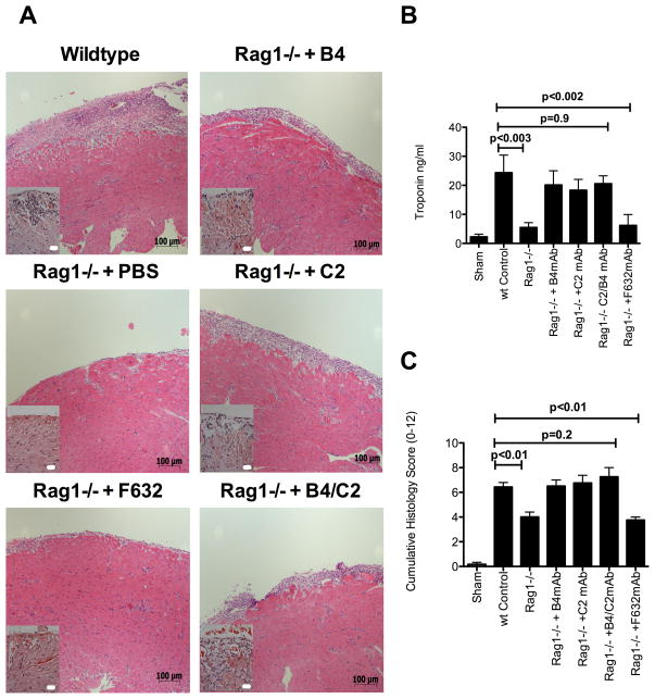
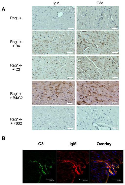
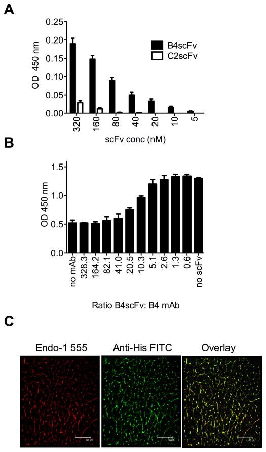
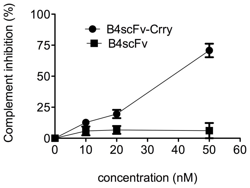
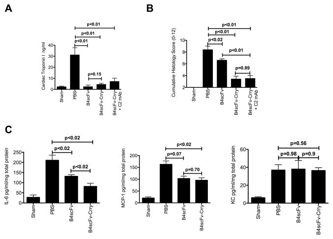
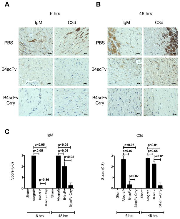
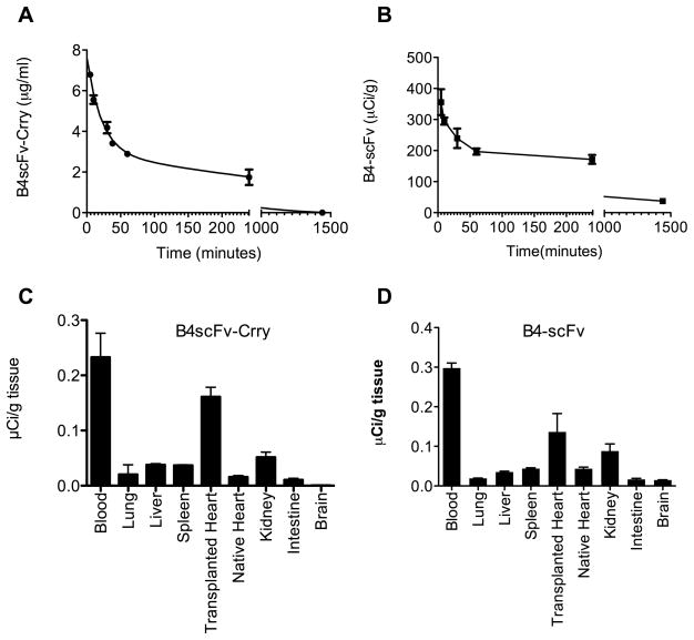
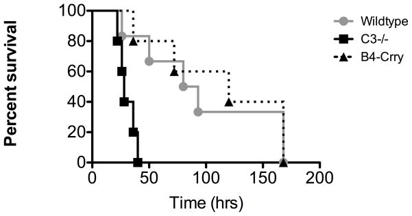
Methods and results: By transplanting hearts from wild-type donor mice into antibody-deficient mice reconstituted with specific self-reactive IgM monoclonal antibodies, we identified neoepitopes expressed after transplantation and demonstrated a key role for IgM recognition of these epitopes in graft injury. With this information, we developed and characterized a therapeutic strategy that exploited the postischemia recognition system of natural antibodies. On the basis of neoepitope identification, we constructed an anti-annexin IV single-chain antibody (scFv) and an scFv linked to Crry, an inhibitor of C3 activation (scFv-Crry). In an allograft transplantation model in which recipients contain a full natural antibody repertoire, both constructs blocked graft IgM binding and complement activation and significantly reduced graft inflammation and injury. Furthermore, scFv-Crry specifically targeted to the transplanted heart and, unlike complement deficiency, did not affect immunity to infection, an important consideration for immunosuppressed transplant recipients.

Conclusions: We identified pathophysiologically important epitopes expressed within the heart after transplantation and described a novel translatable strategy for targeted complement inhibition that has several advantages over currently available approaches.

Keywords: antibodies; complement system proteins; inflammation; ischemia; transplantation.


Figures

Figure 1
Figure 1
Administration of IgM mAbs in Rag1−/− recipients of cardiac isografts. Hearts from Rag1−/− donor mice were transplanted into wt recipients, or into Rag1−/− recipients with or without administration of indicated IgM mAb. Isografts were analyzed 48 hours post-transplantation. A, Representative Hematoxylin and Eosin stained cardiac sections from grafts, showing that epi- and endocardial inflammation and damage is restored by B4, C2, or B4+C2 combined mAbs, but not F632 mAb treatment of otherwise protected Rag1−/− recipients. Scale bars = 20 μm high power insert and 100 μm low power. B, Serum cardiac troponin I levels and C. Semi-quantitative histology injury scores. Pairwise comparisons between wt control vs. Rag1−/−, wt control vs. F632 mAb, wt Control vs B4, C2, B4+C2 mAbs combined were made using non-parametric analysis. Mean ± SEM, n = 4–8.
Figure 2
Figure 2
Deposition of IgM and C3d in cardiac isografts following IgM mAb reconstitution of Rag1−/− recipient mice. A, IgM and C3d binding was assessed by immunohistochemistry in transplanted hearts 6 hours post-transplantation. IgM and C3d deposition is seen in B4, C2, and B4 and C2 combined mAb treated recipients, with staining localized predominantly to endothelial cells of arterioles, capillaries, and microvessels within the graft myocardium. Little to no staining for IgM or C3d seen in grafts from PBS treated Rag1−/− recipients or Rag1−/− recipients treated with F623 control mAb. Representative images, n = 3. Scale bars, 20 μm. B, Colocalization of IgM and C3d in isografts from B4 mAb treated Rag1−/− recipient mice. Binding was assessed by immunofluorescence microscopy of sections from epicardial surface of transplanted hearts isolated 6 hours post-transplantation. IgM and C3d binding colocalized on the vessel endothelium and myocardial tissue. Representative image, n = 3. Scale bars, 53 μm.
Figure 3
Figure 3
Characterization of neoepitope-targeted B4scFv. A, Direct binding of His-tagged B4scFv to annexin IV, as demonstrated by ELISA using anti-His tag detection Ab. C2scFv was used as specificity control. Mean +/− SEM, n = 3. B, Competitive inhibition of B4 mAb binding to annexin IV by B4scFv, as determined by competition ELISA using anti-IgM detection Ab. Mean +/− SEM, n = 3. C, B4scFv targets cardiac allografts post reperfusion. Hearts from Balb/c donors were transplanted into C57BL/6 recipients with administration of B4scFv, and allografts analyzed 6 hours post-transplantation. Images show immunofluorescence staining of section from intra-myocardial space for endothelial cells (red) and His-tagged B4scFv (green), with colocalization of B4scFv and endothelial cells indicated by yellow (composite). B4scFv localized predominantly to the microvasculature of the cardiac allograft. Representative image, n = 3. Scale bars, 68 μm.
Figure 4
Figure 4
Crry activity is retained in the B4scFv-Crry construct. Increasing concentrations of B4scFv-Crry or B4scFv were incubated with activated zymosan particles in mouse serum and C3 deposition assayed by flow cytometry. Mean +/− SD, n = 3.
Figure 5
Figure 5
Effect of B4scFv and B4scFvCrry on allograft ischemia reperfusion injury and inflammation. Recipient mice were treated with PBS, B4scFv, B4scFvCrry, or B4scFvCrry and C2mAb combined, and allografts and serum isolated 48 hours post-transplantation for analysis. A, Serum cardiac troponin I levels. Pairwise comparisons between wt control vs. B4scFv (p<0.01), wt control vs. B4scFv-Crry (p<0.001), wt control vs B4scFv-Crry + C2 mAb (p<0.001), and B4scFv vs. B4scFv-Crry was not significant (p=0.15). Mean ± SEM, n = 6–8. B, Histological assessment of injury from hematoxylin and eosin stained cardiac sections. Non-parametric two tailed analysis demonstrated that wt PBS control vs. B4scFv (p=0.02), wt PBS control vs. B4scFv-Crry (p<0.01), wt control vs B4scFv-Crry + C2 mAb (p<0.01), and B4scFv vs. B4scFv-Crry (p<0.01) were noted. Mean ± SEM, n = 6–8. C, Intragraft expression of IL-6, MCP-1 and KC as determined by by ELISA. Pairwise two tailed non=parametric analysis between wt PBS control vs. B4scFv, wt PBS control vs. B4scFv-Crry, and B4scFv vs. B4scFv-Crry were made for each analyzed cytokine. Mean ± SEM, n = 6–8.
Figure 6
Figure 6
Effect of B4scFv and B4scFvCrry treatment on IgM and C3d deposition in transplanted allografts. Recipient mice were treated with PBS, B4scFv or B4scFvCrry and allografts isolated at 6 hours (A) or 48 hours (B) post-transplantation for analysis. Representative images of IgM and C3d deposition in grafts are shown. Scale bars, 20 μm. C, Semi-quantitative histological scoring of IgM and C3d deposition at 6 and 48 hours post-transplantation. Mean ± SEM, n=6–8.
Figure 7
Figure 7
Serum circulatory half-life and biodistribution of B4scFv-Crry and B4-scFv. A, Plasma levels of B4scFv-Crry and B4-scFv in blood collected at indicated times following i.v. injection were determined by ELISA or radiometry (see methods). Mean ± SEM, n = 3. B, Biodistribution of 125I-labeled B4scFv-Crry and B4-scFv determined 6 hours post-injection and transplantation. Mean ± SEM, n = 3.
Figure 8
Figure 8
Survival analysis in model of polymicrobial sepsis. Survival of C3-deficient mice and wt mice treated with either PBS or 0.2 mg B4scFv-Crry immediately following cecal ligation and puncture. Log-rank tests comparing survival across the three groups demonstrated that survival times were significantly different between groups (p<0.001). All pairwise comparisons demonstrated that survival was significantly greater in PBS and B4scFv-Crry as compared to C3−/− (p<0.01 for all), even after Bonferroni adjustment for multiple comparisons. No significant difference was seen between PBS and B4scFv-Crry (p>0.05). n = 5–6.

Comment in

References

    1. Weiser MR, Williams JP, Moore FD, Jr, Kobzik L, Ma M, Hechtman HB, Carroll MC. Reperfusion injury of ischemic skeletal muscle is mediated by natural antibody and complement. J Exp Med. 1996;183:2343–2348. - PMC - PubMed
    1. Zhang M, Alicot EM, Chiu I, Li J, Verna N, Vorup-Jensen T, Kessler B, Shimaoka M, Chan R, Friend D, Mahmood U, Weissleder R, Moore FD, Carroll MC. Identification of the target self-antigens in reperfusion injury. J Exp Med. 2006;203:141–152. - PMC - PubMed
    1. Zhang M, Austen WG, Jr, Chiu I, Alicot EM, Hung R, Ma M, Verna N, Xu M, Hechtman HB, Moore FD, Jr, Carroll MC. Identification of a specific self-reactive igm antibody that initiates intestinal ischemia/reperfusion injury. PNAS. 2004;101:3886–3891. - PMC - PubMed
    1. Elvington A, Atkinson C, Kulik L, Zhu H, Yu J, Kindy MS, Holers VM, Tomlinson S. Pathogenic natural antibodies propagate cerebral injury following ischemic stroke in mice. J Immunol. 2012;188:1460–1468. - PMC - PubMed
    1. Kulik L, Fleming SD, Moratz C, Reuter JW, Novikov A, Chen K, Andrews KA, Markaryan A, Quigg RJ, Silverman GJ, Tsokos GC, Holers VM. Pathogenic natural antibodies recognizing annexin iv are required to develop intestinal ischemia-reperfusion injury. J Immunol. 2009;182:5363–5373. - PMC - PubMed

Publication types

MeSH terms