. 2015 Jun;197(11):1963-71.
doi: 10.1128/JB.00147-15. Epub 2015 Mar 30.

Role of Bacillus subtilis DNA Glycosylase MutM in Counteracting Oxidatively Induced DNA Damage and in Stationary-Phase-Associated Mutagenesis

Affiliations

Role of Bacillus subtilis DNA Glycosylase MutM in Counteracting Oxidatively Induced DNA Damage and in Stationary-Phase-Associated Mutagenesis

Martha Gómez-Marroquín et al. J Bacteriol. 2015 Jun.

Abstract

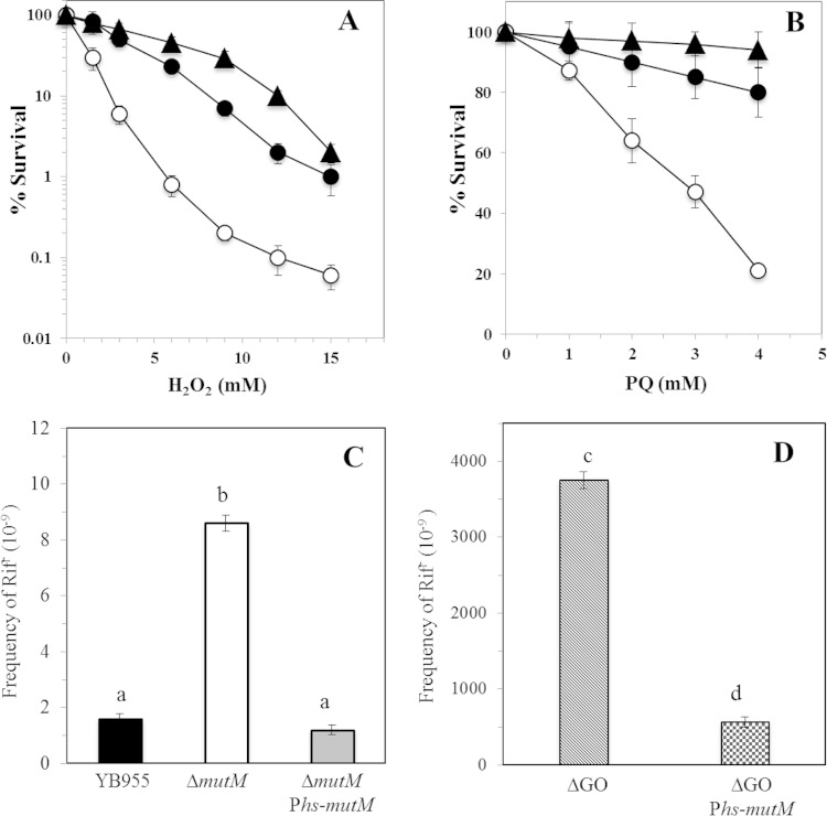
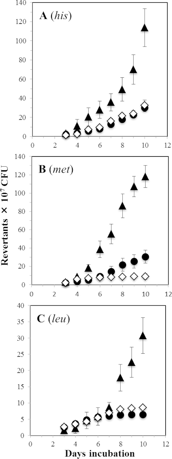
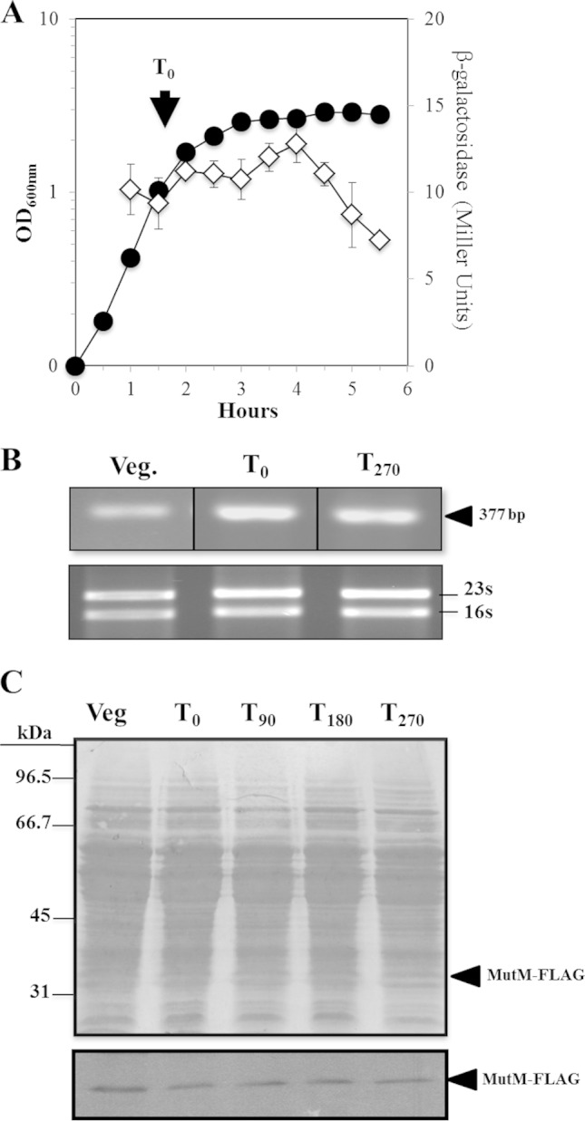
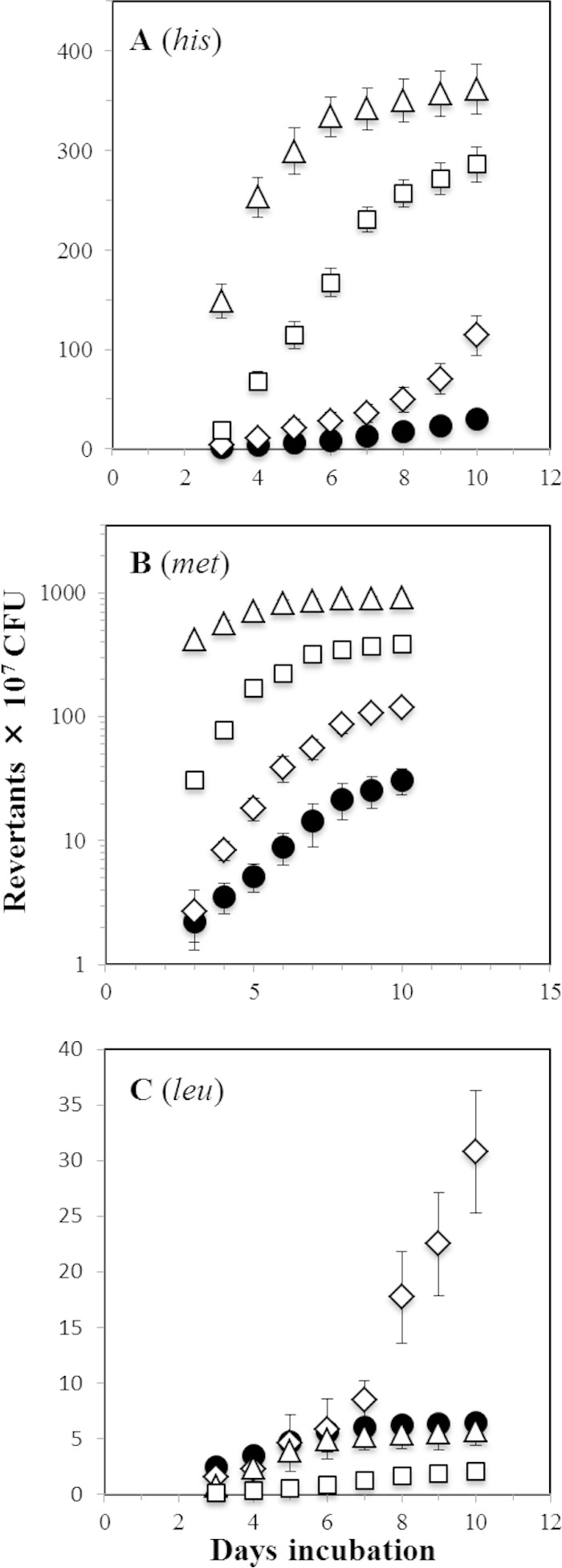
Reactive oxygen species (ROS) promote the synthesis of the DNA lesion 8-oxo-G, whose mutagenic effects are counteracted in distinct organisms by the DNA glycosylase MutM. We report here that in Bacillus subtilis, mutM is expressed during the exponential and stationary phases of growth. In agreement with this expression pattern, results of a Western blot analysis confirmed the presence of MutM in both stages of growth. In comparison with cells of a wild-type strain, cells of B. subtilis lacking MutM increased their spontaneous mutation frequency to Rif(r) and were more sensitive to the ROS promoter agents hydrogen peroxide and 1,1'-dimethyl-4,4'-bipyridinium dichloride (Paraquat). However, despite MutM's proven participation in preventing ROS-induced-DNA damage, the expression of mutM was not induced by hydrogen peroxide, mitomycin C, or NaCl, suggesting that transcription of this gene is not under the control of the RecA, PerR, or σ(B) regulons. Finally, the role of MutM in stationary-phase-associated mutagenesis (SPM) was investigated in the strain B. subtilis YB955 (hisC952 metB5 leuC427). Results revealed that under limiting growth conditions, a mutM knockout strain significantly increased the amount of stationary-phase-associated his, met, and leu revertants produced. In summary, our results support the notion that the absence of MutM promotes mutagenesis that allows nutritionally stressed B. subtilis cells to escape from growth-limiting conditions.

Importance: The present study describes the role played by a DNA repair protein (MutM) in protecting the soil bacterium Bacillus subtilis from the genotoxic effects induced by reactive oxygen species (ROS) promoter agents. Moreover, it reveals that the genetic inactivation of mutM allows nutritionally stressed bacteria to escape from growth-limiting conditions, putatively by a mechanism that involves the accumulation and error-prone processing of oxidized DNA bases.


Figures

FIG 1
FIG 1
Contribution of MutM in the survival of B. subtilis to H2O2 and PQ treatment (A and B) and frequencies of spontaneous mutation to Rifr of different B. subtilis strains (C and D). (A and B) B. subtilis YB955 (parental; ●), PERM751 (ΔmutM; ○), and PERM1199 (ΔmutM amyE::Phs-mutM; ▲) strains were treated with different amounts of H2O2 (A) or PQ (B), and cell viability was determined as described in Materials and Methods. The values shown represent the means and standard deviations from three independent experiments done in triplicate. (C and D) B. subtilis YB955 (parental), PERM751 (ΔmutM), PERM1199 (ΔmutM amyE::Phs-mutM), PERM573 (GO system deletion [ΔGO]), and PERM794 (ΔGO amyE::Phs-mutM) were grown overnight in PAB medium, and frequencies of mutation to Rifr were determined as described in Materials and Methods. Each bar represents the mean of data collected from three independent experiments done in sextuplicate, and the error bars represent standard errors of the means (SEM). The statistical differences (a, b, c, and d) between the mutation frequencies of each strain and condition, as determined by ANOVA (P < 0.05), are shown above each bar.
FIG 2
FIG 2
Stationary-phase-induced reversions to his (A), met (B), and leu (C) of the YB955 (◇), PERM571 (ΔmutM) (▲), and PERM1199 (ΔmutM amyE::Phs-mutM) (●) B. subtilis strains were determined as described in Materials and Methods. Data represent counts from six plates averaged from three separate tests normalized to initial cell titers ± standard deviations (SD).
FIG 3
FIG 3
(A). Levels of β-galactosidase in a mutM-lacZ transcriptional fusion during the vegetative and stationary phases of growth. B. subtilis strain PERM659 was grown in liquid antibiotic (A3) medium. Cell samples were collected at the indicated times and treated with lysozyme, and the extracts were assayed for β-galactosidase as described in Materials and Methods. Data shown are average values from triplicate independent experiments ± SD for β-galactosidase specific activity (◇) and for A600 values (●). (B) RT-PCR analysis of mutM transcription during the vegetative and stationary phases of growth. RNA samples (1 μg) isolated from a B. subtilis YB955 A3 culture, at the steps indicated, were processed for RT-PCR analysis as described in Materials and Methods. The arrowhead shows the size of the expected RT-PCR product. 16S and 23S rRNA bands are shown in the lower panel. (C) Western blot analysis of MutM-FLAG synthesis during the vegetative and stationary phases of growth. B. subtilis strain YB955 was grown in liquid A3 medium. Cell extract samples (∼100 μg of protein; see Materials and Methods), harvested at the steps indicated, were separated by SDS-PAGE and transferred to PVDF membranes (Bio-Rad, Hercules, CA). The blots were stained with Ponceau red (top), probed with a FLAG monoclonal antibody diluted 10,000-fold, and then processed with an ECL Western blot system (bottom). The positions of molecular mass markers are indicated to the left of the stained membrane. T0 is the time point in the culture when the slopes of the logarithmic and stationary phases of growth intercepted. T90, T180, and T270 indicate times in minutes after T0. Veg., vegetative growth.
FIG 4
FIG 4
Stationary-phase-induced reversions to his (A), met (B), and leu (C) of the YB955 (●), PERM571 (ΔmutM) (◇), PERM704 (ΔmutY) (□), and PERM573 (ΔmutM ΔmutY) (△) B. subtilis strains were determined as described in Materials and Methods. Data represent counts from six plates averaged from three separate tests normalized to initial cell titers ± SD.

References

    1. Scandalios JG. 2002. Oxidative stress responses—what have genome-scale studies taught us? Genome Biol 3:REVIEWS1019. doi:10.1186/gb-2002-3-7-reviews1019. - DOI - PMC - PubMed
    1. Saran M, Bors W. 1990. Radical reactions in vivo—an overview. Radiat Environ Biophys 29:249–262. doi:10.1007/BF01210406. - DOI - PubMed
    1. Michaels ML, Miller JH. 1992. The GO system protects organisms from the mutagenic effect of the spontaneous lesion 8-hydroxyguanine (7,8-dihydro-8-oxoguanine). J Bacteriol 174:6321–6325. - PMC - PubMed
    1. Castellanos-Juarez FX, Alvarez-Alvarez C, Yasbin RE, Setlow B, Pedraza-Reyes M. 2006. YtkD and MutT protect vegetative cells but not spores of Bacillus subtilis from oxidative stress. J Bacteriol 188:2285–2289. doi:10.1128/JB.188.6.2285-2289.2006. - DOI - PMC - PubMed
    1. Michaels ML, Cruz C, Grollman AP, Miller JH. 1992. Evidence that MutY and MutM combine to prevent mutations by an oxidatively damaged form of guanine in DNA. Proc Natl Acad Sci U S A 89:7022–7025. doi:10.1073/pnas.89.15.7022. - DOI - PMC - PubMed

Publication types

MeSH terms

LinkOut - more resources