Review
. 2015 Mar 30;208(7):869-80.
doi: 10.1083/jcb.201501101.

A little sugar goes a long way: the cell biology of O-GlcNAc

Review

A little sugar goes a long way: the cell biology of O-GlcNAc

Michelle R Bond et al. J Cell Biol. .

Abstract

Unlike the complex glycans decorating the cell surface, the O-linked β-N-acetyl glucosamine (O-GlcNAc) modification is a simple intracellular Ser/Thr-linked monosaccharide that is important for disease-relevant signaling and enzyme regulation. O-GlcNAcylation requires uridine diphosphate-GlcNAc, a precursor responsive to nutrient status and other environmental cues. Alternative splicing of the genes encoding the O-GlcNAc cycling enzymes O-GlcNAc transferase (OGT) and O-GlcNAcase (OGA) yields isoforms targeted to discrete sites in the nucleus, cytoplasm, and mitochondria. OGT and OGA also partner with cellular effectors and act in tandem with other posttranslational modifications. The enzymes of O-GlcNAc cycling act preferentially on intrinsically disordered domains of target proteins impacting transcription, metabolism, apoptosis, organelle biogenesis, and transport.


Figures
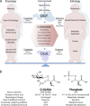
Figure 1.
Figure 1.
O-GlcNAc dynamically impacts biological homeostasis and disease pathologies by integrating environmental and genetic information. (A) The ubiquitous and essential modification of protein serine and threonine residues with O-GlcNAc modulates cellular biology by responding to variable nutrient conditions and integrating cellular programs to respond through nutrient-sensing and -managing networks. By targeting OGT and OGA to discrete intracellular sites, O-GlcNAcylation of diverse proteins (pink hexagon) influences the physiology of processes including memory, metabolism, and immunity. Aberrant O-GlcNAc modification is implicated in pathologies of metabolic and neurodegenertive diseases as well as cancers and autoimmunity. OIP, OGT interacting protein. (B) The O-GlcNAc and O-phosphate modifications share some characteristics but differ in others.
Figure 2.
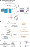
Figure 2.
The nucleotide sugar UDP-GlcNAc resides at the nexus of protein and lipid glycosylation. (A) Dynamic, nutrient-sensitive O-GlcNAc cycling is modulated by metabolite and enzyme availability. Glucose, acetyl-CoA, ATP, glutamine, and uridine are required for the synthesis of UDP-GlcNAc, the ultimate product of the hexosamine biosynthetic pathway (HBP). OGT uses the nucleotide sugar to modify proteins, whereas OGA catalyzes the modification’s removal. hOGT and hOGA splice variants are depicted. OGT and OGA isoforms have unique subcellular distributions and interaction partners, including one another. The O-GlcNAcylation of ID protein domains is known to influence protein secondary structure. MTS, mitochondrial targeting sequence; PPO, phosphoinositide binding domain. (B) Although most cellular glucose (Glc) is metabolized by glycolysis, 2–3% enters the HBP (Marshall et al., 1991a) and is phosphorylated and isomerized in two enzymatic steps to yield fructose-6-phosphate (F-6-P; Aguilera and Zimmermann, 1986; Stachelek et al., 1986). Next, glutamine:fructose-6-phosphate-amidotransferase (GFAT) acts to convert fructose-6-phosphate to glucosamine-6-phosphate (GlcN-6-P) in the HBP’s rate-limiting step (Watzele and Tanner, 1989; Marshall et al., 1991a,b). The installation of an acetyl group (Boehmelt et al., 2000a) is followed by a second isomerization by phosphoglucomutase 3 (PGM3), yielding GlcNAc-1-phosphate (GlcNAc-1-P; Hofmann et al., 1994). In the final step, UDP-GlcNAc pyrophosphorylase utilizes UTP and GlcNAc-1-phosphate to produce UDP-GlcNAc (Mio et al., 1998). Importantly, along with other mechanisms of regulation, GFAT is sensitive to UDP-GlcNAc inhibition, thereby modulating the cellular UDP-GlcNAc available to glycosyltransferases (GlycoTs) at any given time within the cell (McKnight et al., 1992). Salvage pathways can also introduce glucosamine and GlcNAc to enter the HBP directly bypassing GFAT (Bueding and MacKinnon, 1955; Hinderlich et al., 2000). Cells can rapidly take up exogenous glucosamine via the glucose transporter, which can be fully processed to UDP-GlcNAc (Schleicher and Weigert, 2000). Cellular GlcNAc from lysosomal degradation or the degradation of nutritional constituents can be converted by GlcNAc kinase to GlcNAc-6-phosphate (GlcNAc-6-P) and then converted to UDP-GlcNAc (Hinderlich et al., 2000). The diseases associated with deregulation of each HBP enzyme (left) are indicated on the right side of the figure, connected to the enzyme by a line. Glc-6-P, glucose-6-phosphate; GlcNAc-6-P, N-acetylglucosamine-6-phosphate; GK, glucokinase; EMeg32, glucosamine-6-phosphate acetyltransferase; TCA, tricarboxylic acid; NIDDM, noninsulin-dependent diabetes mellitus. (C) The activated nucleotide sugar UDP-GlcNAc is used by concentration-sensitive enzymes in the nucleus, cytoplasm, and on the plasma membrane to glycosylate substrates. Nucleotide sugar transporters actively transport UDP-GlcNAc into cellular organelles including the ER and Golgi. The relative cellular volumes of these organelles that differ in their permeability characteristics make estimates of cytoplasmic and nuclear concentrations of UDP-GlcNAc complicated. The relative abundance of O-GlcNAc is roughly inversely related to the abundance of more complex glycans.
Figure 3.
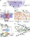
Figure 3.
O-GlcNAc modulates nuclear processes. (A) The nuclear envelope is contiguous with the rough ER and serves to separate the transcriptional machinery in the nucleus from the translation machinery associated with the rough ER and cytoplasm. Among the most heavily O-GlcNAcylated proteins are the nucleoporins (NUPs), 30 of which form the nuclear pore complex (NPC). (B) The NPC is responsible for the exchange of molecules across the nuclear envelope (NE) comprised of the inner and outer nuclear membranes (INM and ONM, respectively). The interaction between NUPs’ phenylalanine-glycine (FG) repeats and nuclear transport factors (NTFs) is required for the transport of cargo through NPCs. In vitro work has suggested that O-GlcNAc addition to FG repeats is critical for nuclear transport events through these “gateways.” (C) O-GlcNAc is implicated in higher-order chromatin structure through its modification of histone tails. In addition, nuclear machinery associated with transcription including transcription factors, transcriptional comodulators (SIN3A), ten eleven translocation proteins (TETs), and the host cell factor 1 (HCF-1) are known to associate with OGT or be O-GlcNAc modified. (D) O-GlcNAc is thought to modulate transcriptional initiation through its modification of the CTD of RNA polymerase II (PolII), which is known to act as a scaffold to recruit numerous transcriptional effectors. Panel D is redrawn and adapted from Phatnani and Greenleaf (2006). BBP, branchpoint-binding protein; PCTD, phosphocarboxy terminal domain; CRF, chromatin remodeling factor; XF, processing/export factor.
Figure 4.
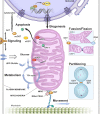
Figure 4.
O-GlcNAc modulates cellular signaling processes, metabolism, mitochondrial trafficking, and function as well as the cell cycle. OGT is intimately involved in transcriptional regulation through its interaction with and modification of SIN3A in the histone deacetylase corepressor complex. It is possible that through this interaction, a subset of regulatory genes is suppressed in a nutrient-sensitive manner yielding downstream consequences. Outside the nucleus, O-GlcNAc-modified cytokeratins are critical mediators of stress-responsive AKT signaling, whose activity is also modulated by O-GlcNAc. O-GlcNAc plays critical roles in both the function and trafficking of mitochondria. Key modified proteins include GTPase dynamin-related protein DRP1 and optical atrophy 1 (OPA1), the consequence of which is increased mitochondrial fission. Furthermore, although it is unclear how the complex of MIRO (mitochondrial Rho GTPase), kinesin-binding protein Milton, and TRAKs (trafficking kinesin protein) regulate mitochondrial trafficking in Drosophila, it is clear that OGT is recruited to the same location and may modulate the movement of the organelle along actin and microtubule networks. Increased levels of O-GlcNAc are also tied to the mitigation of myocardial ischemia-associated mitochondrial Ca2+ and reactive oxygen species generation, loss of membrane potential, and cytochrome c (Cyto c) release. Cell division requires that cellular organelles, including the mitochondria, are properly segregated during mitosis. OGT and OGA are found in a transient complex with Aurora B and phosphatase PP1 during cytokinesis, although the way in which this partitioning occurs is still being studied.

References

    1. Aguilera A., and Zimmermann F.K.. 1986. Isolation and molecular analysis of the phosphoglucose isomerase structural gene of Saccharomyces cerevisiae. Mol. Gen. Genet. 202:83–89 10.1007/BF00330521 - DOI - PubMed
    1. Barnes V.L., Strunk B.S., Lee I., Hüttemann M., and Pile L.A.. 2010. Loss of the SIN3 transcriptional corepressor results in aberrant mitochondrial function. BMC Biochem. 11:26 10.1186/1471-2091-11-26 - DOI - PMC - PubMed
    1. Bauer C., Göbel K., Nagaraj N., Colantuoni C., Wang M., Müller U., Kremmer E., Rottach A., and Leonhardt H.. 2015. Phosphorylation of TET proteins is regulated via O-GlcNAcylation by the O-linked N-acetylglucosamine transferase (OGT). J. Biol. Chem. 290:4801–4812 10.1074/jbc.M114.605881 - DOI - PMC - PubMed
    1. Blatch G.L., and Lässle M.. 1999. The tetratricopeptide repeat: a structural motif mediating protein-protein interactions. BioEssays. 21:932–939 10.1002/(SICI)1521-1878(199911)21:11<932::AID-BIES5>3.0.CO;2-N - DOI - PubMed
    1. Boehmelt G., Fialka I., Brothers G., McGinley M.D., Patterson S.D., Mo R., Hui C.C., Chung S., Huber L.A., Mak T.W., and Iscove N.N.. 2000a. Cloning and characterization of the murine glucosamine-6-phosphate acetyltransferase EMeg32. Differential expression and intracellular membrane association. J. Biol. Chem. 275:12821–12832 10.1074/jbc.275.17.12821 - DOI - PubMed

Publication types

MeSH terms

Substances