. 2015 Mar 30;208(7):881-96.
doi: 10.1083/jcb.201412085.

A novel role of farnesylation in targeting a mitotic checkpoint protein, human Spindly, to kinetochores

Affiliations

A novel role of farnesylation in targeting a mitotic checkpoint protein, human Spindly, to kinetochores

Devinderjit K Moudgil et al. J Cell Biol. .

Abstract

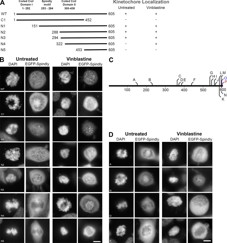
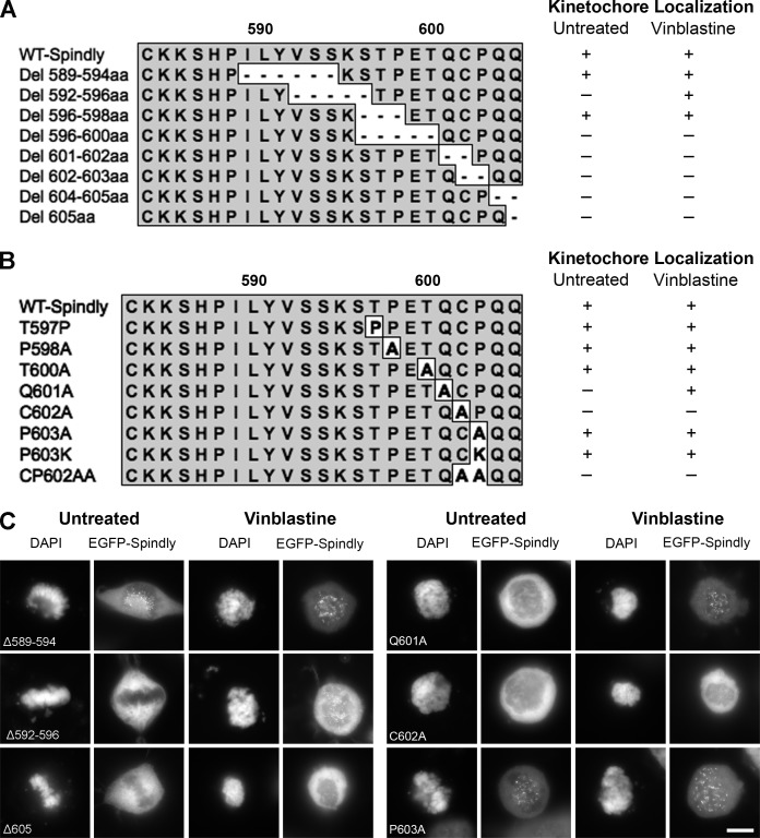
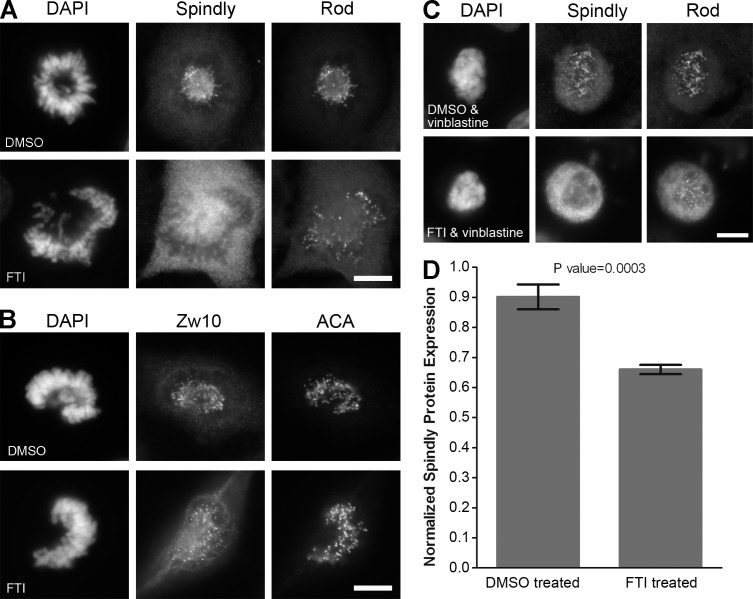
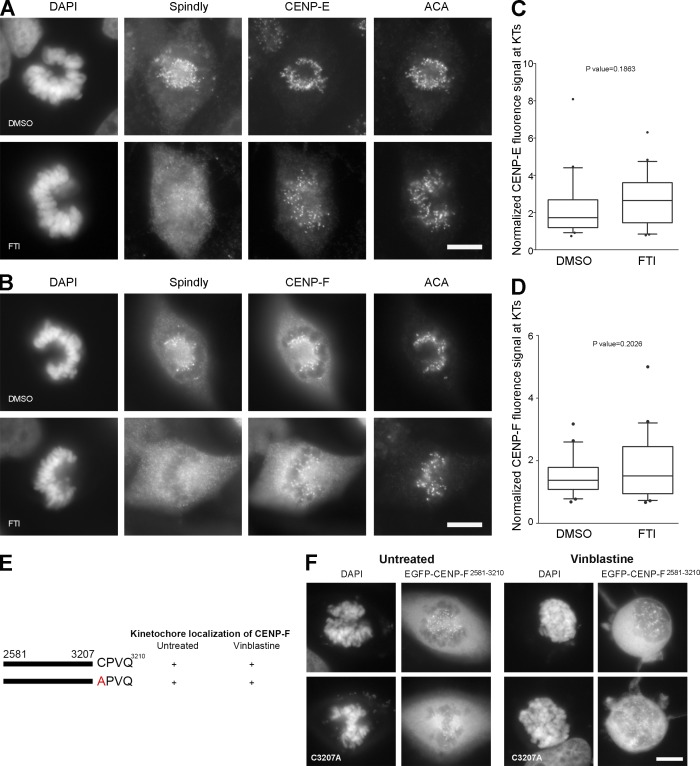
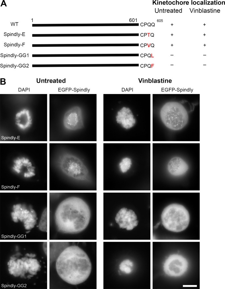
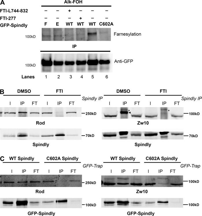
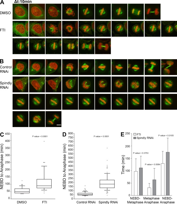
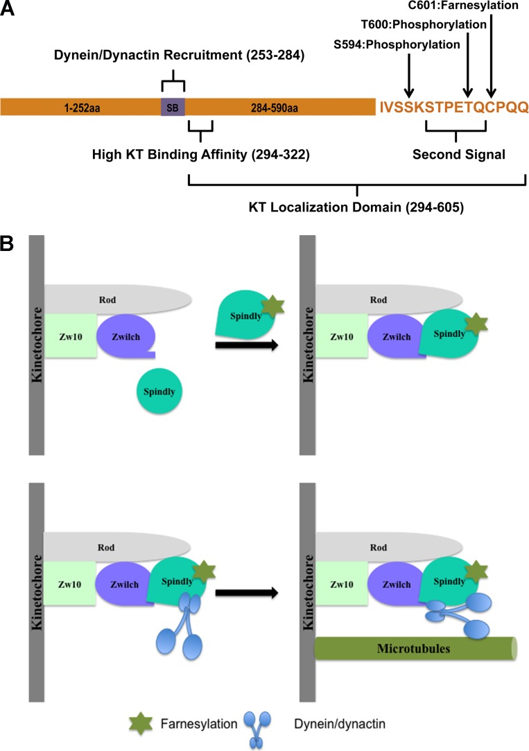
Kinetochore (KT) localization of mitotic checkpoint proteins is essential for their function during mitosis. hSpindly KT localization is dependent on the RZZ complex and hSpindly recruits the dynein-dynactin complex to KTs during mitosis, but the mechanism of hSpindly KT recruitment is unknown. Through domain-mapping studies we characterized the KT localization domain of hSpindly and discovered it undergoes farnesylation at the C-terminal cysteine residue. The N-terminal 293 residues of hSpindly are dispensable for its KT localization. Inhibition of farnesylation using a farnesyl transferase inhibitor (FTI) abrogated hSpindly KT localization without affecting RZZ complex, CENP-E, and CENP-F KT localization. We showed that hSpindly is farnesylated in vivo and farnesylation is essential for its interaction with the RZZ complex and hence KT localization. FTI treatment and hSpindly knockdown displayed the same mitotic phenotypes, indicating that hSpindly is a key FTI target in mitosis. Our data show a novel role of lipidation in targeting a checkpoint protein to KTs through protein-protein interaction.


Figures

Figure 1.
Figure 1.
hSpindly C-terminal is required for KT localization. (A) A schematic diagram of hSpindly depicting truncation mutants (+, KT localization; −, no KT localization). KT localizing capability of each construct was analyzed under vinblastine treatment, which maximally loads checkpoint proteins on KTs. Amino acid numbers are indicated. (B) HeLa cells were transiently transfected with EGFP-hSpindly fusion constructs (shown in A) and KT localization ability of each construct was analyzed using fluorescence microscopy. DAPI stains chromosomes. Representative images show that the C-terminal 294 to 605 aa of hSpindly are required for KT localization. Bar, 10 µm. (C) A schematic diagram depicting the location of the insertion mutants generated in the hSpindly protein. Constructs shown in red were negative for KT localization and constructs shown in blue localized to KTs only under vinblastine treatment. Site of insertion and inserted residues are shown in Table 1. (1D) HeLa cells transiently transfected with EGFP-hSpindly insertion constructs were analyzed for their KT localization ability. Representative images show that the far C terminus is essential for KT localization. Bar, 10 µm.
Figure 2.
Figure 2.
Far C-terminal residues of hSpindly are essential for KT localization. (A) Schematic representation of hSpindly C-terminal deletion mutants (589–605 aa). KT localization of each EGFP-tagged mutant was analyzed through fluorescence microscopy (+, KT localization; −, no KT localization). (B) Schematic representation of hSpindly C-terminal substitution mutants (597–605 aa). KT localization of each EGFP-tagged mutant was analyzed through fluorescence microscopy. (C) Representative images of HeLa cells transfected with EGFP-tagged hSpindly deletion or substitution constructs show that the C terminus of hSpindly is essential for its KT localization. Bar, 10 µm
Figure 3.
Figure 3.
Inhibition of farnesylation abrogates KT localization of hSpindly but not the RZZ complex. (A and B) HeLa cells were treated with 10 µM of L744832 FTI or DMSO for 24 h, fixed, and immunostained for hSpindly, Rod, and Zw10. Complete loss of hSpindly KT localization and normal Rod and Zw10 levels on the KTs were observed in FTI-treated cells. ACA immunostaining labels centromeres and DAPI stains DNA. (C) HeLa cells were treated with FTI or DMSO for 24 h and incubated with 0.5 µM vinblastine for 30 min before harvesting, showing no hSpindly KT localization without affecting Rod KT localization. (D) Normalized hSpindly protein expression from HeLa cells show 25% less expression with FTI treatment as compared with DMSO. n = 3. Error bars are SD from the means. P-value indicated a significant difference. Bars, 10 µm.
Figure 4.
Figure 4.
Inhibition of farnesylation does not affect CENP-E and CENP-F KT localization. (A and B) HeLa cells were treated with 10 µM of L744832 FTI or DMSO for 24 h, fixed, and immunostained for hSpindly, CENP-E or CENP-F, and ACA. CENP-E and CENP-F localized to KTs in FTI-treated HeLa cells, whereas hSpindly did not. (C and D) Normalized fluorescence signals for CENP-E and CENP-F at KTs with 24-h DMSO or FTI treatment. n = 20. Boxes represent interquartile distributions and whiskers represent 10th and 90th percentiles. FTI-treated cells have slight but not significant increase in CENP-E or CENP-F signal as compared with control DMSO as indicated by p-value. (E) A schematic diagram of hCENP-F depicting the minimal KT localization domain (2581–3210 aa) as previously shown by other studies and the C3207A mutation in the C-terminal farnesylation motif (+, KT localization; −, no KT localization). (F) HeLa cells were transiently transfected with EGFP-hCENP-F fusion constructs (shown in Fig. 3 E) and KT localization ability of each construct was analyzed using fluorescence microscopy. Top panel is EGFP-CENP-F2581-3210 and bottom panel is C3207A mutant of the same fragment. The images show that the farnesylation motif of CENP-F is not required for KT localization. Bars, 10 µm.
Figure 5.
Figure 5.
hSpindly farnesylation motif can be substituted with CENP-E or CENP-F farnesylation motif but not a geranylgeranylation motif. (A) A schematic diagram of hSpindly depicting substitution of its farnesylation motif with CENP-E and CENP-F, referred to as Spindly-E and Spindly-F, respectively. The last amino acid of Spindly is changed such that it is a geranylgeranylation motif instead of farnesylation motif referred to as Spindly-GG1 and Spindly-GG2. The KT localization of each construct is shown (+, KT localization; −, non-KT localization). Amino acid numbers are indicated. (B) HeLa cells were transiently transfected with EGFP-hSpindly fusion constructs (shown in A) and KT localization ability of each construct was analyzed using fluorescence microscopy. Representative images show that the farnesylation motif of Spindly can be replaced with the farnesylation motif of known farnesylated proteins CENP-E and CENP-F, but a geranylgeranylation motif cannot target hSpindly to KTs. Bar, 10 µm.
Figure 6.
Figure 6.
hSpindly is farnesylated in vivo and farnesylation regulates its interaction with the RZZ complex. (A) HeLa cells transiently expressing GFP-WT hSpindly, GFP-C602A hSpindly (of CAAX motif), GFP-hSpindly-E, and GFP-hSpindly-F were metabolically labeled with alkynyl-farnesol (prenylation reporter) and HeLa cells expressing WT GFP-Spindly were either treated with FTI or DMSO. (top) Fluorescence detection of farnesylated immunoprecipitated GFP-tagged hSpindly protein as shown in gel. (bottom) Loading control is shown. WT Spindly, Spindly-E, and Spindly-F exhibit bands indicating farnesylation. Farnesylation of WT Spindly is inhibited in the presence of FTIs (lanes 3 and 4). As expected, C602A mutant of hSpindly is not farnesylated in vivo (lane 6). (B) Immunoprecipitation of hSpindly (70 kD) from mitotic HeLa cell lysates followed by Western blot against Zw10 (89 kD) or Rod (250 kD) RZZ complex subunits. I, input; IP, immunoprecipitated; FT, flowthrough. HeLa cells were treated with either DMSO or 10 µM of FTI-L744832 for 24 h before harvesting and arrested in mitosis with nocodazole treatment. Zw10 is indicated by an arrowhead (IP lanes), and an asterisk denotes a nonspecific band. Inhibition of farnesylation leads to loss of hSpindly and RZZ complex interaction. (C) GFP-Trap from mitotic HeLa cells with endogenous hSpindly knockdown and expressing RNAi resistant GFP-WT hSpindly (98.5 kD) or GFP-C602A hSpindly mutant (98.5 kD). GFP-Trap followed by Western blot against Zw10 (89 kD) or Rod (250 kD) subunits of the RZZ complex. Cells were incubated in nocodazole to accumulate in mitosis before harvesting. Cysteine-to-alanine mutant of GFP-hSpindly leads to loss of hSpindly and RZZ complex interaction.
Figure 7.
Figure 7.
Phenocopying of farnesylation inhibition with hSpindly knockdown by RNAi. (A) Representative stills from videos of GFP-tubulin and mCherry H2B expressing HeLa cells treated with either DMSO or 10 µM of FTI-L744832 for 24 h before filming. Time is shown in 10-min intervals. (B) Representative stills from videos of GFP-tubulin and mCherry H2B expressing HeLa cells treated with either control (scrambled) RNAi or hSpindly RNAi for 33 h before filming. Time is shown in 10-min intervals. Bar, 10 µm. (C) Box plots comparing the duration of nuclear envelope breakdown (NEBD) to anaphase between FTI- and DMSO-treated cells show significant mitotic delay in FTI-treated cells. Seven experiments were performed with >60 cells in total analyzed for each treatment. Boxes represent interquartile distributions and whiskers represent 10th and 90th percentiles. (D) Box plots comparing the duration of nuclear envelope breakdown to anaphase between control (scrambled) RNAi and hSpindly RNAi–treated cells show significant mitotic delay in hSpindly RNAi–treated cells. Eight experiments were performed with >80 cells in total analyzed for each treatment. Boxes represent interquartile distributions and whiskers represent 10th and 90th percentiles. (E) Duration of nuclear envelope breakdown to metaphase, metaphase to anaphase, and nuclear envelope breakdown to anaphase in FTI- and hSpindly RNAi–treated HeLa cells shows all are increased in Spindly knockdown cells with metaphase-to-anaphase time difference being significant. n = 18 for FTI and n = 22 for hSpindly RNAi. Error bars are SD from the means.
Figure 8.
Figure 8.
A proposed model of hSpindly KT localization. (A) A schematic representation of hSpindly domains and posttranslational modifications. SB, Spindly box. (B) hSpindly farnesylation induces a conformational change such that it can interact with the RZZ complex subunits Rod and Zwilch, which recruits hSpindly to KTs during mitosis. hSpindly further recruits the dynein–dynactin complex to KTs. Once all the chromosomes are aligned on the metaphase plate, hSpindly and the RZZ complex are transported from the KTs to spindle poles.

References

    1. Ahearn I.M., Haigis K., Bar-Sagi D., and Philips M.R.. 2012. Regulating the regulator: post-translational modification of RAS. Nat. Rev. Mol. Cell Biol. 13:39–51 10.1038/nrm3255 - DOI - PMC - PubMed
    1. Apolloni A., Prior I.A., Lindsay M., Parton R.G., and Hancock J.F.. 2000. H-ras but not K-ras traffics to the plasma membrane through the exocytic pathway. Mol. Cell. Biol. 20:2475–2487 10.1128/MCB.20.7.2475-2487.2000 - DOI - PMC - PubMed
    1. Ashar H.R., James L., Gray K., Carr D., Black S., Armstrong L., Bishop W.R., and Kirschmeier P.. 2000. Farnesyl transferase inhibitors block the farnesylation of CENP-E and CENP-F and alter the association of CENP-E with the microtubules. J. Biol. Chem. 275:30451–30457 10.1074/jbc.M003469200 - DOI - PubMed
    1. Barisic M., and Geley S.. 2011. Spindly switch controls anaphase: spindly and RZZ functions in chromosome attachment and mitotic checkpoint control. Cell Cycle. 10:449–456 10.4161/cc.10.3.14759 - DOI - PubMed
    1. Barisic M., Sohm B., Mikolcevic P., Wandke C., Rauch V., Ringer T., Hess M., Bonn G., and Geley S.. 2010. Spindly/CCDC99 is required for efficient chromosome congression and mitotic checkpoint regulation. Mol. Biol. Cell. 21:1968–1981 10.1091/mbc.E09-04-0356 - DOI - PMC - PubMed

Publication types

MeSH terms

LinkOut - more resources