. 2015 Mar 30;208(7):1003-18.
doi: 10.1083/jcb.201407131.

L1CAM/Neuroglian controls the axon-axon interactions establishing layered and lobular mushroom body architecture

Affiliations

L1CAM/Neuroglian controls the axon-axon interactions establishing layered and lobular mushroom body architecture

Dominique Siegenthaler et al. J Cell Biol. .

Abstract

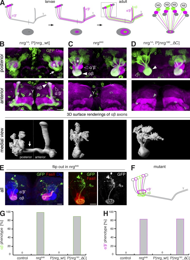
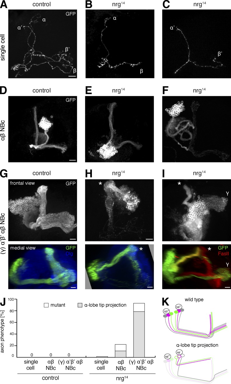
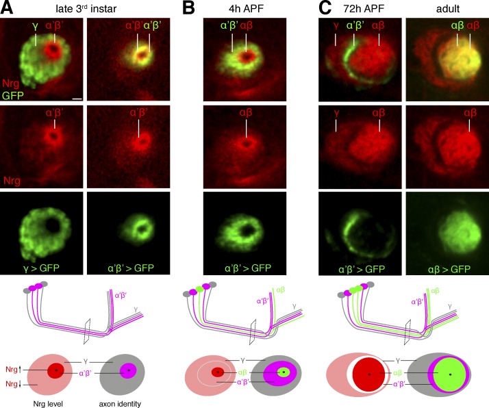
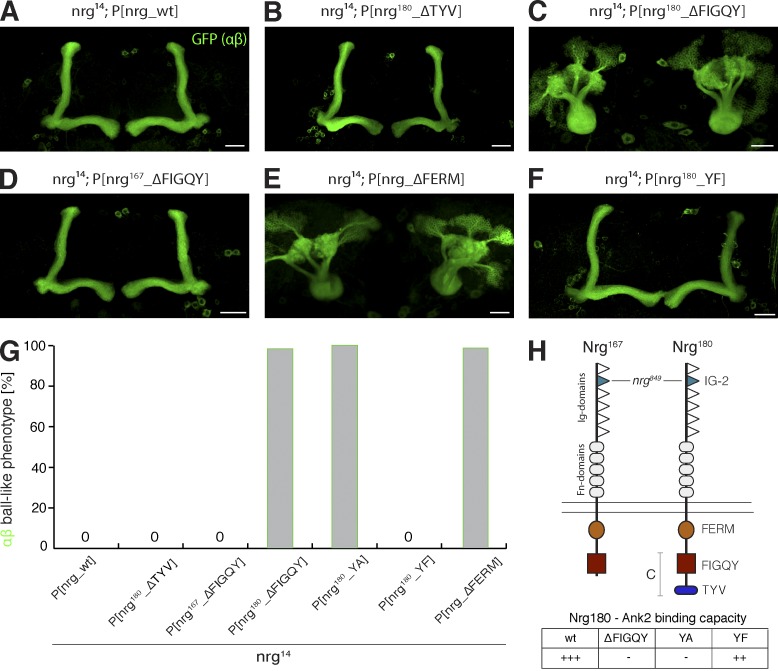
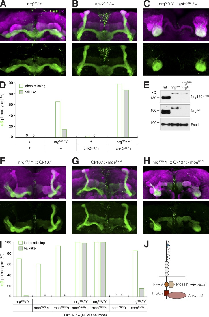
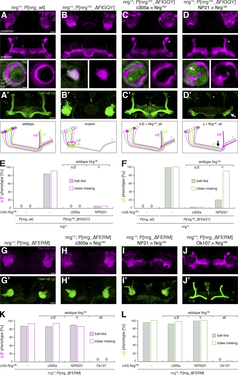
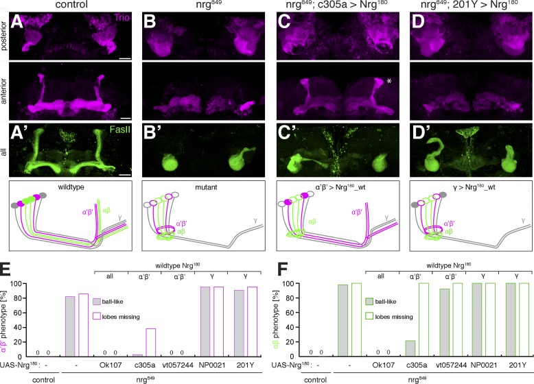
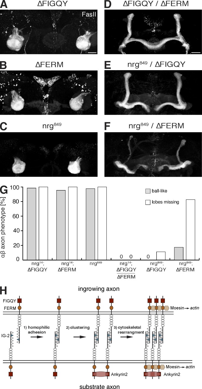
The establishment of neuronal circuits depends on the guidance of axons both along and in between axonal populations of different identity; however, the molecular principles controlling axon-axon interactions in vivo remain largely elusive. We demonstrate that the Drosophila melanogaster L1CAM homologue Neuroglian mediates adhesion between functionally distinct mushroom body axon populations to enforce and control appropriate projections into distinct axonal layers and lobes essential for olfactory learning and memory. We addressed the regulatory mechanisms controlling homophilic Neuroglian-mediated cell adhesion by analyzing targeted mutations of extra- and intracellular Neuroglian domains in combination with cell type-specific rescue assays in vivo. We demonstrate independent and cooperative domain requirements: intercalating growth depends on homophilic adhesion mediated by extracellular Ig domains. For functional cluster formation, intracellular Ankyrin2 association is sufficient on one side of the trans-axonal complex whereas Moesin association is likely required simultaneously in both interacting axonal populations. Together, our results provide novel mechanistic insights into cell adhesion molecule-mediated axon-axon interactions that enable precise assembly of complex neuronal circuits.


Figures

Figure 1.
Figure 1.
Extra- and intracellular Nrg domains contribute to MB axon guidance. (A) Schematic drawings of MB development. Side views of MB axon projections and cross-sections of the pedunculus are shown: γ (gray), α’β’ (magenta), and αβ (green). (B–D) Frontal projections of posterior (top) and anterior (middle) regions of the MBs. αβ neurons are marked by mCD8-GFP expression (c739-Gal4, green), and neurites of all MB are visualized by Dlg (magenta). Bottom panels show medial (side) views of 3D-surface rendered αβ neurons. (B) In nrg14; P[nrg_wt] control animals, axons of all three MB neuron subtypes project through the pedunculus (arrows) into vertical and medial MB lobes (arrowheads). (C and D) In nrg mutant animals carrying either a mutation in the extracellular domain (B; nrg849) or lacking the Nrg180-specific C terminus (C; nrg14; P[nrg180_ΔC]), axons of αβ (green) and α’β’ (magenta/white) neurons fail to project into the pedunculus and accumulate in ball-like structures in the posterior brain ventral to the calyx (asterisks). αβ and α’β’ axons remain segregated (top, arrowheads). Axons of γ neurons (middle, arrowheads) still form a medial lobe that is often thinner compared with controls. (E) Frontal projections of entire MBs of an nrg849 mutant in which individual αβ neurons are labeled by mCD8-GFP (201Y-Gal4) using a flip-out approach. αβ axons are marked with FasII (red), neuropil with Dlg (blue). Bars, 20 µm. (F) Schematic model of axonal projections in nrg14; P[nrg180_ΔC] mutant animals. (G and H) Quantification of aberrant ball-like projections of αβ (G) or α’β’ (H) neurons. Phenotypes were assayed using FasII (αβ) or Trio (α’β’). n = 60, 46, 44, and 54 (G) and n = 26, 28, 24, and 34 (H), in the respective order of the genotypes indicated.
Figure 2.
Figure 2.
Nrg controls MB axon tract choice. (A–I) MARCM analysis of nrg14 mutants. Bars, 10 µm. (A–C) Frontal projections of control and nrg14 mutant single-cell clones. Absence of nrg14 in individual MB neurons does not cause obvious alteration of axonal projections. (D–F) Frontal projections of control and nrg14 αβ NB clones (NBc). The majority of nrg14 αβ NBc do not show an axonal phenotype (E). (F) Example of an nrg14 αβ NBc in which axons fail to project into the pedunculus and form circular projections in the posterior brain. (G–I) Large control and nrg14 NBc that include either α’β’ and αβ (H) or all three MB subtypes (G and I). Top panels show frontal projections of the entire MB. Bottom panels show medial (side) views of the NBc marked by GFP (green) and Dlg (blue; G and H) or FasII (red; I). In contrast to controls, nrg14 mutant α’β’ and αβ axons but not γ axons (I) project aberrantly straight to the α lobe tip, bypassing the MB pedunculus and lobes (asterisks). (J) Quantification of MARCM phenotypes (n = 31, 11, 5, 131, 9, and 14, in the respective order of the genotypes indicated). (K) Schematic drawing of wild-type and nrg14 mutant axon trajectories in G–I.
Figure 3.
Figure 3.
Nrg is dynamically expressed during MB development. (A–E) Analysis of Nrg180 expression (red) in MB pedunculus cross-sections at the position indicated in the schematics using cell type–specific Gal4 lines driving mCD8-GFP (green). The following Gal4-driver lines were used: NP0021 (γ neurons), c305a (α’β’ neurons), and c739 (αβ neurons). Bar, 2.5 µm. (A) In late third instar larvae, high levels of Nrg are present in α’β’ axons, which are surrounded by γ axons expressing lower Nrg levels. (B) At early pupal stages (4 h after puparium formation [APF]), Nrg is down-regulated in α’β’ axons and now highly expressed in c305a-Gal4–negative αβ axons. (C) In late pupal and adult stages, Nrg is down-regulated in α’β’ axons but remains expressed at high levels in αβ and at lower levels in γ axons.
Figure 4.
Figure 4.
Intracellular FIGQY and FERM domains are required for MB development. (A–F) αβ neurons marked by mCD8-GFP expression (c739-Gal4). Frontal projections of the anterior (A, B, D, and F) or of the entire brain are shown (C and E). Bars, 20 µm. (A) In nrg14; P[nrg_wt] control animals, αβ axons form medial and vertical lobes in the anterior brain. (B) The binding motif for PDZ domain containing proteins (TYV) of Nrg180 is not required for MB axon pathfinding. (C and D) Deletion of the FIGQY motif of Nrg180 but not of Nrg167 results in aberrant axonal accumulations in the posterior brain and absence of anterior αβ lobes. (E) Deletion of the FERM protein–interacting domain results in aberrant projections and an absence of αβ lobes. (F) A YF mutation within the FIGQY motif of Nrg180 restores MB development but β lobes were fused at the midline (see also Fig. S2). (G) Quantification of aberrant ball-like projections of αβ axons analyzed using FasII or c739>mCD8-GFP (n = 44, 66, 80, 81, 78, 84, and 56, in the respective order of the genotypes indicated). (H) Schematic model of the domain structure of the Nrg isoforms Nrg180 and Nrg167. The positions of the extracellular mutation nrg849 and of intracellular domains are indicated. A summary of the in vitro Nrg–Ank2 interaction data from Enneking et al. (2013) is displayed.
Figure 5.
Figure 5.
Nrg–Ank2 association controls MB axon guidance. (A–C and F–H) Frontal projections of the anterior (A, B, F, and G), posterior (C), or entire (H) region of the MBs visualized using FasII (green, αβ axons) and Dlg (magenta, neuropil). Bars, 20 µm. (A) In hemizygous nrg305 mutant animals, αβ axons display branching and lobe formation defects but rarely fail to project through the pedunculus. (B) Heterozygous mutations of ank2 (ank2518/+) do not affect MB development. (C) Removal of one copy of ank2 in hemizygous nrg305 mutant animals severely enhances the MB axon phenotype, with MB axons failing to enter the pedunculus and forming aberrant ball-like structures in the posterior brain. (D) Quantification of the αβ axon phenotype assayed using FasII (n = 60, 43, 42, and 44, respectively, in the order of the genotypes given). (E) Western blot analysis of Nrg expression in larval brain extracts. The nrg305 GFP-trap mutation reduces protein expression of both Nrg180 and Nrg167. (F) nrg305 mutant animals display mild axonal defects including branching and lobe formation defects. (G) Knockdown of Moesin in MB neurons causes defects in αβ axon branching and lobe formation but does not lead to aberrant axonal accumulations in the posterior brain. (H) Knockdown of Moesin in MB neurons in nrg305 mutant animals results in a dramatic enhancement of the phenotype compared with both individual genotypes, with MB axons now forming aberrant ball-like structure in the posterior brain. (I) Quantification of αβ axon phenotype using FasII (n = 43, 38, 30, 72, 26, 26, and 26, respectively, in the order of the genotypes indicated). (J) Schematic model indicating essential Nrg interaction partners.
Figure 6.
Figure 6.
Trans-axonal control of pedunculus and lobe formation. (A–D) Frontal projections of posterior (top) and anterior regions (middle) of MBs marked by Trio (magenta; α’β’, high; and γ, low). Bottom panels show cross-sections of the pedunculus stained for Trio (magenta) and Dlg (green). Bars: (top) 20 µm; (bottom) 2.5 µm. (A’–D’) Top panels show frontal projections of entire MBs marked by FasII (green; αβ, high; and γ, low). Schematics summarize axonal projection phenotypes. Bars, 20 µm. (A) In control nrg14; P[nrg_wt] animals, Trio-positive axons of α’β’ and γ neurons project into anterior lobes. Within the pedunculus, γ, α’β’, and αβ axons are clearly segregated into distinct concentric layers. (A’) αβ axons form medial and vertical lobes. (B) In nrg14; P[nrg180_ΔFIGQY] mutant animals, α’β’ axons fail to project into the pedunculus and form aberrant ball-like projections in the posterior brain. Only γ neurons (also Trio positive; imaged at higher gain settings compared with controls) form anterior lobes. (B’) αβ axons fail to form anterior lobes and form aberrant projections in the posterior brain. (C) Expression of wild-type Nrg180 in α’β’ neurons of nrg14; P[nrg180_ΔFIGQY] mutants restores anterior projections of α’β’ neurons. Minor perturbations of axonal layer organization are evident in the pedunculus. (C’) In these animals, projections of αβ mutant axons are also efficiently rescued and αβ lobes form next to the wild-type Nrg180-expressing α’β’ lobes (asterisk in C). (D) Expression of wild-type Nrg180 in γ neurons of nrg14; P[nrg180_ΔFIGQY] mutants also rescues α’β’ projections. Pedunculus cross-sections reveal aberrant organization of axonal layers, with mutant αβ axons inappropriately in contact with γ axons (arrow). (D’) In these animals, αβ axons grow into the pedunculus to the pedunculus divide (heel, arrow) but fail to form medial or vertical lobes (note the altered appearance of α’β’ lobes in D due to the absence of αβ lobes, indicated by the asterisk). (E) Quantification of α’β’ phenotypes (n = 24, 55, 18, and 30, respectively, in the order of the genotypes indicated). (F) Quantification of αβ phenotypes (n = 44, 61, 69, and 36, respectively, in the order of the genotypes indicated). (G–J and G’–J’) Frontal projections of entire MBs. (G and G’) In nrg14; P[nrg_ΔFERM] mutant animals, axons of α’β’ and αβ neurons form aberrant ball-like projections in the posterior brain and fail to form anterior lobes. (H and H’) Expression of wild-type Nrg180 in α’β’ neurons of nrg14; P[nrg_ΔFERM] mutants does not rescue the MB phenotype. (I and I’) Expression of wild-type Nrg180 in γ neurons of nrg14; P[nrg_ΔFERM] mutants does not rescue the MB phenotype. (J and J’) Expression of wild-type Nrg180 in all MB neurons efficiently rescues axonal projections. Bars, 20 µm. (K) Quantification of the α’β’ phenotypes (n = 44, 61, 28, 31, and 24, respectively, in the order of the genotypes indicated). (L) Quantification of the αβ phenotypes (n = 102, 82, 65, 48, and 30, respectively, in the order of the genotypes indicated).
Figure 7.
Figure 7.
Extracellular adhesion controls axonal intercalation. (A–D) Anterior and posterior projections of α’β’ and γ neurons marked by Trio (magenta) are shown. (A’–D’) Top panels show entire MB projections of αβ axons marked by FasII (green). Schematics summarize the axonal phenotypes. (A and B) In contrast to control animals, in nrg849 mutant animals α’β’ axons fail to enter the pedunculus and form ball-like aggregates in the posterior brain. Medial γ lobe projections show minor defects. (B’) In mutant animals, αβ axons also fail to enter the pedunculus. (C) Cell type–specific expression of wild-type Nrg180 in α’β’ neurons of nrg849 mutant animals restores α’β’ lobular projections. (C’) No rescue of αβ projections was observed when using vt057244-Gal4; however, we frequently observed partial rescue of αβ axons into the pedunculus but no rescue of lobe formation despite presence of α’β’ lobes when using c305a-Gal4. (D and D’) Expression of wild-type Nrg180 in γ neurons of nrg849 mutant animals does not rescue α’β’ or αβ projections. (E and F) Quantification of α’β’ (E) and αβ (F) axon phenotypes. Rescue data are presented using Gal4 drivers expressing wild-type Nrg180 in all MB neurons (Ok107-Gal4), α’β’ neurons (c305a-Gal4 and vt057244-Gal4), or γ neurons (NP0021-Gal4, 201Y-Gal4; n = 26, 28, 20, 34, 36, 21, and 43 for E and n = 60, 46, 20, 28, 38, 28, and 44 for F, in the respective order of the genotypes indicated). Bars, 20 µm.
Figure 8.
Figure 8.
Cooperative control of Nrg-mediated trans-axonal interactions. (A–F) Frontal projections of the entire MBs (A–C) or only anterior regions (D–F). αβ axons are marked by FasII (white). Bars, 20 µm. (A–C) All hypomorphic nrg mutations result in identical αβ axon projection defects. (D–F) Transheterozygous combinations of two mutations almost completely restore MB projections. Aberrant β-lobe fusions were present in some ΔFIGQY/ΔFERM mutant animals (D), and severe perturbations of lobe formation were evident in animals transheterozygous for ΔFERM and nrg849 (F). (G) Quantification of the αβ phenotype (n = 63, 64, 46, 40, 36, 46, respectively, in the order of the genotypes indicated). (H) Model of the formation of functional Nrg clusters during trans-axonal interactions. Trans-axonal Nrg interactions are stabilized by Ank2-mediated clustering. Interactions with Moesin provide a link to the actin cytoskeleton that enables formation of stable complexes providing cellular adhesion.

Similar articles

Cited by

References

    1. Adams M.D., Celniker S.E., Holt R.A., Evans C.A., Gocayne J.D., Amanatides P.G., Scherer S.E., Li P.W., Hoskins R.A., Galle R.F., et al. . 2000. The genome sequence of Drosophila melanogaster. Science. 287:2185–2195 10.1126/science.287.5461.2185 - DOI - PubMed
    1. Aso Y., Grübel K., Busch S., Friedrich A.B., Siwanowicz I., and Tanimoto H.. 2009. The mushroom body of adult Drosophila characterized by GAL4 drivers. J. Neurogenet. 23:156–172 10.1080/01677060802471718 - DOI - PubMed
    1. Bates K.E., Sung C.S., and Robinow S.. 2010. The unfulfilled gene is required for the development of mushroom body neuropil in Drosophila. Neural Dev. 5:4 10.1186/1749-8104-5-4 - DOI - PMC - PubMed
    1. Bieber A.J., Snow P.M., Hortsch M., Patel N.H., Jacobs J.R., Traquina Z.R., Schilling J., and Goodman C.S.. 1989. Drosophila neuroglian: a member of the immunoglobulin superfamily with extensive homology to the vertebrate neural adhesion molecule L1. Cell. 59:447–460 10.1016/0092-8674(89)90029-9 - DOI - PubMed
    1. Bischof J., Maeda R.K., Hediger M., Karch F., and Basler K.. 2007. An optimized transgenesis system for Drosophila using germ-line-specific phiC31 integrases. Proc. Natl. Acad. Sci. USA. 104:3312–3317 10.1073/pnas.0611511104 - DOI - PMC - PubMed

Publication types

MeSH terms