Review
. 2014 Jan 27:2:2.
doi: 10.1186/2047-2501-2-2. eCollection 2014.

Retinal blood vessels extraction using probabilistic modelling

Affiliations
Review

Retinal blood vessels extraction using probabilistic modelling

Djibril Kaba et al. Health Inf Sci Syst. .

Abstract

The analysis of retinal blood vessels plays an important role in detecting and treating retinal diseases. In this review, we present an automated method to segment blood vessels of fundus retinal image. The proposed method could be used to support a non-intrusive diagnosis in modern ophthalmology for early detection of retinal diseases, treatment evaluation or clinical study. This study combines the bias correction and an adaptive histogram equalisation to enhance the appearance of the blood vessels. Then the blood vessels are extracted using probabilistic modelling that is optimised by the expectation maximisation algorithm. The method is evaluated on fundus retinal images of STARE and DRIVE datasets. The experimental results are compared with some recently published methods of retinal blood vessels segmentation. The experimental results show that our method achieved the best overall performance and it is comparable to the performance of human experts.

Keywords: Expectation maximisation; Retinal images; Vessel segmentation.


Figures

Figure 1
Figure 1
Bias correction results. (a) STARE image with intensity inhomogeneity. (b) Bias field. (c) Bias corrected image. (d) DRIVE image with intensity inhomogeneity. (e) Bias field. (f) Bias corrected image.
Figure 2
Figure 2
Adaptive histogram equalisation results. (a) r=3, h=45. (b) r=6, 45. (c) r=3, h=81. (d) r=6, h=81.
Figure 3
Figure 3
Distance map images. (a) STARE image. (b) STARE distance map. (c) DRIVE image. (d) DRIVE distance map.
Figure 4
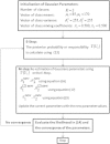
Figure 4
The EM algorithm summary. The steps of EM algorithm.
Figure 5
Figure 5
The EM algorithm and length filter results. (a) Fundus retinal image. (b) The EM algorithm output image. (c) the Length filter output image.
Figure 6
Figure 6
The sample results of our method. (a) STRARE fundus image. (b) Our method result. (c) Ground truth. (d) DRIVE fundus image. (e) Our method result. (f) Ground truth.

References

    1. Fritzsche K, Can A, Shen H, Tsai C, Turner J, Stewart C, Roysam B. Automated model based segmentation, tracing and analysis of retinal vasculature from digital fundus images. State-of-The-Art Angiography Appl Plaque Imaging Using MR, CT, Ultrasound X-rays. 2003;2003(6):225–298.
    1. Hoover A, Kouznetsova V, Goldbaum M. Locating blood vessels in retinal images by piecewise threshold probing of a matched filter response. Med Imaging IEEE Trans. 2002;19(3):203–210. doi: 10.1109/42.845178. - DOI - PubMed
    1. Mendonça AM, Campilho A. Segmentation of retinal blood vessels by combining the detection of centerlines and morphological reconstruction. IEEE Trans Med Imaging. 2006;25(9):1200–1213. doi: 10.1109/TMI.2006.879955. - DOI - PubMed
    1. Zhang B, Zhang L, Zhang L, Karray F. a vessel extraction by matched filter with first-order derivative of Gaussian. Comput Biol Med. 2010;40(4):438–445. doi: 10.1016/j.compbiomed.2010.02.008. - DOI - PubMed
    1. Xu L, Luo S. A novel method for blood vessel detection from retinal images. Biomed Eng Online. 2010;9:1–10. doi: 10.1186/1475-925X-9-14. - DOI - PMC - PubMed

LinkOut - more resources