. 2015 Apr 14;112(15):4624-9.
doi: 10.1073/pnas.1420833112. Epub 2015 Mar 30.

Autoinhibition of MDMX by intramolecular p53 mimicry

Affiliations

Autoinhibition of MDMX by intramolecular p53 mimicry

Lihong Chen et al. Proc Natl Acad Sci U S A. .

Abstract

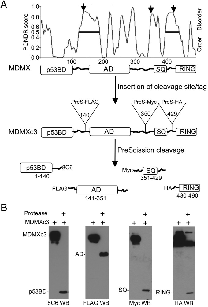
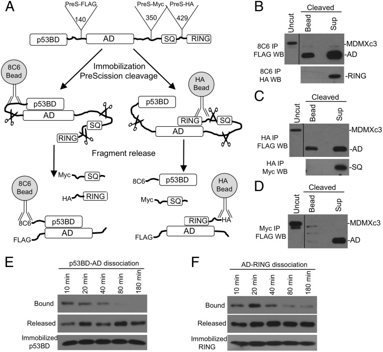
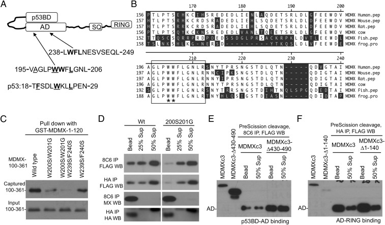
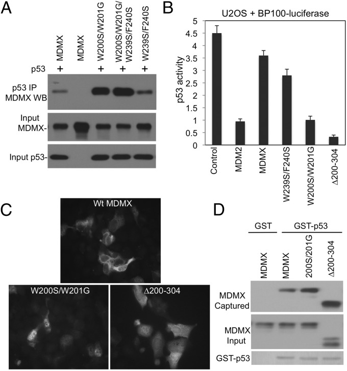
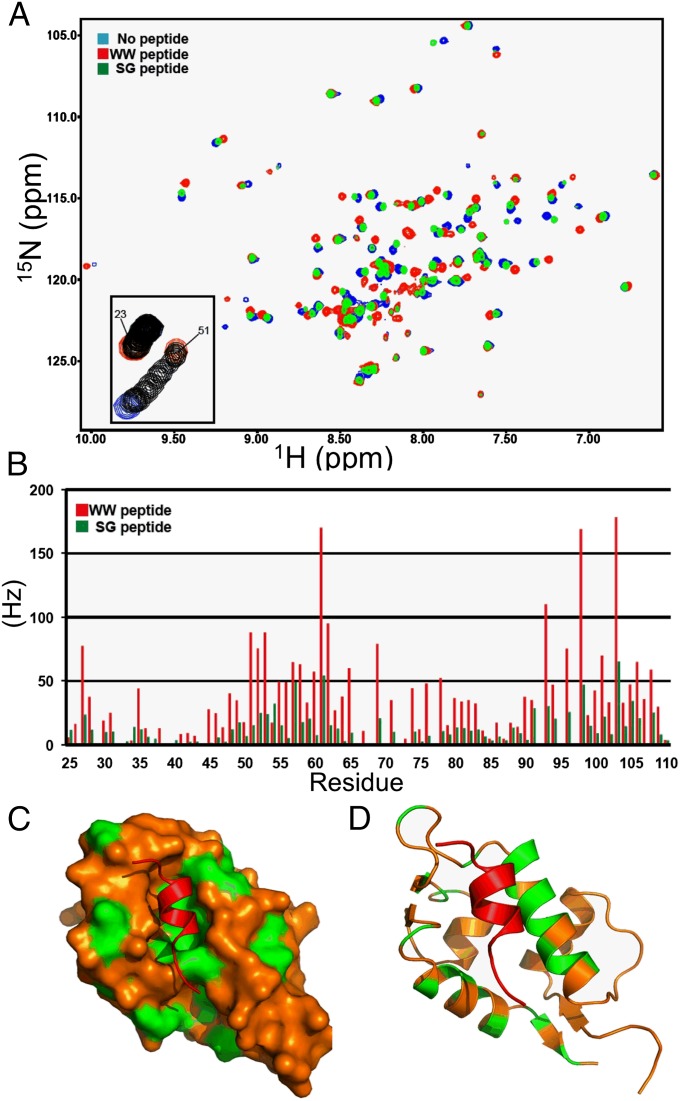
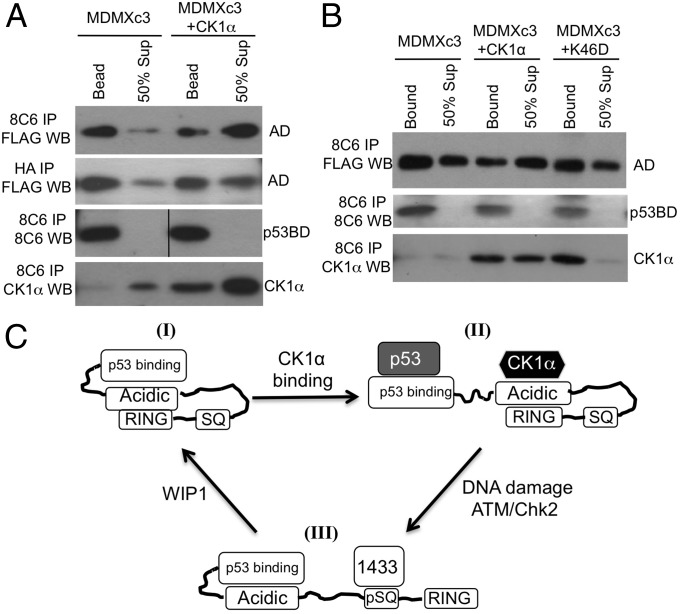
The p53 inhibitor MDMX is controlled by multiple stress signaling pathways. Using a proteolytic fragment release (PFR) assay, we detected an intramolecular interaction in MDMX that mechanistically mimics the interaction with p53, resulting in autoinhibition of MDMX. This mimicry is mediated by a hydrophobic peptide located in a long disordered central segment of MDMX that has sequence similarity to the p53 transactivation domain. NMR spectroscopy was used to show this hydrophobic peptide interacts with the N-terminal domain of MDMX in a structurally analogous manner to p53. Mutation of two critical tryptophan residues in the hydrophobic peptide disrupted the intramolecular interaction and increased p53 binding, providing further evidence for mechanistic mimicry. The PFR assay also revealed a second intramolecular interaction between the RING domain and central region that regulates MDMX nuclear import. These results establish the importance of intramolecular interactions in MDMX regulation, and validate a new assay for the study of intramolecular interactions in multidomain proteins with intrinsically disordered regions.

Keywords: MDMX; acidic domain; intramolecular; p53; protease.


Conflict of interest statement

The authors declare no conflict of interest.

Figures

Fig. 1.
Fig. 1.
Design of protease cleavable MDMXc3. (A) Identification of MDMX disordered regions for the insertion of cleavage sites. PreScission cleavage site and epitope tags were inserted after residues 140, 350, and 429 to create MDMXc3. Cleavage by PreScission protease produces four fragments each containing a unique epitope. (B) Lysate of H1299 transfected with MDMXc3 was digested with 0.1 µg/µL PreScission for 10 min at 4 °C and analyzed by Western blot to detect the production of individual fragments.
Fig. 2.
Fig. 2.
Proteolytic fragment release (PFR) assay. (A) Diagram of PFR assay for MDMX intramolecular interactions. MDMXc3 was immobilized on beads by using different antibodies and cleaved by PreScission for <40 min. The beads and supernatant were analyzed by Western blot to detect fragment release from the beads. (B) MDMXc3 expressed in H1299 was immobilized on 8C6 beads (captures MDMXc3 through the p53BD fragment) and incubated with PreScission for 40 min. The beads and supernatant were analyzed by FLAG and HA Western blot. The presence of AD fragment in the bead fraction indicates slow dissociation from the immobilized p53BD. Absence of RING fragment in the bead fraction indicates lack of binding to the p53BD. (C) Immobilization of MDMXc3 through the RING domain revealed RING binding to AD but not SQ fragment. (D) Immobilization of MDMX through the SQ fragment revealed lack of binding to the AD. (E) MDMXc3 captured on 8C6 beads was cleaved with PreScission in <5 min. Beads and supernatants were separated at indicated time points and analyzed by FLAG Western blot to detect the dissociation of AD from p53BD. (F) MDMXc3 immobilized using HA antibody was analyzed for the dissociation of AD from RING after cleavage at indicated time points.
Fig. 3.
Fig. 3.
Identification of an internal p53 mimetic sequence in MDMX. (A) Proposed model of p53BD–AD intramolecular interaction and the presence of two potential pocket-binding sequences in the AD. (B) Alignment of MDMX WW sequence from various species. (C) Indicated substitutions were introduced into MDMX-100–361 fragment and expressed in H1299 cells. The cell lysate was incubated with beads loaded with GST-MDMX-1-120, the captured 100–361 fragment was detected by 8C6 Western blot. (D) The SG mutations were introduced into MDMXc3, expressed in H1299 cells, and analyzed for p53BD–AD and AD–RING interactions using the PFR assay. (E) MDMXc3 was subcloned to delete the RING domain and analyzed for p53BD–AD binding using PFR assay. (F) MDMXc3 was subcloned to delete the p53BD and analyzed for AD–RING binding by using PFR assay.
Fig. 4.
Fig. 4.
Regulation of p53 binding and localization by the p53 mimetic sequence. (A) Mutations of the putative p53 mimetic sequences were introduced into full-length MDMX and coexpressed with p53 in H1299 cells. MDMX-p53 binding was analyzed by IP-Western blot. (B) U2OS was cotransfected with p53-response reporter BP100-luciferase and MDMX mutants. Endogenous p53 activity was measured by luciferase assay and normalized to cotransfected CMV-lacZ. (C) U2OS cells were transiently transfected with MDMX mutants and stained for MDMX localization by using 8C6 immunofluorescence. (D) Beads loaded with GST-p53 were incubated with H1299 lysate expressing MDMX mutants. p53 binding to MDMX mutants was determined by Western blot.
Fig. 5.
Fig. 5.
Interaction of MDMX WW and SG peptides with the p53-binding pocket. (A) Overlay of 1H-15N HSQC spectra for 15N-MDMX (blue), 15N-MDMX+WW peptide (red), and 15N-MDMX+SG peptide (green). (B) Chemical shift changes for MDMX p53BD residues binding to the WW peptide (red bars) or the SG peptide (green bars). (C) Surface image of the MDMX p53BD structure. The residues that have combined chemical shifts close to or greater than 50 Hz (upon binding the WW peptide) are highlighted in green. (D) Cartoon showing the chemical shifts on the back side of two helices obscured on the surface image.
Fig. 6.
Fig. 6.
Regulation of MDMX intramolecular interactions by CK1α. (A and B) MDMXc3 was cotransfected with CK1α and CK1α-K46D kinase-dead mutant into U2OS cells, and the changes in intramolecular interactions were analyzed by using PFR assay. (C) A model of MDMX intramolecular interactions. MDMX can assume multiple conformational states. I, a closed state of weak p53 binding and cytoplasmic localization due to intramolecular interactions. II, interaction with CK1α opens the N terminus for p53 binding. III, DNA damage recruits 14-3-3 and disrupts binding to CK1α, inhibits the N terminus, and exposes the RING to mediate nuclear import and heterodimerization with MDM2. ATM/Chk2 and WIP1-mediated phosphorylation and dephosphorylation alter the balance between the conformational states.

References

    1. Vousden KH, Lane DP. p53 in health and disease. Nat Rev Mol Cell Biol. 2007;8(4):275–283. - PubMed
    1. Grier JD, Xiong S, Elizondo-Fraire AC, Parant JM, Lozano G. Tissue-specific differences of p53 inhibition by Mdm2 and Mdm4. Mol Cell Biol. 2006;26(1):192–198. - PMC - PubMed
    1. Li B, Cheng Q, Li Z, Chen J. p53 inactivation by MDM2 and MDMX negative feedback loops in testicular germ cell tumors. Cell Cycle. 2010;9(7):1411–1420. - PMC - PubMed
    1. Terzian T, et al. Haploinsufficiency of Mdm2 and Mdm4 in tumorigenesis and development. Mol Cell Biol. 2007;27(15):5479–5485. - PMC - PubMed
    1. Wang YV, Leblanc M, Wade M, Jochemsen AG, Wahl GM. Increased radioresistance and accelerated B cell lymphomas in mice with Mdmx mutations that prevent modifications by DNA-damage-activated kinases. Cancer Cell. 2009;16(1):33–43. - PMC - PubMed

Publication types

MeSH terms