Review
. 2015 Apr;12(4):307-18.
doi: 10.1038/nmeth.3324.

Integrative, dynamic structural biology at atomic resolution--it's about time

Affiliations
Review

Integrative, dynamic structural biology at atomic resolution--it's about time

Henry van den Bedem et al. Nat Methods. 2015 Apr.

Abstract

Biomolecules adopt a dynamic ensemble of conformations, each with the potential to interact with binding partners or perform the chemical reactions required for a multitude of cellular functions. Recent advances in X-ray crystallography, nuclear magnetic resonance (NMR) spectroscopy and other techniques are helping us realize the dream of seeing--in atomic detail--how different parts of biomolecules shift between functional substates using concerted motions. Integrative structural biology has advanced our understanding of the formation of large macromolecular complexes and how their components interact in assemblies by leveraging data from many low-resolution methods. Here, we review the growing opportunities for integrative, dynamic structural biology at the atomic scale, contending there is increasing synergistic potential between X-ray crystallography, NMR and computer simulations to reveal a structural basis for protein conformational dynamics at high resolution.


Conflict of interest statement

Competing financial interests statement

We declare no competing financial interests.

Figures

Box 1 Figure
Box 1 Figure
Examples of synergistic insights from NMR and X-ray. (a) Allosteric mutations to WT ecDHFR:NADP+:FOL (left, mutations shown as cyan spheres in inset) abrogated the chemical step of catalysis. RD measurements indicated that millisecond exchange in the active site was absent, except for one amino acid (inset, red sphere). CONTACT network analysis revealed that FG loop amino acids led to frustration of active site functional motions (right) (b) Ground state (blue) and excited state (red) of the C-terminal domain of L99A T4L. The mutation results in a cavity, shown in yellow contour. Fragments 100—120 and 132—146 were remodeled with CS-Rosetta from CPMG RD chemical shifts (inset), starting from the ground state crystal structure. Helices F and G adopt different conformations between the ground and excited states. (c) Excited state of HIV-1 TAR characterized from RDC measurements. A sample-and-select procedure (KGSrna) identified a ten-member ensemble from 20,000 samples that agreed with RDC measurements to within experimental error. A representative from the ensemble resembling the excited state was further optimized to obtain base-pairs consistent with the RDCs (inset). (d) Twenty-four amino acids in the NMR bundle of mouse γ-Glutamylamine Cyclotransferase have per-residue displacements exceeding the mean values for the entire polypeptide chain, but low B-factors in the crystal structure, while exhibiting millisecond conformational exchange. These amino acids include six of nine catalytic residues (in red), and surround the active site. This suggests that sites with elevated structural disorder and slow exchange in solution, while ordered in the crystal structure can indicate functional relevance.
Box 2 Figure
Box 2 Figure
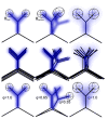
in X-ray crystallography, resolution and model selection interact to affect the interpretation of conformational heterogeneity. Electron density (blue) at progressively worse resolution can be fit by different classes of models (black lines).
Figure 1
Figure 1
Protein dynamics across temporal (x-axis) and spatial (y-axis) scales. Proteins exhibit conformational dynamics ranging from atomic vibrational motions around average positions on the pico-second timescale, (bond vibrations, leftmost cartoon at the bottom) to exchanging conformational substates of rotameric side-chains, to loop motions, to collective exchanges and increasingly larger substructures at millisecond or even longer timescales (right-most cartoon at bottom). Experimental techniques to probe structure and dynamics are highlighted in blue, and methods to represent protein conformations or conformational ensembles are highlighted in red. Conventional, synchrotron-based X-ray data can result in different structural characterizations (highlighted in red), which can additionally provide a structural basis of NMR observables. Picosecond dynamics are commonly modeled with a harmonic B-factor (local) or TLS (global) model in crystal structures. Nanosecond to microsecond motions result in conformational (anharmonic) substates, which require multi-conformer or ensemble models for visualization. Time-resolved X-ray experiments depend on conformational substates frozen into the crystal. NMR order parameters derived from spin relaxation experiments have established a link between fast protein dynamics in solution and the crystalline state. Chemical shift and residual dipolar coupling (RDC) data measures dynamics spanning nine orders of magnitude. MD simulations or conformational sampling algorithms can aid in interpreting RDC data. R and CPMG RD experiments report on exchanging substates at millisecond timescales. Serial Femtosecond Crystallography (SFX) in particular enables access to conformational ensembles across many orders of magnitude of timescales.
Figure 2
Figure 2
NMR experiments report on motions across different timescales. The structural basis of these motions and the fitting procedures govern the conversion of these experimental observables into structural restraints.
Figure 3
Figure 3
At physiological temperatures, crystalline environments mildly affect biomolecular motions. (a) R1 relaxation rates for methyl side-chains of R-spectrin SH3 in solid (horizontal axis) and solution (vertical axis) state. The correlation coefficient between the data obtained from the two environments is 0.95, suggesting highly similar motions. Data points are expected to lie along a 45° line if there are no differences between the crystalline and the solution state. (b) Solution structure of human Ubiquitin exhibiting a type I conformation β-turn. c) A type II conformation β-turn in microcrystalline human Ubiquitin. A peptide flip of D52 is stabilized by a hydrogen bond to E24, and by a water-mediated hydrogen bond to crystal contact K63 (not shown). Furthermore, E24 is stabilized by crystal-contact E64.
Figure 4
Figure 4
Cryocooling of protein crystals irregularly selects conformational sub-states. Isomorphous Fo-Fo maps of two independently collected pairs (in 2005 and 2013) of room and cryogenic temperature data sets of ecDHFR:NADP+:FOL are shown, contoured at 0.4e/Å. a) RT05-CRYO05 show widespread positive difference peaks (green, red indicates negative peaks), indicating that RT data sets exhibit elevated conformational heterogeneity. The CRYO05-CRYO13 difference map (b) shows peaks of both signs, which are absent in the RT05-RT13 data (c), pointing to irregular and unpredictable changes in structure and dynamics owing to cryocooling. d) The Fenwick-Wright framework relates crystallographic, isotropic atomic displacement parameters obtained from anharmonic sub-states to order parameters in solution. The angular order parameter, S2, reports on angular diffusion between discrete states of atoms ui and uj through angle θij. The orthogonal order parameter, S2, reports on angular diffusion of bond vectors within states through, for instance, angle αj. The method revealed excellent agreement for atomic displacements measured with X-ray crystallography and NMR in solution.
Figure 5
Figure 5
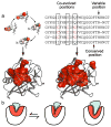
Networks of conformational exchange are evolution’s engines. (a) CONTACT networks are identified from clash-and-relieve pathways of alternate main- and side-chain conformers in a multi-conformer qFit model (top left). Pathways that share residues are grouped into networks. A CONTACT network representing conformational exchange in the enzyme Cyclophilin A is shown in red surface on the molecule (bottom left). Sectors are networks composed of co-evolving amino acids identified from Statistical Coupling Analysis (right). The similarities between the CONTACT network and the sector are striking, suggesting that conformational exchange may possibly be a phylogenetic instrument that enables members of the family to evolve towards specific functions and accommodating a wide variety of ligands. (b) This view allows two distinct scenarios. Within species, networks (cartoon, red) are optimized to evolve new function by enabling conformational exchange between substates to bind to (functionally) different and new partner molecules. Across species, networks could be further specialized, enabling new functions and/or losing their ability to exchange with substates associated with previous functions (rightmost cartoon).
Figure 6
Figure 6
Illustration of Serial Femtosecond Crystallography (SFX). Nanocrystals are extruded from a jet into the X-ray Free Electron Laser beam. In time resolved studies, a “pump” laser beam is placed in the path of the crystal. The laser pulse can uncage a substrate or excite a naturally occurring chromophore, starting a chemical process. The small crystal size can also allow rapid mixing of substrates, enabling the possibility to monitor enzymatic reactions. Varying the distance between the laser and X-ray pulses would intercept the process at different times, resulting in a molecular movie. We depict four possible scenarios of the conformational transitions, represented by colors, after excitation by the laser. In the top scenario, all unit cells are synchronized through a series of conformational changes represented by the different colors. This scenario gives a straightforward interpretation: as the distance is varied, the electron density map changes from one state to the next. Next, we show a two state system where some of the unit cells switch to the dark state with no detectible intermediates. Here, occupancies refinement at high resolution can determine the relative populations. Complications arise when conformational changes are asynchronous, as depicted in the third scenario. Prior knowledge of the conformational landscape is essential to determine the shifting occupancies of different states. In the fourth scenario, the lattice becomes disordered as the conformational changes occur resulting in a loss of diffraction resolution (pink crystals). If the lattice stabilizes in a new conformation, information about the kinetics, but not the intermediate structures can be extracted from the experiment.

References

    1. Boehr DD, McElheny D, Dyson HJ, Wright PE. The dynamic energy landscape of dihydrofolate reductase catalysis. Science. 2006;313:1638–42. CPMG relaxation dispersion experiments characterize the structural dynamics of the catalytic cycle of DHFR as a sequence of ‘linked’ ground and excited states. - PubMed
    1. Henzler-Wildman KA, et al. Intrinsic motions along an enzymatic reaction trajectory. Nature. 2007;450:838–44. - PubMed
    1. Fleishman SJ, et al. Computational design of proteins targeting the conserved stem region of influenza hemagglutinin. Science. 2011;332:816–21. - PMC - PubMed
    1. Whitehead TA, et al. Optimization of affinity, specificity and function of designed influenza inhibitors using deep sequencing. Nat Biotechnol. 2012;30:543–8. - PMC - PubMed
    1. Linder M, Johansson AJ, Olsson TSG, Liebeschuetz J, Brinck T. Computational design of a Diels-Alderase from a thermophilic esterase: the importance of dynamics. J Comput Aided Mol Des. 2012;26:1079–95. - PubMed

Publication types

Substances

LinkOut - more resources