. 2015 Mar 31;5(3):e535.
doi: 10.1038/tp.2015.24.

Electrical stimulation alleviates depressive-like behaviors of rats: investigation of brain targets and potential mechanisms

Affiliations

Electrical stimulation alleviates depressive-like behaviors of rats: investigation of brain targets and potential mechanisms

L W Lim et al. Transl Psychiatry. .

Abstract

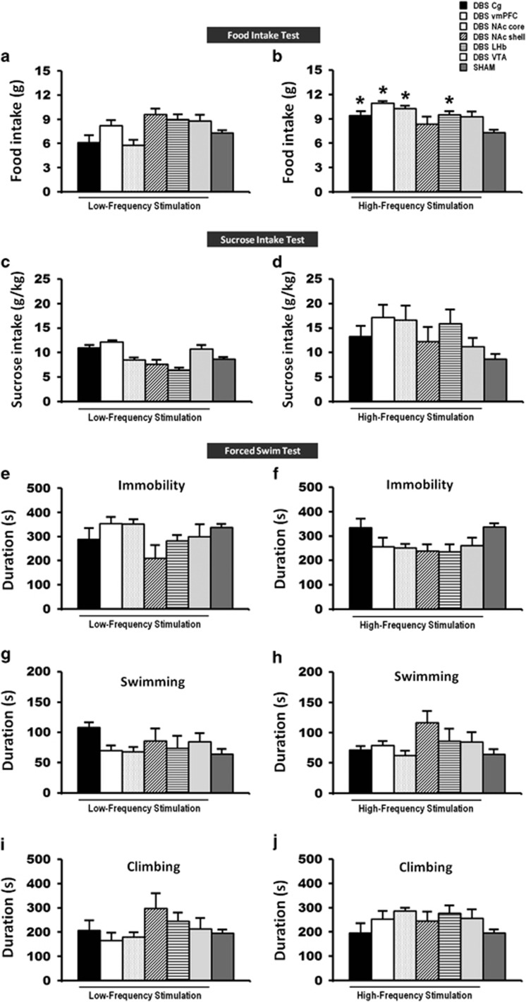
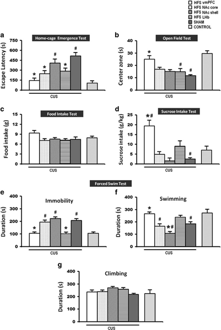
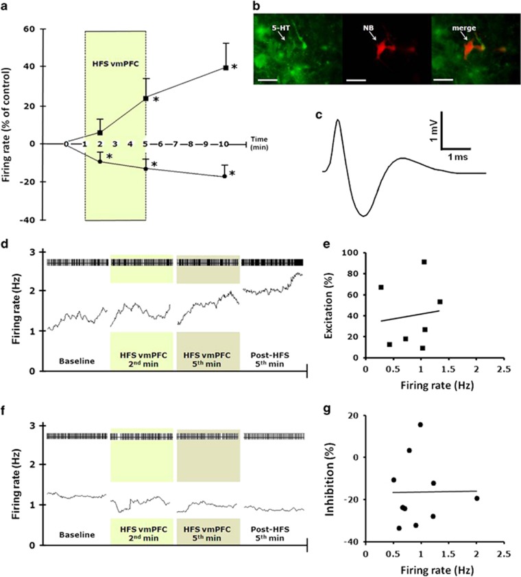
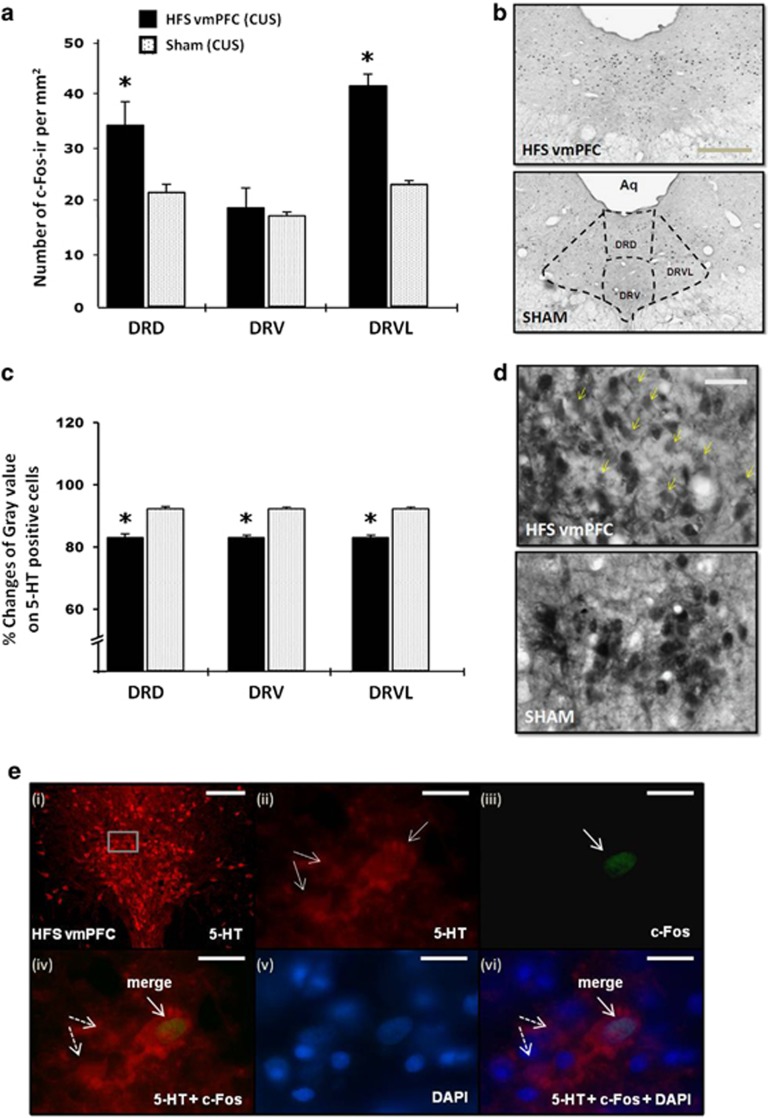
Deep brain stimulation (DBS) is a promising therapy for patients with refractory depression. However, key questions remain with regard to which brain target(s) should be used for stimulation, and which mechanisms underlie the therapeutic effects. Here, we investigated the effect of DBS, with low- and high-frequency stimulation (LFS, HFS), in different brain regions (ventromedial prefrontal cortex, vmPFC; cingulate cortex, Cg; nucleus accumbens (NAc) core or shell; lateral habenula, LHb; and ventral tegmental area) on a variety of depressive-like behaviors using rat models. In the naive animal study, we found that HFS of the Cg, vmPFC, NAc core and LHb reduced anxiety levels and increased motivation for food. In the chronic unpredictable stress model, there was a robust depressive-like behavioral phenotype. Moreover, vmPFC HFS, in a comparison of all stimulated targets, produced the most profound antidepressant effects with enhanced hedonia, reduced anxiety and decreased forced-swim immobility. In the following set of electrophysiological and histochemical experiments designed to unravel some of the underlying mechanisms, we found that vmPFC HFS evoked a specific modulation of the serotonergic neurons in the dorsal raphe nucleus (DRN), which have long been linked to mood. Finally, using a neuronal mapping approach by means of c-Fos expression, we found that vmPFC HFS modulated a brain circuit linked to the DRN and known to be involved in affect. In conclusion, HFS of the vmPFC produced the most potent antidepressant effects in naive rats and rats subjected to stress by mechanisms also including the DRN.


Figures

Figure 1
Figure 1
Schematic illustration of the anatomical placement of stimulating electrode in the vmPFC (a), LHb (b), Cg (c), NAc (d) and the VTA (e). Photomicrograph of a 30-μm-thick coronal section from the brain of a rat showing the histological verification of the electrode location in the vmPFC (scale bar, 1 mm). The symbol (formula image
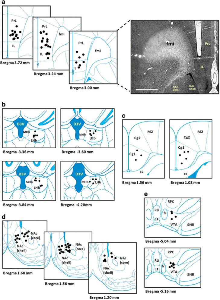
) indicates the tips of all electrode localization. cc, corpus callosum; Cg1 and Cg2, cingulate cortex 1 and cingulate cortex 2; D3V, dorsal third ventricle; fmi, forceps minor of corpus callosum; fr, fasciculus retroflexus; IF, interfascicular nucleus; IL, infralimbic; LHb, lateral habenular nucleus; MHb, medial habenular nucleus; M2, secondary motor cortex; NAc core, nucleus accumbens core; NAc shell, nucleus accumbens shell; PrL, prelimbic; RLi, rostral linear nucleus of the raphe; RPC, parvicellular part of the red nucleus; SNR, reticular part of the substantia nigra; vmPFC, ventromedial prefrontal cortex; VTA, ventral tegmental area.
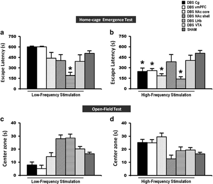
Figure 2
Figure 2
A set of bar graphs showing the measures of anxiety-like behavior by home-cage emergence (a and b) and open-field (c and d) tests in naive animal experiments. Before testing, animals were stimulated for 15 min and continuously for another 5 min during both the anxiety tasks. LFS of the LHb, and HFS of the Cg, vmPFC, NAc core and LHb reduced the escape latency from the home-cage emergence test, indicating anxiolytic behavior. No significant difference was found in the open-field test of both LFS and HFS groups. Data represent means±s.e.m. Significant difference from the sham animals, *P<0.05. Cg, cingulate cortex; HFS, high-frequency stimulation; LFS, low-frequency stimulation; LHb, lateral habenular nucleus; NAc, nucleus accumbens; vmPFC, ventromedial prefrontal cortex.
Figure 3
Figure 3
A set of bar graphs showing the measures of food motivation by food intake test (a and b), levels of anhedonia by sucrose intake test (c and d) and forced-swim behavior test (ej) in naive animal experiments. Before testing, animals were stimulated for 15 min and continuously for 2 h in the food intake test, 1 h in the sucrose intake test and 10 min in the forced-swim test. HFS of the Cg, vmPFC, NAc core and LHb significantly increased the levels of motivation for food intake. No significant difference was found in the sucrose intake and forced-swim behaviors of both LFS and HFS groups, as well as the food intake test of LFS group. Data represent means±s.e.m. Significant difference from the sham animals, (*P<0.05). Cg, cingulate cortex; HFS, high-frequency stimulation; LFS, low-frequency stimulation; LHb, lateral habenular nucleus; NAc, nucleus accumbens; vmPFC, ventromedial prefrontal cortex.
Figure 4
Figure 4
A set of bar graphs showing the measures of anxiety-like behavior by home-cage emergence (a) and open-field (b) tests, food motivation by food intake test (c), measures of anhedonia by sucrose intake test (d) and the forced-swim behavior test (eg) after HFS in the CUS rat model of depression. Animals were similarly stimulated and tested as in the naive animal experiment. Note: HFS of the vmPFC, NAc core, and the LHb reduced anxiety-like behavior in the home-cage emergence test. However, in the open-field and sucrose intake tests, vmPFC HFS, but not other DBS targets, significantly increased time spent of rats in the open-field center zone and increased sucrose intake, indicating anxiolytic and alleviation of anhedonic-like behavior as compared with CUS sham rats. Finally, both the LHb and vmPFC HFS reduced forced-swim immobility. Data represent means±s.e.m. Significant difference from the CUS sham animals, *P<0.05; significant difference from the non-CUS control animals, #P<0.05. CUS, chronic unpredictable stress; DBS, deep brain stimulation; HFS, high-frequency stimulation; LHb, lateral habenular nucleus; NAc, nucleus accumbens; vmPFC, ventromedial prefrontal cortex.
Figure 5
Figure 5
Effects of vmPFC HFS on DRN neuronal firing. (a) Excitatory and inhibitory effects on putative 5-HT DRN neurons (n=18) firing rate changes before, during and after vmPFC HFS. Darkened zone area indicates stimulation period (100 Hz, 100 μA, 100 μs). Firing rate was quantified during the final 60 s before stimulation as control baseline, during the second and fifth minute of vmPFC HFS, and on the fifth minute following cessation of vmPFC stimulation. Note, both excitatory and inhibitory effects on the firing rates of 5-HT neurons were observed by vmPFC HFS. (b) Photomicrographs showing immunohistochemical characterization of a neurobiotin (NB) juxtacellular-labeled 5-HT immunopositive neuron in the rat DRN. Scale bar, 20 μm at × 40 magnification. (c) A triphasic extracellular waveform shape with a broad spike width typically used to indicate for 5-HT neuron. (d and f) Spike trains and mean firing rate (15-s bins) of 5-HT putative neurons, before, during and after 5 min HFS of the vmPFC. Darkened zone areas indicate stimulation period (second and fifth minute of vmPFC HFS). Plot of percentage excitation (e) and inhibition (g) during the fifth minute after vmPFC HFS against baseline firing rates. Significant difference from the baseline levels *P<0.05. 5-HT, 5-hydroxytryptamine, serotonin; DRN, dorsal raphe nucleus; HFS, high-frequency stimulation; vmPFC, ventromedial prefrontal cortex.
Figure 6
Figure 6
Effects of vmPFC HFS on the c-Fos neural activity (a) and the percentage changes of mean gray value on 5-HT positive cells (c) in the DRN region of CUS animal model of depression. Representative photomicrographs of c-Fos (b; scale bar, 500 μm at × 4 magnification) and 5-HT (d; scale bar, 50 μm at × 40 magnification) histochemical staining of 30-μm-thick sections in the DRN. Note, vmPFC HFS resulted in higher levels of neuronal activation (that is, increased c-Fos positive cells) in the DRN areas as compared with the CUS sham animals. The small dots represent c-Fos-ir cells per mm2. Meanwhile, vmPFC HFS caused a remarkable reduction of optical density of 5-HT containing cells in the DRN area, indicating a local release of 5-HT, which increases in extracellular 5-HT in the DRN and forebrain projection areas. Representative high-power confocal images (e) are shown for the localization of 5-HT (red; e-i and -ii) and c-Fos (green; e-iii) immunofluorescence positive cells. DAPI (blue; e-v) was used as a nuclear stain. Merged images demonstrated the co-localization (arrows) of 5-HT and c-Fos (e-iv), as well as counterstained with DAPI (e-vi) in the DRD. Dotted arrows demonstrated non-c-Fos 5-HT positive cells (e-iv and -vi). Scale bar for fluorescent images: 100 μm (e-i) at low-power magnification; and 20 μm (e-iivi) at × 100 magnification. Significant difference from the CUS sham animals, *P<0.05. 5-HT, 5-hydroxytryptamine, serotonin; Aq, aqueduct; CUS, chronic unpredictable stress; DAPI, 4',6-diamidino-2-phenylindole; DRN, dorsal raphe nucleus dorsal part; DRV, dorsal raphe nucleus ventral part; DRVL, dorsal raphe nucleus ventrolateral part; HFS, high-frequency stimulation; vmPFC, ventromedial prefrontal cortex.

References

    1. Kessler RC, Berglund P, Demler O, Jin R, Koretz D, Merikangas KR, et al. The epidemiology of major depressive disorder: results from the National Comorbidity Survey Replication (NCS-R) JAMA. 2003;289:3095–3105. - PubMed
    1. Alonso J, Angermeyer MC, Bernert S, Bruffaerts R, Brugha TS, Bryson H, et al. Prevalence of mental disorders in Europe: results from the European Study of the Epidemiology of Mental Disorders (ESEMeD) project. Acta Psychiatr Scand Suppl. 2004;420:21–27. - PubMed
    1. American Psychiatric Association Practice guideline for the treatment of patients with major depressive disorder (revision) Am J Psychiatry. 2002;157:1–45. - PubMed
    1. Temel Y, Hescham SA, Jahanshahi A, Janssen ML, Tan SK, van Overbeeke JJ, et al. Neuromodulation in psychiatric disorders. Int Rev Neurobiol. 2012;107:283–314. - PubMed
    1. Temel Y, Lim LW. Neurosurgical treatments of depression. Curr Top Behav Neurosci. 2013;14:327–339. - PubMed

Publication types