. 2015 Mar 27;5(2):1141-71.
doi: 10.3390/life5021141.

Advances in Understanding Carboxysome Assembly in Prochlorococcus and Synechococcus Implicate CsoS2 as a Critical Component

Affiliations

Advances in Understanding Carboxysome Assembly in Prochlorococcus and Synechococcus Implicate CsoS2 as a Critical Component

Fei Cai et al. Life (Basel). .

Abstract

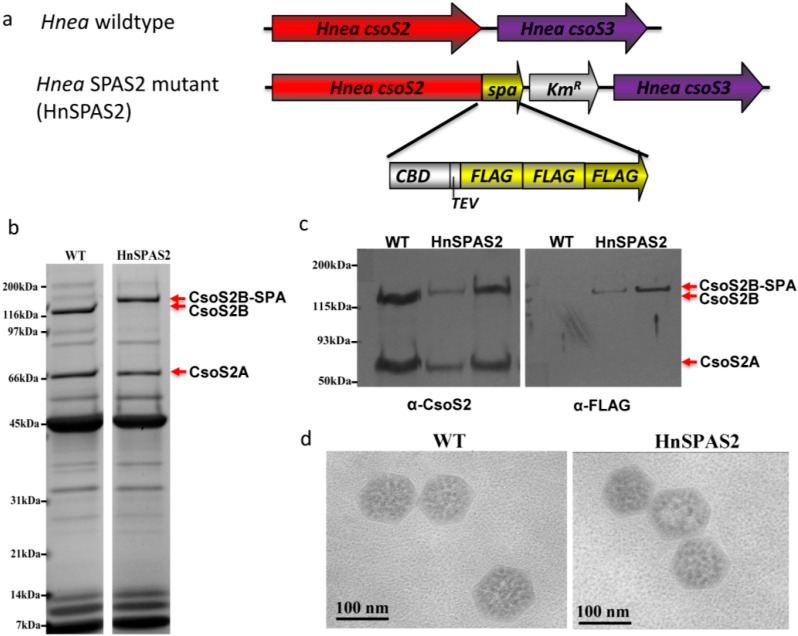
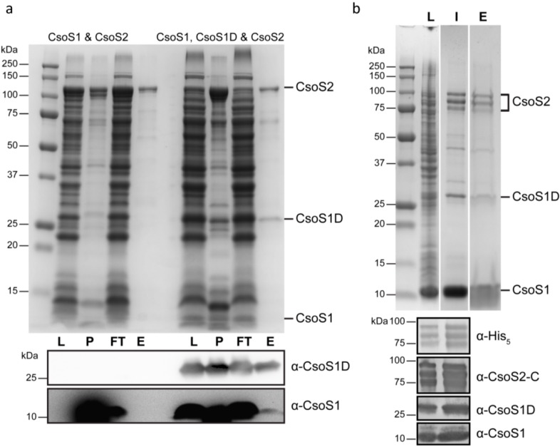
The marine Synechococcus and Prochlorococcus are the numerically dominant cyanobacteria in the ocean and important in global carbon fixation. They have evolved a CO2-concentrating-mechanism, of which the central component is the carboxysome, a self-assembling proteinaceous organelle. Two types of carboxysome, α and β, encapsulating form IA and form IB d-ribulose-1,5-bisphosphate carboxylase/oxygenase, respectively, differ in gene organization and associated proteins. In contrast to the β-carboxysome, the assembly process of the α-carboxysome is enigmatic. Moreover, an absolutely conserved α-carboxysome protein, CsoS2, is of unknown function and has proven recalcitrant to crystallization. Here, we present studies on the CsoS2 protein in three model organisms and show that CsoS2 is vital for α-carboxysome biogenesis. The primary structure of CsoS2 appears tripartite, composed of an N-terminal, middle (M)-, and C-terminal region. Repetitive motifs can be identified in the N- and M-regions. Multiple lines of evidence suggest CsoS2 is highly flexible, possibly an intrinsically disordered protein. Based on our results from bioinformatic, biophysical, genetic and biochemical approaches, including peptide array scanning for protein-protein interactions, we propose a model for CsoS2 function and its spatial location in the α-carboxysome. Analogies between the pathway for β-carboxysome biogenesis and our model for α-carboxysome assembly are discussed.


Figures

Figure 1
Figure 1
Schematic of α-carboxysome gene organization in three model organisms. Locus boundaries are based on the LoClass algorithm for BMCclassification [4]. Conserved cso genes are color-coded: Bacterial Microcompartment domain (BMC; pfam00936)-containing genes (csoS1s) in orange; RuBisCO large and small subunits (cbbL/S) in dark and light green, respectively; csoS2 in red; carbonic anhydrase (csoS3) in purple; genes belong to pfam03319 (csoS4A/B) in yellow; and genes encoding pterin-4 alpha-carbinolamine dehydratase-like protein (PCD-like) in magenta. Gray-blue genes are shared within the BMC locus subtype; gray genes are shared with at least one other BMC locus type; white genes indicate that this gene is not considered part of the locus [4]. Annotations for gray-blue or gray coded genes are as following: 1. por (protochlorophyllide oxidoreductase); 2. chlL (light-independent protochlorophyllide reductase iron-sulfur ATP-binding protein); 3. chlB (light-independent protochlorophyllide reductase subunit B); 4. chlN (light-independent protochlorophyllide reductase subunit N); 5. HAM1; 6. sbtA (high-affinity bicarbonate transporter); 7. sbtB (or annotated as nitrogen regulatory protein P-II); 8. cbiA (cobyrinic acid a,c-diamide synthase); 9. vwfA (von Willebrand factor type A); 10. nuoL (NADH-quinone oxidoreductase subunit L); 11. conserved gene with unknown function DUF2309 and 12. cbbQ (a putative catalytic chaperone of RuBisCO). Details on the gene organization of the subtypes of the α-carboxysome among all sequenced cyanobacterial genomes are also reviewed in Roberts et al. 2012 [8].
Figure 2
Figure 2
Expression of Hnea rCsoS2 in E. coli with an N- or a C-terminal tag. (a) Schematic of the short and long form of rCsoS2 proteins produced by E. coli when codons for either an N- or a C-terminal tag are genetically fused to the open reading frame of Hnea csoS2 gene. In the case of a C-terminal tag, the short form (boxed by dotted lines) cannot be purified via affinity chromatography because of lack of the C-terminal tag. (b) Purified rCsoS2 in comparison with CsoS2A and CsoS2B from native source. The left lane shows the short (CsoS2A) and long (CsoS2B) form of CsoS2 protein in purified Hnea carboxysomes. When expressed in E. coli with an N-terminal tag, both the short and long forms can be purified using an affinity column (middle lane). When expressed in E. coli with a C-terminal tag, only the long form can be recovered after affinity purification followed by self-cleavage of the tag.
Figure 3
Figure 3
Knockout of CsoS2 abolishes carboxysome formation in Hnea. When the csoS2 gene is interrupted by the insertion of a KmR cassette (a); no carboxysomes are apparent in mutant cells comparing to wildtype Hnea cells with carboxysomes (indicated by red arrows) under the same growth condition (b).
Figure 4
Figure 4
A Hnea mutant with SPA-tagged CsoS2. (a) The SPA tag fused to the C-terminus of CsoS2 contains a 3x FLAG epitope and a calmodulin binding domain (CBD) separated by a TEV protease site. A kanamycin resistance gene (KmR) cassette follows the SPA tag to allow for selection. (b) Both wildtype and mutant carboxysomes can be purified and their polypeptide separation patterns are similar, except SPA-tagged CsoS2B is slightly larger than untagged CsoS2B. (c) Western blots of wildtype and mutant cells blocked against α-CsoS2 antisera and α-FLAG antibodies. Only the long form of CsoS2 has a FLAG epitope tag in HnSPAS2. No cross-reactivity with small polypeptides was observed. (d) Purified wildtype and HnSPAS2 mutant carboxysomes are indistinguishable in TEM images.
Figure 5
Figure 5
A Hidden Markov model (HMM) logo for all α-Cyanobacterial CsoS2 orthologs. The Y-axis represents the information content (aka relative entropy), and the letters divide the stack height according to their estimated probability at a given position. MED4 and MIT9313 CsoS2 sequences are aligned to the corresponding position on the logo, and the predicted secondary structural motifs are colored red and orange for α-helices and β-strands, respectively. N- and M-region repeats are indicated by cyan and green underlining, respectively. Short repeats (3 amino acids) that occur three units per group (except in the last group) are outlined in light-gray boxes. Relatively conserved residues of the C-region are underlined in red. Putative transition areas between three regions are indicated by brown arrows. For demonstration purposes only, a simplified presentation of results from the protein-binding assay against MIT9313 CsoS2 peptide array (see Section 2.12) are mapped onto the logo. The starting position of peptides among all positive hits is marked with RuBisCO, CsoS1 or CsoS1D symbols only if the averaged signal intensity (1) ranks in the top 10 out of all positive hits or (2) is a local maximum with >5 sequential positive hits. The saturation of each symbol is relative to its fraction ratio to the maximum signal intensity (as 100% saturation) of all positive hits from a given binding assay.
Figure 6
Figure 6
Repetitive motifs found in the three representative CsoS2 proteins. Repetitive motif found in the N-region (cyan) and M-region (green) of MED4 (a); MIT9313 (b); and Hnea (c); CsoS2 proteins; (d) MEME motif for the N-repeats; (e) long and short form of MEME motif for the M-repeats.
Figure 7
Figure 7
A CsoS2 phylogram. CsoS2 orthologs (Table S2) found in α-Cyanobacteria (green), α-Proteobacteria (blue), β-Proteobacteria (orange), γ-Proteobacteria (black), Actinobacteria (red) and Nitrospirae (magenta) are shown in the phylogram. Purple phototrophs, which belong to the γ-Proteobacteria, are shown in purple. Bootstrap values were obtained from 100 replicates; nodes receiving bootstrap values greater than 75 or between 50 and 74 are indicated by filled circles or filled triangles, respectively. Numbers correspond to organism ID numbers given in Table S2.
Figure 8
Figure 8
Folding predictions for MIT9313 CsoS2. (a) Fold-Index prediction; (b) PONDR prediction; (c) ribbon presentation of ab initio folding prediction by QUARK for each M-repeat, shown in a rainbow spectrum from N-terminus (blue) to C-terminus (red).
Figure 9
Figure 9
Biophysical characterizations of Prochlorococcus CsoS2. (a) SAXS analysis of MED4 CsoS2 and (b) the pair distribution function of the same SAXS data; (c) Near UV Circular Dichroism spectroscopy of the same protein; (d) The pair distribution functions of SAXS data measured on MIT9313 CsoS2 in isolation and after mixing with MIT9313 CsoS1 and MIT9313 RuBisCO.
Figure 10
Figure 10
Native agarose gel electrophoresis of Hnea recombinant carboxysome protein and Hnea RuBisCO mixtures. Lanes from top to bottom: rCsoS2 (20 µL at 0.6 mg/mL), RuBisCO (20 µL at 1.1 mg/mL), rCsoS1A (20 µL at 0.7 mg/mL), BSA (20 µL at 1.0 mg/mL), rCsoS2 w. RuBisCO and rCsoS1A (20 µL each), rCsoS2 w. RuBisCO, rCsoS1A and BSA (20 µL each), and rCsoS2 w. BSA and rCsoS1A (20 µL each). By itself, the positively charged rCsoS2 migrates to the negative electrode; rCsoS1A and RuBisCO migrate to the positive electrode. When mixed, rCsoS2 drags its interaction partners, but not BSA, towards the negative electrode.
Figure 11
Figure 11
Pull-down assay and purification of α-carboxysome interaction complexes. (a) SDS-PAGE and Western blots (α-CsoS1 and α-CsoS1D) of lysate (L) from cells co-expressing CsoS1 and CsoS2 or CsoS1, CsoS1D and CsoS2 and the corresponding cell debris after breaking the cells (P), flow-through fractions (FT) and elutions (E) after pull-down assay using Glutathione-Sepharose magnetic beads; (b) SDS-PAGE of lysate (L) from cells co-expressing CsoS1, CsoS1D and CsoS2 from a single construct, the isolated complex (I) and the elution (E) from pull-down assay using Ni-NTA-agarose magnetic beads. Western blots for samples L and I are shown underneath using four different antibodies: α-His5, α-CsoS2-C, α-CsoS1 and α-CsoS1D. All three bands that have higher MW (between 75–100 kDa) are identified as CsoS2 with intact C-termini.
Figure 12
Figure 12
A working model for the location and function of CsoS2 in α-carboxysome assembly. Prior to α-carboxysome formation, RuBisCO and shell proteins such as CsoS1 are recruited by CsoS2. Subsequently, CsoS1 hexamers tile together and form shells anchored by CsoS2 via its C-region, and RuBisCO line up while associated with CsoS2. As a result, the carboxysome is assembled during the simultaneous formation of the shell and packing of RuBisCOs. CsoS2 may adapt different conformations in the final stage; a network of CsoS2 is formed based on inter-molecular interactions among CsoS2 proteins, which may be mediated through disulfide bonds formed between conserved Cysteine residues found in M-repeats. Short forms of CsoS2 (CsoS2A) will only organize RuBisCO but not provide anchoring to the shell. The tail of C-region may be exposed on the surface of the carboxysome and accessible from cytoplasm.

References

    1. Goericke R., Welschmeyer N.A. The marine prochlorophyte Prochlorococcus contributes significantly to phytoplankton biomass and primary production in the Sargasso Sea. Deep Sea Res. 1993;40:2283–2294. doi: 10.1016/0967-0637(93)90104-B. - DOI
    1. Liu H., Nolla H.A., Campbell L. Prochlorococcus growth rate and contribution to primary production in the equatorial and subtropical North Pacific Ocean. Aquat. Microb. Ecol. 1997;12:39–47. doi: 10.3354/ame012039. - DOI
    1. Liu H., Campbell L., Landry M.R., Nolla H.A., Brown S.L., Constantinou J. Prochlorococcus and Synechococcus growth rates and contributions to production in the Arabian Sea during the 1995 Southwest and Northeast Monsoons. Deep Sea Res. 1998;45:2327–2352. doi: 10.1016/S0967-0645(98)00073-3. - DOI
    1. Axen S.D., Erbilgin O., Kerfeld C.A. A taxonomy of bacterial microcompartment loci constructed by a novel scoring method. PLoS Comput. Biol. 2014;10 doi: 10.1371/journal.pcbi.1003898. - DOI - PMC - PubMed
    1. Marin B., Nowack E.C., Glockner G., Melkonian M. The ancestor of the Paulinella chromatophore obtained a carboxysomal operon by horizontal gene transfer from a Nitrococcus-like gamma-proteobacterium. BMC Evol. Biol. 2007;7 doi: 10.1186/1471-2148-7-85. - DOI - PMC - PubMed