. 2015;12(3):343-53.
doi: 10.1080/15476286.2015.1017205.

miR-214 promotes osteoclastogenesis by targeting Pten/PI3k/Akt pathway

Affiliations

miR-214 promotes osteoclastogenesis by targeting Pten/PI3k/Akt pathway

Chenyang Zhao et al. RNA Biol. 2015.

Abstract

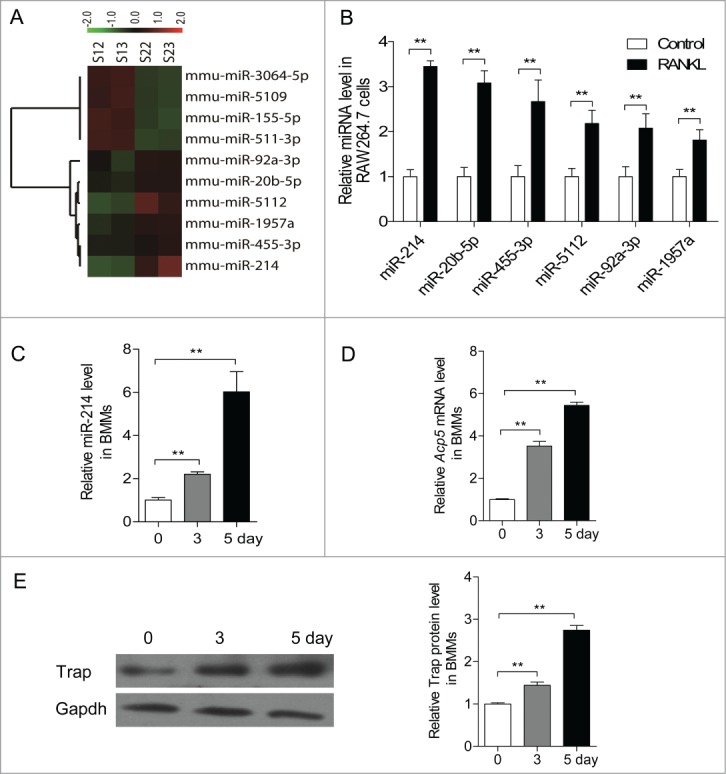
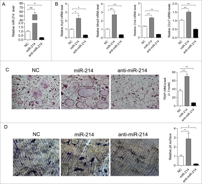
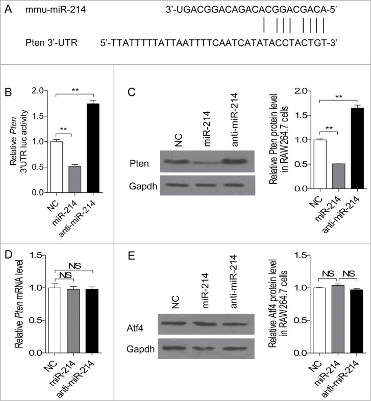
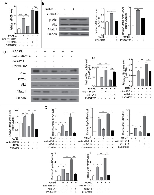
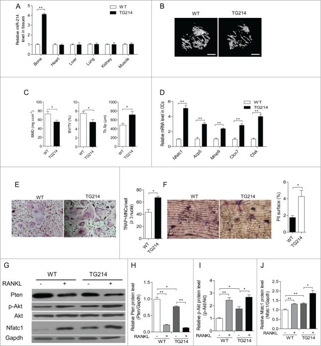
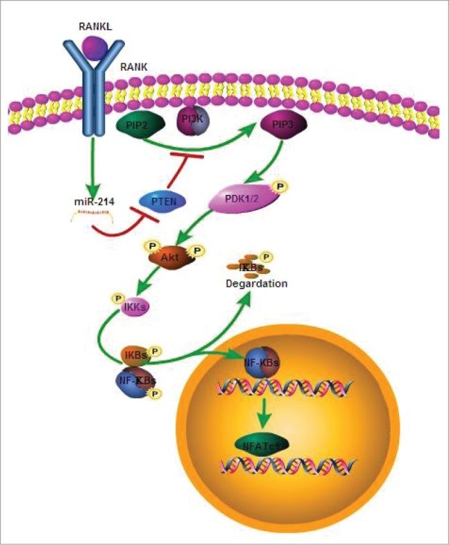
microRNA is necessary for osteoclast differentiation, function and survival. It has been reported that miR-199/214 cluster plays important roles in vertebrate skeletal development and miR-214 inhibits osteoblast function by targeting ATF4. Here, we show that miR-214 is up-regulated during osteoclastogenesis from bone marrow monocytes (BMMs) with macrophage colony stimulating factor (M-CSF) and receptor activator of nuclear factor-κB ligand (RANKL) induction, which indicates that miR-214 plays a critical role in osteoclast differentiation. Overexpression of miR-214 in BMMs promotes osteoclastogenesis, whereas inhibition of miR-214 attenuates it. We further find that miR-214 functions through PI3K/Akt pathway by targeting phosphatase and tensin homolog (Pten). In vivo, osteoclast specific miR-214 transgenic mice (OC-TG214) exhibit down-regulated Pten levels, increased osteoclast activity, and reduced bone mineral density. These results reveal a crucial role of miR-214 in the differentiation of osteoclasts, which will provide a potential therapeutic target for osteoporosis.

Keywords: BMD, bone mineral density; BMMs, bone marrow monocytes; BV/TV, ratio of bone volume to tissue volume; Dnm3os, Dnm3 opposite strand; M-CSF, macrophage colony stimulating factor; NFATc1, nuclear factor of activated T-cells cytoplasmic; OC-TG214, osteoclast specific miR-214 transgenic mice; PI 3-kinase; PTEN; Pten, phosphatase and tensin homolog; RANKL, receptor activator of nuclear factor-κB ligand; TRAP, tartrate-resistant acid phosphatase; Tb.Sp, trabecular spacing; WT, wild-type; miRNA; micro CT, Micro computed tomography; osteoclast; osteoporosis; qRT-PCR, quantitative real-time PCR.


Figures

Figure 1.
Figure 1.
miR-214 was upregulated during osteoclastogenesis. (A) Microarray assays were performed in RAW 264.7 cells with or without RANKL induction for 2 d Red and green indicate high expression levels and low expression levels, respectively. Shown are the miRNAs that changed more than 1.fold5-. (B) Relative miRNA levels in RANKL-induced RAW 264.7 cells was analyzed by qRT-PCR, miRNA levels were normalized to U6. (C) BMMs were cultured with M-CSF (10 ng/ml) alone for 1 day, and followed with M-CSF (30 ng/ml) and RANKL (50 ng/ml) for indicated times. Relative miR-214 levels in M-CSF+RANKL-induced BMMs were analyzed by qRT-PCR, miR-214 levels were normalized to U6. (D) Relative Acp5 mRNA levels in M-CSF+RANKL-induced BMMs were analyzed by qRT-PCR and normalized to Gapdh. (E) Trap protein levels in BMMs with RANKL-induction for indicated times were determined by western blot analysis and normalized to Gapdh. The data represent the mean ± SEM of 3 experiments in triplicate. **P < 0.01.
Figure 2.
Figure 2.
miR-214 regulates osteoclastogenesis from BMMs. (A) BMMs were induced with M-CSF and RANKL for 5 d Relative miR-214 levels were determined by qRT-PCR after treatment with miR-214 mimics or anti-miR-214 or negative control (NC) and miR-214 levels were normalized to U6. (B) Relative Acp5, Mmp9, Ctsk and Clcn7 mRNA levels were determined by qRT-PCR and normalized to Gapdh. (C) Representative photographs of OC formation (original magnification 200ċ) and the number of TRAP-positive multinucleated cells (MNCs) are shown. (D) Bone resorption areas on bovine bone slices were measured by image analysis. Representative images of resorbed bone slices (original magnification 4ċ). The data represent the mean ± SEM of 3 experiments in triplicate. *P < 0.05, **P < 0.01.
Figure 3.
Figure 3.
miR-214 directly targets Pten in osteoclasts. (A) Sequence alignment of mouse miR-214 with 3'-UTR of Pten. The seed sequences of miR-214 (up) and 3'-UTR of Pten (down) are complementary. (B) The effect of miR-214, anti-miR-214 and NC on luciferase activity in RAW 264.7 cells transfected with the Pten 3' UTR reporter. (C) Effect of miR-214, anti-miR-214 and NC on Pten protein level by protein gel blot analysis. miR-214 mimics, anti-miR-214 or NC were transfected into RANKL-induced RAW 264.7 cells. The protein levels were normalized to Gapdh. (D) Pten mRNA level was determined by qRT-PCR. The level of Pten were normalized to Gapdh. (E) Atf4 protein level was determined by western blot analysis and normalized to Gapdh. The data represent the mean ± SEM of 3 experiments in triplicate. *P < 0.05, **P < 0.01, NS, not significant.
Figure 4.
Figure 4.
miR-214 promotes osteoclast differentiation through Pten involved pathway. (A) miR-214 levels were determined by qRT-PCR in RAW 264.7 cells after treatment with RANKL, miR-214 mimics, anti-miR-214 and PI3K inhibitor LY294002. miR-214 levels were normalized to U6. (B) Effect of RANKL and PI3K inhibitor LY294002 on the protein levels of Nfatc1 and phosphorylated Akt in RAW 264.7 cells. Nfatc1 and phosphorylated Akt levels were analyzed by protein gel blot, and Gapdh was used as the internal control. (C) Effect of miR-214, anti-miR-214 and PI3K inhibitor LY294002 on the protein levels of Pten, Nfatc1 and phosphorylated Akt in RAW 264.7 cells. Pten, Nfatc1 and phosphorylated Akt levels were analyzed by western blot, and Gapdh was used as the internal control. (D) Effect of miR-214, anti-miR-214 and PI3K inhibitor LY294002 on NFATc1, Acp5, Ctsk, Mmp9, Clcn7 mRNA levels in RAW 264.7 cells. The mRNA levels were analyzed by qRT-PCR, and products were normalized to Gapdh. The data represent the mean ± SEM of 3 independent experiments in triplicate. *P < 0.05, **P < 0.01, NS, not significant.
Figure 5.
Figure 5.
Promotion of osteoclast activity by miR-214 in vivo. (A) qRT-PCR analysis of miR-214 levels in bone and other tissues (normalized to those in WT mice) from WT and Acp5-miR-214 transgenic (OC-TG214) mouse lines. miR-214 levels were normalized to U6. (B) MicroCT examination and representative images showing The 3-dimensional trabecular architecture in OC-TG214 and WT mice. Scale bars, 1 mm. (C) Micro CT measurements for BMD, BV/TV and Tb.Sp in the proximal tibia of WT and OC-TG214 mice. n=5. (D) Nfatc1, Acp5, Mmp9, Clcn7 and Ctsk mRNA levels in Oscar+ separated by cell sorting with flow cytometry were analyzed by qRT-PCR. The expression levels were normalized to Gapdh. (E) TRAP staining (original magnification 200ċ) and the number of TRAP-positive MNCs of RANKL-induced BMMs isolated from bone marrow of WT and OC-TG214 mice. (F) Bone resorption areas on bovine bone slices were measured by image analysis. Representative images of resorbed bone slices (original magnification 4ċ). (G, H, I, J) Pten, Nfatc1 and phosphorylated Akt levels in M-CSF and RANKL induced BMMs from WT and OC-TG214 mice for 4 d were measured by protein gel blot and expressed as the ratios of densitometry to Gapdh. Gapdh was used as the internal control. The data represent the mean ± SEM of 3 experiments in triplicate. *P < 0.05, **P < 0.01.
Figure 6.
Figure 6.
The model for effect of miR-214 on osteoclastogenesis. MiR-214 regulates RANKL-activated PI3K/Akt signaling pathway for osteoclast differentiation through targeting Pten.

References

    1. Del Fattore A, Teti A, Rucci N. Osteoclast receptors and signaling. Arch Biochem Biophys 2008; 473:147–60; http://dx.doi.org/ 10.1016/j.abb.2008.01.011. - DOI - PubMed
    1. Ross FP, Teitelbaum SL. alphavbeta3 and macrophage colony-stimulating factor: partners in osteoclast biology. Immunol Rev 2005; 208:88–105; PMID:16313343; http://dx.doi.org/ 10.1111/j.0105-2896.2005.00331.x. - DOI - PubMed
    1. Miyazaki T, Tanaka S, Sanjay A, Baron R. The role of c-Src kinase in the regulation of osteoclast function. Mod Rheumatol 2006; 16: 68–74; PMID:16633924; http://dx.doi.org/ 10.3109/s10165-006-0460-z. - DOI - PubMed
    1. Edwards CM, Mundy GR. Eph receptors and ephrin signaling pathways: a role in bone homeostasis. Int J Med Sci 2008; 5:263–72; PMID:18797510; http://dx.doi.org/ 10.7150/ijms.5.263. - DOI - PMC - PubMed
    1. Hattersley G, Owens J, Flanagan AM, Chambers TJ. Macrophage colony stimulating factor (M-CSF) is essential for osteoclast formation in vitro. Biochem Biophys Res Commun 1991; 177:526–31; PMID:2043138; http://dx.doi.org/ 10.1016/0006-291X(91)92015-C. - DOI - PubMed

Publication types

MeSH terms