

Review of spectral domain-enhanced depth imaging optical coherence tomography of tumors of the retina and retinal pigment epithelium in children and adults
- PMID: 25827543
- PMCID: PMC4399121
- DOI: 10.4103/0301-4738.154384
Review of spectral domain-enhanced depth imaging optical coherence tomography of tumors of the retina and retinal pigment epithelium in children and adults
Abstract
Background: Spectral domain (SD) enhanced depth imaging optical coherence tomography (EDI-OCT) is a useful tool for anatomic, cross-sectional imaging of retinal conditions.
Aims: The aim was to identify characteristic patterns of retinal and retinal pigment epithelial tumors on EDI-OCT in children and adults.
Settings and design: Retrospective review.
Materials and methods: Analysis of published reports and personal observations using office-based EDI-OCT for adults and portable hand-held SD OCT for infants and children.
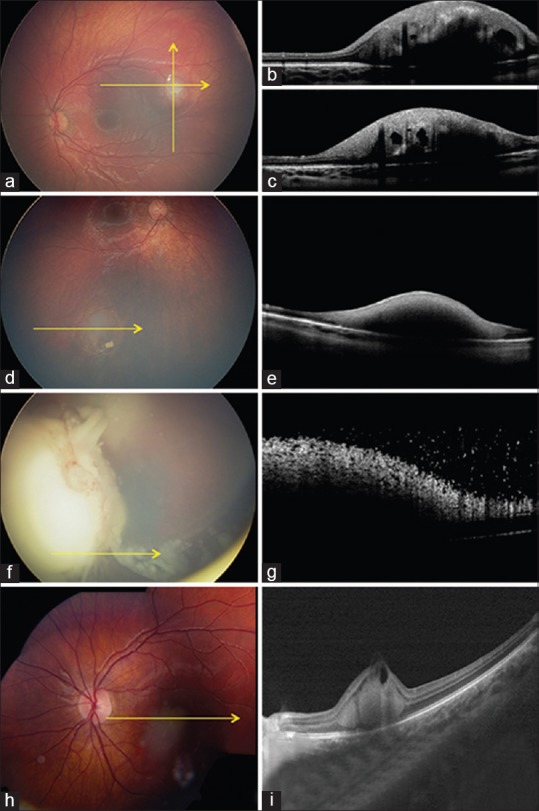
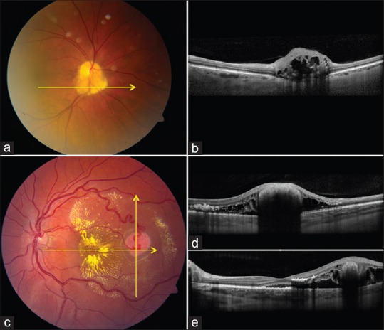
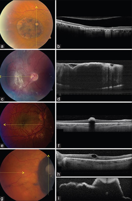
Results: Using EDI-OCT, retinal tumors such as small retinoblastoma, astrocytic hamartoma, and hemangioblastoma arose abruptly from the retina, immediately adjacent to normal retina. Small exophytic retinoblastoma and retinal hemangioblastoma showed the full-thickness, homogeneous retinal disorganization with surrounding normal retina "draping" over the margins. Retinoblastoma occasionally had intralesional cavities and surrounding subretinal fluid. Hemangioblastoma often had adjacent intraretinal edema and subretinal fluid. Astrocytic hamartoma arose within the nerve fiber layer and sometimes with a "moth-eaten" or cavitary appearance. Retinal pigment epithelial (RPE) lesions such as congenital hypertrophy of RPE appeared flat with shadowing, occasional subretinal cleft, and abrupt photoreceptor loss. Congenital simple hamartoma showed an abrupt elevation from the inner retina with crisp, dark posterior shadowing. Combined hamartoma of the retina/RPE showed vitreoretinal traction causing "sawtooth mini-peak" or gently "maxi-peak" folding of the retina. RPE adenoma often produces remote macular edema or epiretinal membrane and the tumor has an irregular, "rugged" surface with deep shadowing.
Conclusions: Enhanced depth imaging optical coherence tomography shows characteristic patterns that are suggestive of certain retinal and RPE tumors.
Conflict of interest statement
Figures
References
-
- Shields CL, Materin MA, Shields JA. Review of optical coherence tomography for intraocular tumors. Curr Opin Ophthalmol. 2005;16:141–54. - PubMed
-
- Spaide RF, Koizumi H, Pozzoni MC. Enhanced depth imaging spectral-domain optical coherence tomography. Am J Ophthalmol. 2008;146:496–500. - PubMed
-
- Shields CL, Mashayekhi A, Luo CK, Materin MA, Shields JA. Optical coherence tomography in children: Analysis of 44 eyes with intraocular tumors and simulating conditions. J Pediatr Ophthalmol Strabismus. 2004;41:338–44. - PubMed
Publication types
MeSH terms
LinkOut - more resources
Full Text Sources
Other Literature Sources
