. 2015 Jun;106(6):672-686.
doi: 10.1111/cas.12663. Epub 2015 May 12.

Inflammatory features of pancreatic cancer highlighted by monocytes/macrophages and CD4+ T cells with clinical impact

Affiliations

Inflammatory features of pancreatic cancer highlighted by monocytes/macrophages and CD4+ T cells with clinical impact

Takuya Komura et al. Cancer Sci. 2015 Jun.

Abstract

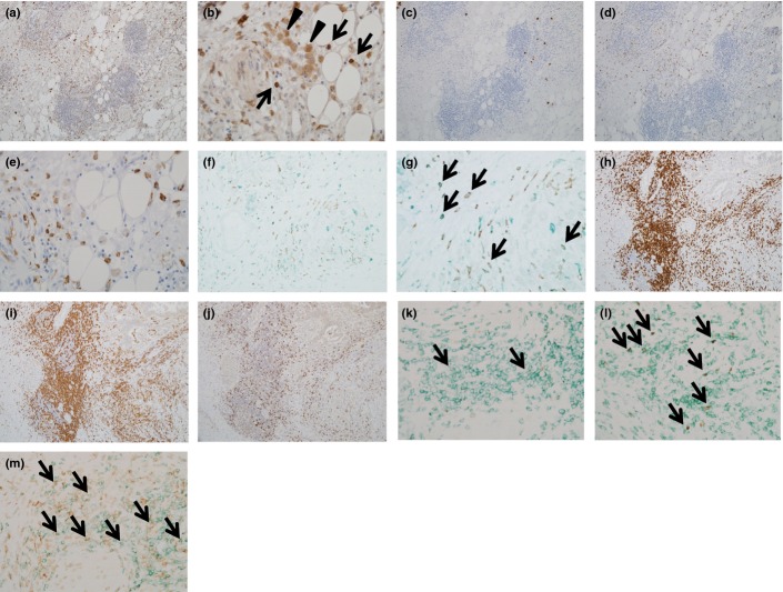
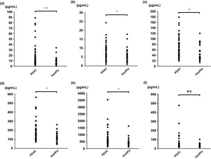
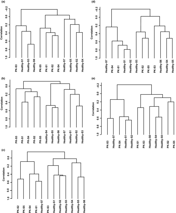
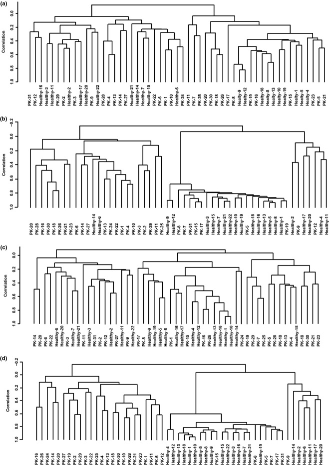
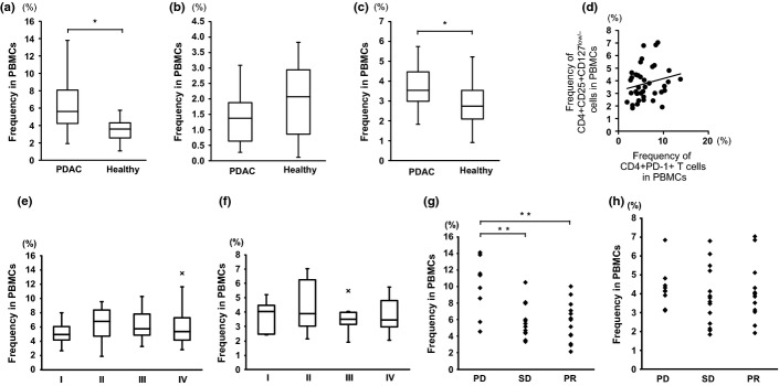
Pancreatic ductal adenocarcinoma (PDAC) is among the most fatal of malignancies with an extremely poor prognosis. The objectives of this study were to provide a detailed understanding of PDAC pathophysiology in view of the host immune response. We examined the PDAC tissues, sera, and peripheral blood cells of PDAC patients using immunohistochemical staining, the measurement of cytokine/chemokine concentrations, gene expression analysis, and flow cytometry. The PDAC tissues were infiltrated by macrophages, especially CD33+CD163+ M2 macrophages and CD4+ T cells that concomitantly express programmed cell death-1 (PD-1). Concentrations of interleukin (IL)-6, IL-7, IL-15, monocyte chemotactic protein-1, and interferon-γ-inducible protein-1 in the sera of PDAC patients were significantly elevated. The gene expression profile of CD14+ monocytes and CD4+ T cells was discernible between PDAC patients and healthy volunteers, and the differentially expressed genes were related to activated inflammation. Intriguingly, PD-1 was significantly upregulated in the peripheral blood CD4+ T cells of PDAC patients. Correspondingly, the frequency of CD4+PD-1+ T cells was increased in the peripheral blood cells of PDAC patients, and this increase correlated to chemotherapy resistance. In conclusion, inflammatory conditions in both PDAC tissue and peripheral blood cells in PDAC patients were prominent, highlighting monocytes/macrophages as well as CD4+ T cells with influence of the clinical prognosis.

Keywords: CD4+ T cells; macrophages; monocytes; pancreatic ductal adenocarcinoma; programmed cell death-1.


Figures

Figure 1
Figure 1
Immunohistochemical analysis of pancreatic ductal adenocarcinoma (PDAC) tissues. Surgically resected PDAC tissues were immunohistochemically stained. (a, b) CD33: several positive cells were scattered in the fibroadipose area around PDAC, which were mainly composed of a monocyte (arrows in b) and macrophage (arrowheads in b) morphology. (c) Neutrophil elastase: a few positive cells were observed. (d, e) CD14: several positive cells highlighting the monocyte morphology. (f, g) Double immunostaining of CD163 (brown) and CD33 (green). Most of the CD163+ cells were also positive for CD33. (h) CD3: lymphoid aggregation around PDAC was mainly composed of CD3+ T cells. (i) CD4: the majority of lymphoid aggregation was CD4+ T cells. (j) CD8: the number of CD8+ cells was small compared to that of CD4+ cells shown in (i). (k) Double immunostaining of T-bet (brown, nuclear expression) and CD4 (green). Double-positive cells were observed (arrows), but the number was very small. (l) Double immunostaining of FoxP3 (brown, nuclear expression) and CD4 (green). Several double-positive cells were scattered (arrows). (m) PD-1 (brown) and CD4 (green): several double-positive cells were found (arrows). Magnification: a, c, d, f, h, i, and j, ×40; b, e, g, k, l, and m, ×100.
Figure 2
Figure 2
Concentration of cytokines and chemokines in sera obtained from pancreatic ductal adenocarcinoma patients (n = 50) prior to treatment and from healthy volunteers (n = 27). The serum concentration of cytokines and chemokines was measured using a multiplex bead immunoassay system. (a) Interleukin (IL)-6, (b) IL-7, (c) monocyte chemotactic protein-1, (d) IL-15, (e) interferon-γ-inducible protein-1, and (f) IL-8. *P < 0.05, **P < 0.01.
Figure 3
Figure 3
Unsupervised clustering analysis of gene expression profiles of subfractions of peripheral blood cells from patients with pancreatic ductal adenocarcinoma (PK; n = 7) and healthy volunteers (n = 5). RNA was isolated from entire blood cells or each subfraction of peripheral blood cells, followed by gene expression analysis using DNA microarray. Genes that passed the quality check control were used in each clustering analysis. (a) Entire blood cells, 7039 genes; (b) CD14+ cells, 6602 genes; (c) CD4+ cells, 6770 genes; (d) CD8+ cells, 7621 genes; and (e) CD15+ cells, 9728 genes.
Figure 4
Figure 4
Unsupervised clustering analysis of the gene expression profile of CD14+ monocytes and CD4+ T cells in the peripheral blood of pancreatic ductal adenocarcinoma (PDAC) patients (PK) and healthy volunteers. RNA was isolated from all blood cells or each subfraction of peripheral blood cells from 31 PDAC patients and 22 healthy volunteers, followed by gene expression analysis using DNA microarray. (a, b) Hierarchical analysis of gene expression for isolated CD4+ cells in peripheral blood using all 10 868 filtered genes (a) or 266 genes whose expression was significantly altered between PDAC patients and healthy volunteers ≥1.5-fold with P < 0.001 (b). (c, d) Hierarchical analysis of gene expression for isolated CD14+ cells in peripheral blood using all 11 947 filtered genes (c) or 126 genes whose expression was significantly altered between PDAC patients and healthy volunteers ≥1.5-fold at P < 0.01 (d).
Figure 5
Figure 5
Frequency of CD4+PD-1+ cells and CD4+CD25+CD127low/− cells in PBMCs of pancreatic ductal adenocarcinoma (PDAC) patients (n = 50) and healthy volunteers (n = 27). The frequencies of CD4+PD-1+ cells (a), CD8+PD-1+ cells (b), and CD4+CD25+CD127low/− cells (c) were assessed by flow cytometry. (d) Scattergram of the frequencies of CD4+PD-1+ cells and CD4+CD25+CD127low/− cells in PDAC patients. (e, f) The frequency of CD4+PD-1+ cells (e) and CD4+CD25+CD127low/− cells (f) in the PBMCs of PDAC patients in the context of each clinical stage. (g, h) The chemotherapy responsiveness and frequency of CD4+PD-1+ cells (g) and CD4+CD25+CD127low/− cells (h). PD, progressive disease; PR, partial responsiveness; SD, stable disease. *< 0.05; **P < 0.01.

References

    1. Matsuda T, Ajiki W, Marugame T, et al. Population-based survival of cancer patients diagnosed between 1993 and 1999 in Japan: a chronological and international comparative study. Jpn J Clin Oncol. 2011;41:40–51. - PubMed
    1. Vincent A, Herman J, Schulick R, Hruban RH, Goggins M. Pancreatic cancer. Lancet. 2011;378:607–20. - PMC - PubMed
    1. Paulson AS, Tran Cao HS, Tempero MA, Lowy AM. Therapeutic advances in pancreatic cancer. Gastroenterology. 2013;144:1316–26. - PubMed
    1. Reni M, Cordio S, Milandri C, et al. Gemcitabine versus cisplatin, epirubicin, fluorouracil, and gemcitabine in advanced pancreatic cancer: a randomised controlled multicentre phase III trial. Lancet Oncol. 2005;6:369–76. - PubMed
    1. Fridman WH, Pagès F, Sautès-Fridman C, Galon J. The immune contexture in human tumours: impact on clinical outcome. Nat Rev Cancer. 2012;12:298–306. - PubMed

Publication types