. 2015 Jun 15;23(12):2828-38.
doi: 10.1016/j.bmc.2015.03.027. Epub 2015 Mar 17.

Rational design of allosteric-inhibition sites in classical protein tyrosine phosphatases

Affiliations

Rational design of allosteric-inhibition sites in classical protein tyrosine phosphatases

Cynthia M Chio et al. Bioorg Med Chem. .

Abstract

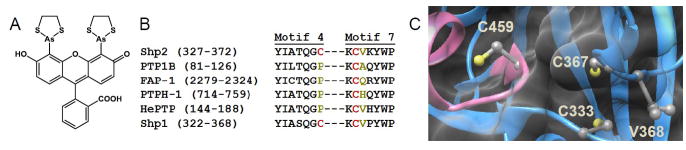
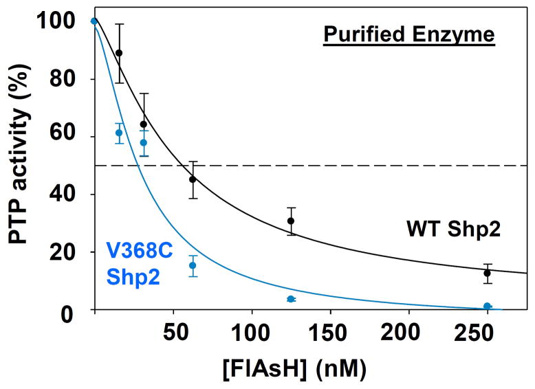
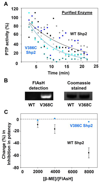
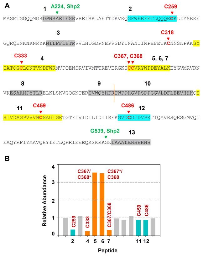
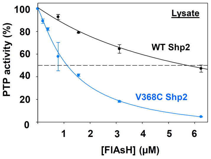
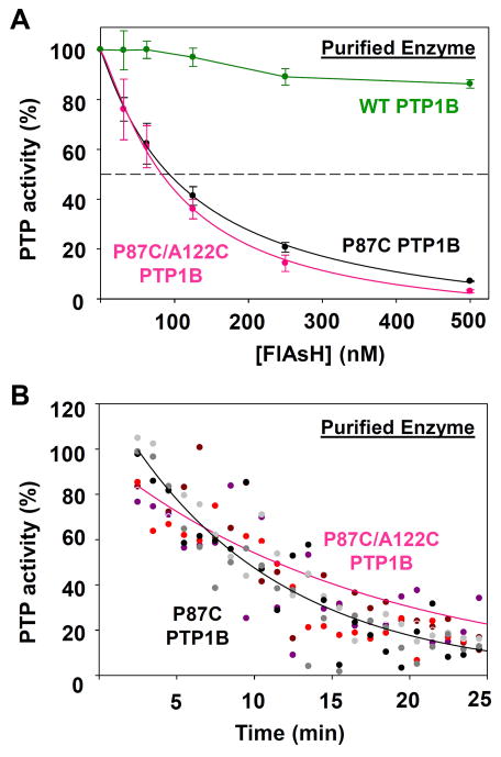
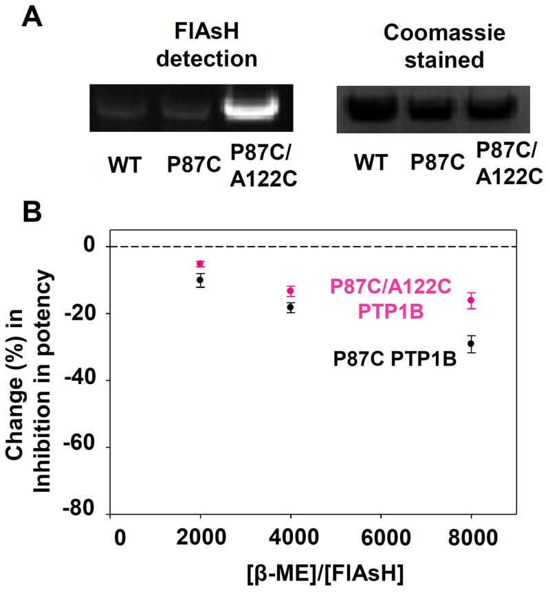
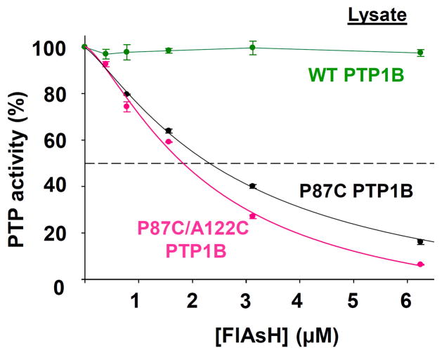
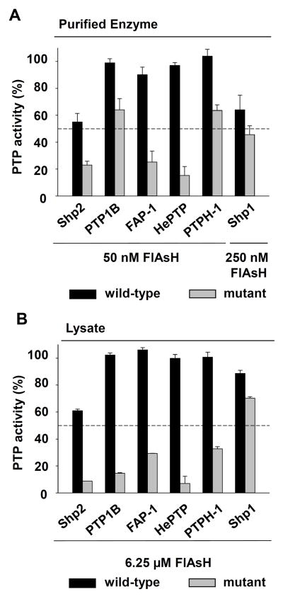
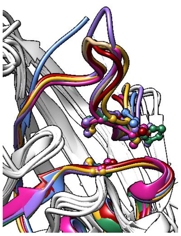
Protein tyrosine phosphatases (PTPs), which catalyze the dephosphorylation of phosphotyrosine in protein substrates, are critical regulators of metazoan cell signaling and have emerged as potential drug targets for a range of human diseases. Strategies for chemically targeting the function of individual PTPs selectively could serve to elucidate the signaling roles of these enzymes and would potentially expedite validation of the therapeutic promise of PTP inhibitors. Here we report a novel strategy for the design of non-natural allosteric-inhibition sites in PTPs; these sites, which can be introduced into target PTPs through protein engineering, serve to sensitize target PTPs to potent and selective inhibition by a biarsenical small molecule. Building on the recent discovery of a naturally occurring cryptic allosteric site in wild-type Src-homology-2 domain containing PTP (Shp2) that can be targeted by biarsenical compounds, we hypothesized that Shp2's unusual sensitivity to biarsenicals could be strengthened through rational design and that the Shp2-specific site could serve as a blueprint for the introduction of non-natural inhibitor sensitivity in other PTPs. Indeed, we show here that the strategic introduction of a cysteine residue at a position removed from the Shp2 active site can serve to increase the potency and selectivity of the interaction between Shp2's allosteric site and the biarsenical inhibitor. Moreover, we find that 'Shp2-like' allosteric sites can be installed de novo in PTP enzymes that do not possess naturally occurring sensitivity to biarsenical compounds. Using primary-sequence alignments to guide our enzyme engineering, we have successfully introduced allosteric-inhibition sites in four classical PTPs-PTP1B, PTPH-1, FAP-1, and HePTP-from four different PTP subfamilies, suggesting that our sensitization approach can likely be applied widely across the classical PTP family to generate biarsenical-responsive PTPs.

Keywords: Allostery; Biarsenicals; FlAsH; Inhibitor sensitization; Protein engineering; Protein tyrosine phosphatases.


Figures

Figure 1
Figure 1
Design of classical PTP mutants that possess unnatural sensitivity to biarsenical compounds. (A) Chemical structure of the biarsenical compound FlAsH. (B) Partial amino-acid sequence alignment of the human PTPs discussed in this study, showing only structural motifs 4 and 7 of the PTP domain (as assigned by Anderson et al). Highlighted in red are Shp2’s C333 and C367 and their cysteine counterparts in other PTPs. Highlighted in dark yellow are the amino acid residues substituted with cysteine in the present study. (The PTP-domain primary-sequence numbering varies widely due to the diversity in protein size and structure of the PTP family outside of the conserved PTP domain.) (C) Three-dimensional structure of Shp2’s catalytic domain (PDB ID: 3B7O). Shp2 is shown as a blue ribbon, with the conserved active-site motif highlighted in pink and featuring the catalytic cysteine C459. The side chains of C333, C367, V368, and C459 are colored by atom type. The enzyme’s surface is rendered transparently so that the buried residues C333 and C367 can be visualized.
Figure 2
Figure 2
V368C Shp2 is inhibited more potently by FlAsH than wild-type Shp2. The phosphatase activities of purified wild-type and V368C Shp2 (50 nM) were measured with pNPP in the presence of the indicated FlAsH concentrations after 120-minute pre-incubations. The activities were normalized to DMSO-only controls for the corresponding enzymes.
Figure 3
Figure 3
V368C Shp2 displays enhanced allosteric-site interactions with FlAsH. (A) Wild-type and V368C Shp2’s rates of dephosphorylating pNPP in the presence of 500 nM FlAsH over 1-minute windows were normalized to a DMSO-only control. The results of three independent experiments (grayscale for wild-type Shp2, bluescale for V368C Shp2) were averaged and the averaged data was fitted as single-variable exponential decays. (B) Purified wild-type and V368C Shp2 were treated with FlAsH, run on an SDS/PAGE gel, and visualized both under UV light and by Coomassie staining. (C) After incubation with 125 nM FlAsH and varying concentrations of β-mercaptoethanol (β-ME), PTP activities of purified wild-type and V368C Shp2 were measured with pNPP and normalized to no-FlAsH controls.
Figure 4
Figure 4
FlAsH protects cysteines 333, 367, and 368 in V368C Shp2 from modification by iodoacetic acid. V368C Shp2 (2.7 μM) was incubated with DMSO or FlAsH (27 μM), followed by addition of iodoacetic acid (50 mM). The labeled protein was trypsinized and the abundances of the resulting peptides were quantitated by LC/MS/MS. Relative abundances indicate the normalized intensities of the indicated peptides in a FlAsH-treated sample as compared to its no-FlAsH control. Peptides containing positions 333, 367, and 368 are highlighted in orange; peptides containing other carboxymethylated cysteines are highlighted in blue; other non-cysteine-containing peptides appear in gray. The sequences of peptides 1–13 are provided in panel A. Asterisks in panel B indicate non-carboxymethylated (free) cysteine residues detected in the LC/MS/MS experiment.
Figure 5
Figure 5
V368C Shp2 is targeted more potently by FlAsH than wild-type Shp2 in a complex proteomic mixture. Crude lysates from bacterial cells expressing wild-type or V368C Shp2 were treated with the indicated FlAsH concentrations. Total PTP activities of the lysates were then measured with pNPP and normalized to DMSO-only controls for the corresponding enzymes.
Figure 6
Figure 6
Purified P87C PTP1B and P87C/A122C PTP1B exhibit marginal differences in FlAsH sensitivity. (A) The phosphatase activities of purified wild-type and engineered PTP1B enzymes (50 nM) were measured with pNPP in the presence of the indicated FlAsH concentrations after 120-minute pre-incubations. The activities were normalized to DMSO-only controls for the corresponding enzymes. (B) The PTP1B mutants’ rates of dephosphorylating pNPP in the presence of 500 nM FlAsH over 1-minute windows were normalized to a DMSO-only control. The results of three independent experiments (grayscale for P87C PTP1B, redscale for P87C/A122C PTP1B) were averaged and the averaged data was fitted as single-variable exponential decays.
Figure 7
Figure 7
The tricysteine allosteric site of P87C/A122C PTP1B binds FlAsH more tightly than the dicysteine allosteric site of P87C PTP1B. (A) Purified wild-type and engineered PTP1B variants were treated with FlAsH, run on an SDS/PAGE gel, and visualized both under UV light and by Coomassie staining. (B) After incubation with 125 nM FlAsH and varying concentrations of β-mercaptoethanol (β-ME), PTP activities of purified PTP1B variants were measured with pNPP and normalized to no-FlAsH controls.
Figure 8
Figure 8
FlAsH-induced inhibition of P87C PTP1B and P87C/A122C PTP1B in a complex proteomic mixture. Crude lysates from bacterial cells expressing wild-type PTP1B, P87C PTP1B, or P87C/A122C PTP1B were treated with the indicated FlAsH concentrations. Total PTP activities of the lysates were then measured with pNPP and normalized to DMSO-only controls for the corresponding enzymes.
Figure 9
Figure 9
Allosteric-site engineering confers unnatural FlAsH sensitivity on PTPs from multiple subfamilies. (A) The phosphatase activities of purified wild-type and engineered PTPs (50 nM) were measured with pNPP in the presence of the indicated FlAsH concentrations after 120-minute pre-incubations. The activities were normalized to DMSO-only controls for the corresponding enzymes. (B) Crude lysates from bacterial cells expressing wild-type or engineered PTPs were treated with the indicated FlAsH concentrations. Total PTP activities of the lysates were then measured with pNPP and normalized to DMSO-only controls for the corresponding enzymes.
Figure 10
Figure 10
Structural conservation of PTP motifs 4 and 7. The PTP-domain structures of Shp2 (red, PDB ID: 3B7O), PTPH-1 (sea green, PDB ID: 2B49), FAP-1 (violet, PDB ID: 1WCH), Shp1 (magenta, PDB ID: 1GWZ), HePTP (blue, PDB ID: 2A3K), and PTP1B (gold, PDB ID: 1SUG) were aligned and superimposed using the UCSF Chimera package. For clarity, only the ribbons corresponding to residues 327–373 (human Shp2 numbering) are colored.

References

    1. Andersen JN, Mortensen OH, Peters GH, Drake PG, Iversen LF, Olsen OH, Jansen PG, Andersen HS, Tonks NK, Moller NP. Mol Cell Biol. 2001;21:7117. - PMC - PubMed
    1. Hendriks W, Pulido R. Biochim Biophys Acta. 2013;1832:1673. - PubMed
    1. Hardy S, Julien SG, Tremblay ML. Anticancer Agents Med Chem. 2012;12:4. - PubMed
    1. Blaskovich MA. Curr Med Chem. 2009;16:2095. - PubMed
    1. Tiganis T, Bennett AM. Biochem J. 2007;402:1. - PMC - PubMed

Publication types

MeSH terms

Substances

LinkOut - more resources