Clinical Trial
. 2015 Apr;45(4):593-605.
doi: 10.1007/s00247-014-3210-y. Epub 2015 Apr 1.

Common normal variants of pediatric vertebral development that mimic fractures: a pictorial review from a national longitudinal bone health study

Collaborators, Affiliations
Clinical Trial

Common normal variants of pediatric vertebral development that mimic fractures: a pictorial review from a national longitudinal bone health study

Jacob L Jaremko et al. Pediatr Radiol. 2015 Apr.

Abstract

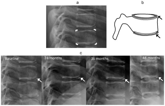
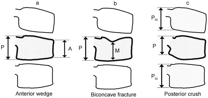
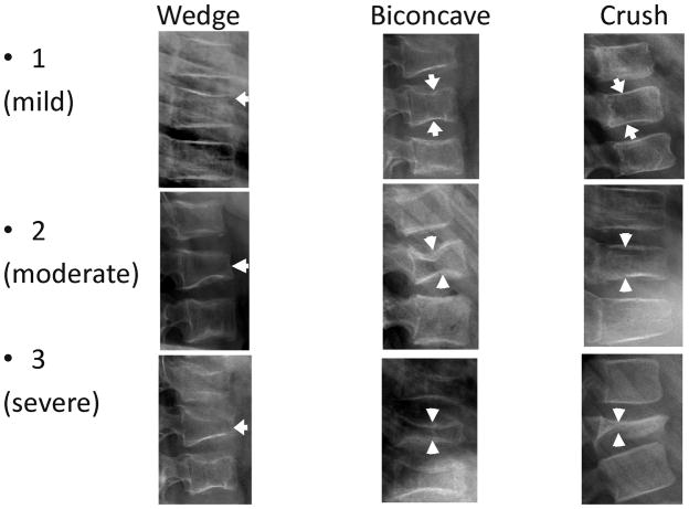
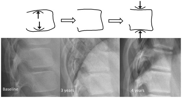
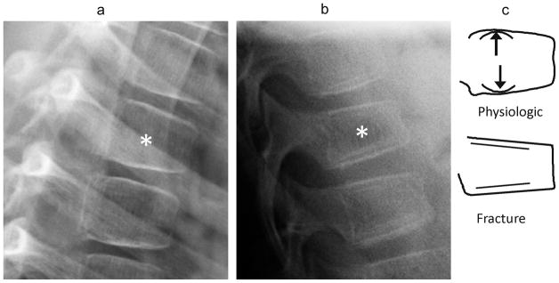
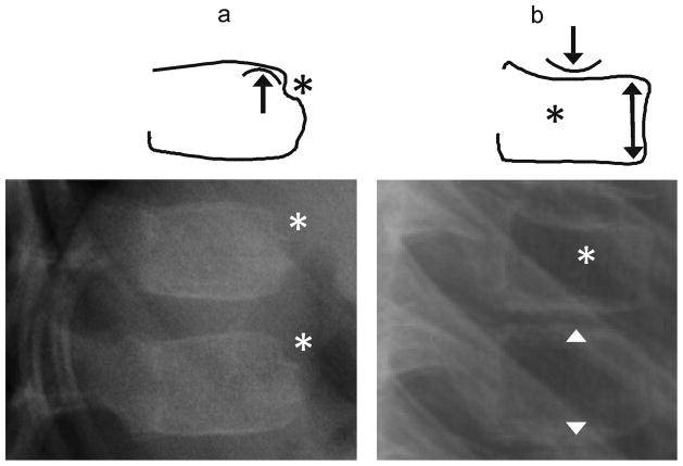
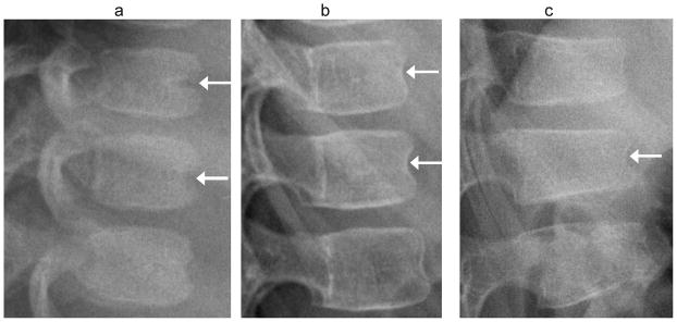
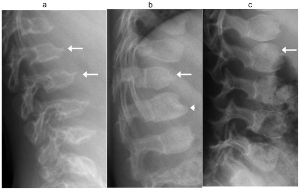
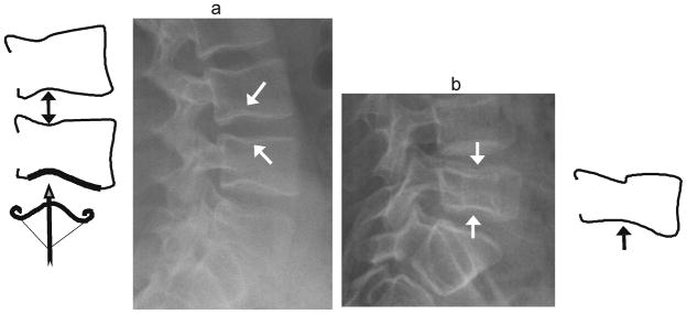
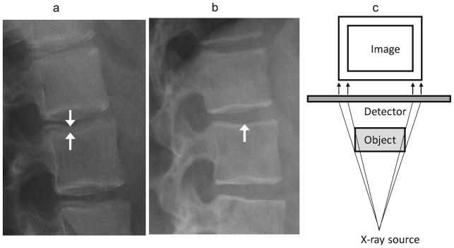
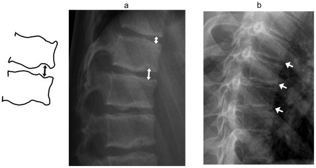
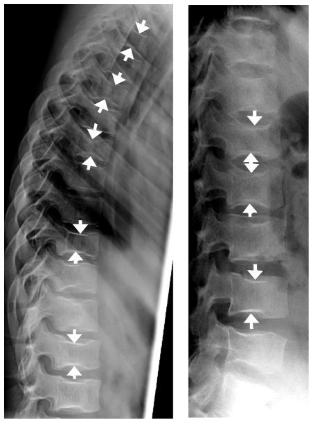
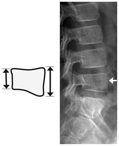
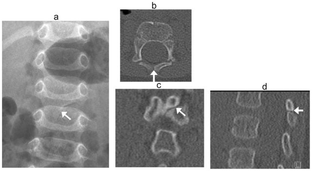
Children with glucocorticoid-treated illnesses are at risk for osteoporotic vertebral fractures, and growing awareness of this has led to increased monitoring for these fractures. However scant literature describes developmental changes in vertebral morphology that can mimic fractures. The goal of this paper is to aid in distinguishing between normal variants and fractures. We illustrate differences using lateral spine radiographs obtained annually from children recruited to the Canada-wide STeroid-Associated Osteoporosis in the Pediatric Population (STOPP) observational study, in which 400 children with glucocorticoid-treated leukemia, rheumatic disorders, and nephrotic syndrome were enrolled near glucocorticoid initiation and followed prospectively for 6 years. Normal variants mimicking fractures exist in all regions of the spine and fall into two groups. The first group comprises variants mimicking pathological vertebral height loss, including not-yet-ossified vertebral apophyses superiorly and inferiorly, which can lead to a vertebral shape easily over-interpreted as anterior wedge fracture, physiological beaking, or spondylolisthesis associated with shortened posterior vertebral height. The second group includes variants mimicking other radiologic signs of fractures: anterior vertebral artery groove resembling an anterior buckle fracture, Cupid's bow balloon disk morphology, Schmorl nodes mimicking concave endplate fractures, and parallax artifact resembling endplate interruption or biconcavity. If an unexpected vertebral body contour is detected, careful attention to its location, detailed morphology, and (if available) serial changes over time may clarify whether it is a fracture requiring change in management or simply a normal variant. Awareness of the variants described in this paper can improve accuracy in the diagnosis of pediatric vertebral fractures.


Conflict of interest statement

Conflicts of Interest: None.

Figures

Fig. 1
Fig. 1
Ring apophyses. (a) Lateral radiograph ossification of ring apophyses in a 10 year-old girl on glucocorticoid treatment for chronic renal disease. (b) Corresponding diagram of development of ring apophyses at endplate margins (arrows). (c) Lateral radioography shows ossification and fusion of ring apophyses (arrows) over a four year period in the same girl
Fig. 2
Fig. 2
Diagram of the Genant semi-quantitative grading system for vertebral compression fractures. (a) Anterior wedging: measure the anterior height loss relative to posterior height (i.e., ratio = (P−A)/P × 100%). (b) Biconcave fracture: measure the minimum central vertebral body height relative to posterior height (i.e., ratio = (P−M)/P × 100%). Note that this fracture description is a misnomer because in many cases only one endplate is involved; the term is retained for fidelity with the original Genant grading system. (c) Posterior crush: because the posterior body wall is altered in this type of fracture and cannot serve as its own reference, this height should be measured relative to the posterior height of the adjacent vertebrae. The lower of the two ratios (Phi–P)/Phi and (Plo–P)/Plo is recorded as a percentage. Because L4 is the lowest level scored in Genant, at L4 only the L3 posterior height is used for comparison. Genant grades are: 0, height loss 20% or less; 1 (mild), >20–25%; 2 (moderate), >25–40%; 3 (severe), >40%. (Conceptually similar to figures in Genant 1993, [23], and Siminoski 2014, [24])
Fig. 3
Fig. 3
Examples of actual fractures of each subtype in the Genant semi-quantitative grading system. All images are sagittal radiographs of thoracolumbar vertebrae oriented with anterior to the right. Top row: 5 year-old girl with acute lymphoblastic leukemia; 9 year-old girl with acute lymphoblastic leukemia; 6 year-old boy with acute lymphoblastic leukemia. Middle row: 10 year-old girl with acute lymphoblastic leukemia; 10 year-old girl with osteogenesis imperfecta; 5 year-old girl with acute lymphoblastic leukemia. Bottom row: 9 year-old girl with acute lymphoblastic leukemia; 8 year-old girl with acute lymphoblastic leukemia; 10 year-old boy with histiocytosis. Arrows indicate the fracture in each image. In the image of grade 2 crush fracture, note also the grade 1 wedge fracture at the vertebra below
Fig. 4
Fig. 4
Normal progression from convex to concave endplates at T12 and L1 over 4 years, in a girl on glucocorticoid treatment for nephrotic syndrome, first imaged at age 9 years. Note also the ossification and fusion of ring apophyses during this time, which contributes to the final apparent concavity of endplates, as seen on lateral radiography.
Fig. 5
Fig. 5
(a) Anterior lateral radiographs shows exaggerated anterior wedging (near 25% height loss) in a mid-thoracic vertebra (*), in a 7 year-old girl treated with glucocorticoids for linear scleroderma. This was present and unchanged on all scans for 3 years. (b) Lateral radiograph shows a wedge compression fracture with near 25% anterior height loss at T11 (*), in a 9 year-old girl treated with glucocorticoids for acute lymphoblastic leukemia. (c) Diagram distinguishes between these. In both cases the endplates appear uninterrupted such that this criterion is not helpful. Note that in the upper schematic vertebra, drawn from image (a), the posterior portions of the endplates retain their normal childhood convexity, while in the lower vertebra, drawn from image (b), the endplates are flattened and linear. Also, (b) shows a double endplate contour (bone-in-bone), which, although nonspecific, can be seen during fracture healing
Fig. 6
Fig. 6
Indentation at an anterior ring apophysis vs. fracture. (a) Step deformities (*) at anterior superior endplates of T12 and L1 in a 5 year-old boy treated with glucocorticoids for nephrotic syndrome represent prominent notches at the as yet un-ossified anterior ring apophysis. A clue that this is a normal variant is the preserved outward convexity of the endplates immediately posterior to this. The anterior cortex should be stronger than the trabecular bone just posterior to it, so a fracture involving anterior cortex ought to also depress the endplate posterior to it. (b) Typical lateral radiographic appearance of a thoracic compression fracture (*), in a 10 year-old boy treated with glucocorticoids for acute lymphoblastic leukemia. Although there is some anterior height loss, the greatest height loss is in the mid-vertebra, where little vertically oriented cortical bone is available to protect from compressive loading. The endplate is concave upward just posterior to the anterior cortex, rather than convex as in image (a). Note the normal convex-outward appearance of the lower vertebra seen in image (b) (arrowheads)
Fig. 7
Fig. 7
Anterior vertebral wall vascular groove vs. fracture. (a) Lateral radiograph shows a typical linear indentation (arrows) at mid-anterior vertebral body wall representing a vertebral artery groove, in a 1.4 year-old girl treated with glucocorticoids for juvenile inflammatory arthritis. This is unlikely to be confused with fracture. (b) Lateral radiograph shows residual indentation (arrows) at anterior vertebral body walls from ossifying vertebral artery grooves in a 5 year-old girl, with. (c) In this anterior wedge compression fracture (arrow) in a 7 year-old boy with linear flattening of endplates, the indentation of anterior cortex is similar to image (b) and may be incidental. The children in (b) and (c) were both treated with glucocorticoids for acute lymphoblastic leukemia
Fig. 8
Fig. 8
Anterior vertebral beaking at upper lumbar vertebrae in mucopolysaccharidoses, for comparison with normal variation. (a) Lateral radiograph in a 5 year-old boy with Morquio syndrome shows typical central beaking most pronounced at L1 and L2 (arrows). (b) Lateral radiograph in a 4 year-old boy with Hurler syndrome shows hypoplastic anterior aspect of T12 with gibbus deformity (arrow), and inferior beaking at L1 (arrowhead). (c) Lateral radiograph in a 4 year-old boy with Hunter syndrome shows exaggerated convexity of vertebral body contours, which appear rounded, with prominent impressions at un-ossified ring apophyses (arrow)
Fig. 9
Fig. 9
Cupid’s bow vs. biconcave fracture. (a) Diagram and lateral radiograph depict Cupid’s bow or balloon disk at multiple levels in a 17 year-old boy treated with glucocorticoids for vasculitis. This normal variant is a curved indentation centred at the posterior third of the endplate (arrows). The shape resembles Cupid’s bow (diagram, bottom), and when present at adjacent endplates, it gives the impression of disk expansion, hence it is also known as ‘balloon disk’. (b) Biconcave endplate fracture at L5, in a 10 year-old boy treated with glucocorticoids for acute lymphoblastic leukemia. There is interruption of the superior endplate of L5, the endplate concavities are centered at the mid-disk rather than the posterior third, there is overall height loss, and adjacent levels are not affected
Fig. 10
Fig. 10
Parallax effect. (a) Lateral radiographs of the upper lumbar spine in a 12 year-old girl treated with glucocorticoids for mixed connective tissue disease shows non-overlapping endplates, especially at L2 (arrows), due to parallax. (b) A follow-up image obtained 3 years later shows overlapping endplates without parallax effect (arrow). (c) Diagram of parallax effect. The side of the object closest to the X-ray source is magnified and eccentrically positioned compared to the other side. For vertebrae, this gives an appearance of bone-in-bone, or endplate interruption. This is minimized by using a source as far away as possible, and centering the beam appropriately
Fig. 11
Fig. 11
Normal variants involving irregular endplates. (a) Diagram and lateral radiograph shows multiple Schmorl nodes near thoracolumbar junction (arrows) in a 16 year-old boy treated with glucocorticoids for vasculitis. The endplates are irregular with areas of focal depression and sclerosis implying sites of intraosseous disk herniation. There is only minimal anterior wedging, not meeting criteria for wedge compression fracture. (b) Lateral radiograph shows Scheuermann kyphosis in a 14 year-old girl treated with glucocorticoids for dermatomyositis. Endplate irregularity and mild anterior wedging are seen at multiple mid-thoracic levels, resulting in increased kyphosis
Fig. 12
Fig. 12
Sickle cell anemia. Lateral radiographs (a, b) show central endplate deformity at multiple levels in 13 year-old girl with sickle cell anemia, mimicking biconcave endplate compression fractures. Nearly all levels are involved to variable extent, with arrows showing some of the most prominent endplate depressions in this girl
Fig. 13
Fig. 13
Trapezoidal L5. Diagram and lateral radiograph shows a shorter posterior wall than anterior of L5 (arrow), giving a trapezoidal vertebral body shape in a 7- year-old girl with mixed connective tissue disease. This does not represent a posterior crush fracture, but is a normal variation
Fig. 14
Fig. 14
Incomplete midline fusion. Developmental cleft mimics fracture in a 6 –month- old boy transferred to tertiary hospital after fall from bed. Radiographs were reported as showing a vertebral fracture. (a) Anteroposterior radiograph shows oblique lucency at posterior elements of L2 (arrow). (b) Axial, (c) coronal and (d) sagittal CT images in bone windows demonstrate smooth, sclerotic margins at the oblique developmental cleft (arrows)

References

    1. Leroux J, Vivier PH, Ould Slimane M, Foulongne E, Abu-Amara S, Lechevallier J, Griffet J. Early diagnosis of thoracolumbar spine fractures in children. A prospective study. Orthop Traumatol Surg Res. 2013;99:60–65. - PubMed
    1. Halton J, Gaboury I, Grant R, et al. Advanced vertebral fracture among newly diagnosed children with acute lymphoblastic leukemia: results of the Canadian Steroid-Associated Osteoporosis in the Pediatric Population (STOPP) research program. J Bone Miner Res. 2009;24:1326–1334. - PMC - PubMed
    1. Alos N, Grant RM, Ramsay T, et al. High incidence of vertebral fractures in children with acute lymphoblastic leukemia 12 months after the initiation of therapy. J Clin Oncol. 2012;30:2760–2767. - PMC - PubMed
    1. Huber AM, Gaboury I, Cabral DA, et al. Prevalent vertebral fractures among children initiating glucocorticoid therapy for the treatment of rheumatic disorders. Arthritis Care Res (Hoboken) 2010;62:516–526. - PMC - PubMed
    1. Rodd C, Lang B, Ramsay T, et al. Incident vertebral fractures among children with rheumatic disorders 12 months after glucocorticoid initiation: a national observational study. Arthritis Care Res (Hoboken) 2012;64:122–131. - PMC - PubMed

Publication types

MeSH terms

Substances

Grants and funding