Review
. 2016 Jan;275 Pt 3(0 3):305-315.
doi: 10.1016/j.expneurol.2015.03.020. Epub 2015 Mar 28.

Astrocyte roles in traumatic brain injury

Affiliations
Review

Astrocyte roles in traumatic brain injury

Joshua E Burda et al. Exp Neurol. 2016 Jan.

Abstract

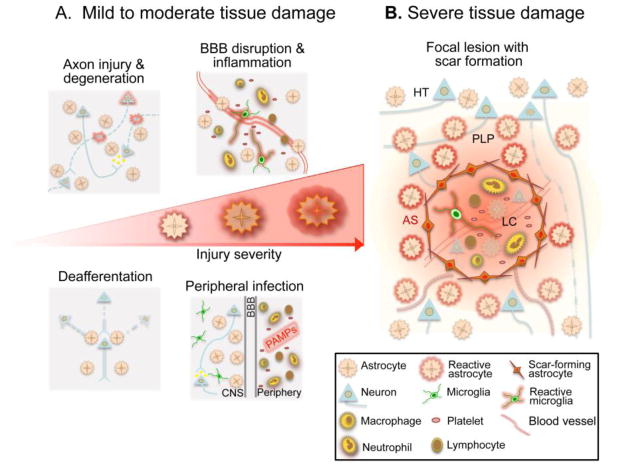
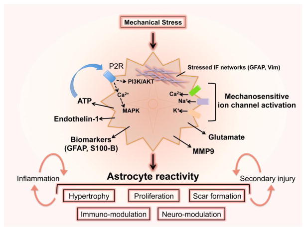
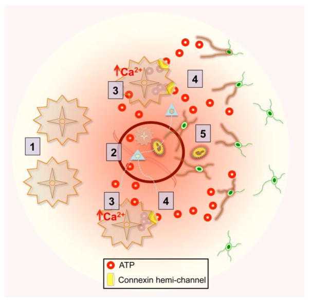
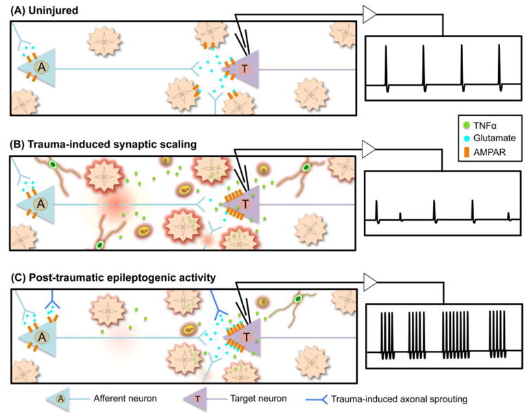
Astrocytes sense changes in neural activity and extracellular space composition. In response, they exert homeostatic mechanisms critical for maintaining neural circuit function, such as buffering neurotransmitters, modulating extracellular osmolarity and calibrating neurovascular coupling. In addition to upholding normal brain activities, astrocytes respond to diverse forms of brain injury with heterogeneous and progressive changes of gene expression, morphology, proliferative capacity and function that are collectively referred to as reactive astrogliosis. Traumatic brain injury (TBI) sets in motion complex events in which noxious mechanical forces cause tissue damage and disrupt central nervous system (CNS) homeostasis, which in turn trigger diverse multi-cellular responses that evolve over time and can lead either to neural repair or secondary cellular injury. In response to TBI, astrocytes in different cellular microenvironments tune their reactivity to varying degrees of axonal injury, vascular disruption, ischemia and inflammation. Here we review different forms of TBI-induced astrocyte reactivity and the functional consequences of these responses for TBI pathobiology. Evidence regarding astrocyte contribution to post-traumatic tissue repair and synaptic remodeling is examined, and the potential for targeting specific aspects of astrogliosis to ameliorate TBI sequelae is considered.

Keywords: Astrocyte; Astrogliosis; CNS; Inflammation; Neuroplasticity; Scar; Traumatic brain injury.


Figures

Figure 1
Figure 1. Reactive astrogliosis following TBI is a graded and heterogeneous response that reflects the severity of CNS tissue damage
A, In response to mild or moderate tissue damage, astrocytes undergo hypertrophic reactive astrogliosis that includes molecular, structural and functional changes. Different forms of tissue pathology, such as local axonal injury and degeneration, blood brain barrier (BBB) disruption with inflammatory cell extravasation, deafferentation and synapse degeneration due to distal axon injury, or exposure to PAMPs associated with peripheral bacterial or viral infection, can all uniquely influence astrocyte function and in different combinations can drive specific forms of astrogliosis. These hypertrophic reactive astrocytes are intermingled among viable neural cells in areas of injured, but surviving and functioning neural tissue. B, Severe tissue damage elicits neural and glial cell degeneration, vascular breakdown and a robust innate and adaptive immune response, leading to the formation of tissue compartments with distinct forms of reactive astrogliosis. Immediately adjacent to the injury, astrocytes proliferate and intertwine to form an astroglial scar (AS) that surrounds and restricts the spread of the intense inflammatory response in the lesion core. These scar forming astrocytes are present in areas that contain few if any surviving neural cells, and their main interactions are with non-neural cells in tissue lesions. Adjacent to the astrocyte scar, features characteristic of mild or moderate brain trauma are present and taper with distance from the lesion core (LC). In these areas reactive astrocytes undergo changes in morphology and function characteristic of hypertrophic reactive astrogliosis as described in A, and these reactive astrocytes interact with injured but surviving cells in the perilesion perimeter (PLP). Astrocyte reactivity in the PLP may also influence neurons and glia in the healthy tissue (HT) distal to the injury.
Figure 2
Figure 2. Astrocytes sense and respond to mechanical strain after TBI
Physical strain deforms flexible networks of intermediate filaments within astrocytes and activates ion influx through mechanosensitvie cation channels. Rises in intracellular calcium cause astrocyte ATP release that signals in an autocrine or paracrine manner, driving multiple intra- and inter-cellular signaling pathways and inducing the release of endothelin-1, MMP9 and glutamate. Depending on the severity of the mechanical insult, astrocyte reactivity may involve complex changes in phenotype and function that respond to and influence neuroinflammatory responses to injury as well as mechanisms of secondary TBI pathogenesis. Trauma also causes astrocytes to release GFAP and calcium-binding S100B that may serve as biomarkers of TBI severity.
Figure 3
Figure 3. ATP gradients from astrocytes direct the initial innate immune response to TBI
(1) Non-reactive astrocytes have the capacity to sense and release ATP. (2) Local trauma triggers ATP release from injured cells. (3) ATP signaling to other astrocytes causes a rapid and persistent rise in intracellular calcium in the surrounding astroglial network. (4) Calcium-induced release of ATP via astrocyte connexin hemi-channels generates an ATP gradient that signals to innate immune cells (5) activating and recruiting them to the site of injury.
Figure 4
Figure 4. Contribution of astrocyte-mediated synaptic scaling to post-traumatic epileptogenesis. A
, In the uninjured brain, astrocytes buffer ions and neurotransmitters from the synaptic space and are actively involved in maintenance of neural activity and firing rate. B, TBI induced neuronal and axonal injury reduces afferent input to target neurons. The resulting reduction in synaptic activity elicits excitatory synaptic scaling, in part, by reactive astrocyte-derived TNFα, which stimulates AMPAR insertion into the postsynaptic membrane of target neurons, thereby increasing excitability. Infiltrating inflammatory cells also produce local gradients of TNFα, further amplifying post-synaptic excitability. C, Partial or complete recovery of traumatized afferent axonal input, and/or newly formed connections formed through trauma-induced axonal sprouting can drive epileptogenic burst firing in the post-traumatic hyperexcitable target neuron. Lingering inflammation and associated TNFα can continue to potentiate the synaptic scaling and hyperexcitability.

References

    1. Abbott NJ, Ronnback L, Hansson E. Astrocyte-endothelial interactions at the blood-brain barrier. Nat Rev Neurosci. 2006;7:41–53. - PubMed
    1. Ahmed SM, Rzigalinski BA, Willoughby KA, Sitterding HA, Ellis EF. Stretch-induced injury alters mitochondrial membrane potential and cellular ATP in cultured astrocytes and neurons. J Neurochem. 2000;74:1951–1960. - PubMed
    1. Akassoglou K, Probert L, Kontogeorgos G, Kollias G. Astrocyte-specific but not neuron-specific transmembrane TNF triggers inflammation and degeneration in the central nervous system of transgenic mice. J Immunol. 1997;158:438–445. - PubMed
    1. Allen NJ, Bennett ML, Foo LC, Wang GX, Chakraborty C, Smith SJ, Barres BA. Astrocyte glypicans 4 and 6 promote formation of excitatory synapses via GluA1 AMPA receptors. Nature. 2012;486:410–414. - PMC - PubMed
    1. Alvarez JI, Katayama T, Prat A. Glial influence on the blood brain barrier. Glia. 2013;61:1939–1958. - PMC - PubMed

Publication types