

4D hyperspherical harmonic (HyperSPHARM) representation of surface anatomy: a holistic treatment of multiple disconnected anatomical structures
- PMID: 25828650
- PMCID: PMC4405486
- DOI: 10.1016/j.media.2015.02.004
4D hyperspherical harmonic (HyperSPHARM) representation of surface anatomy: a holistic treatment of multiple disconnected anatomical structures
Abstract
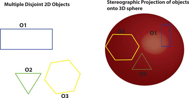
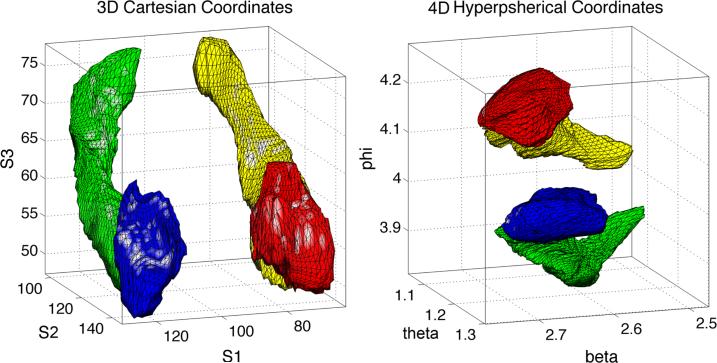
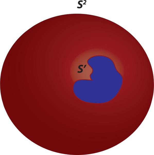
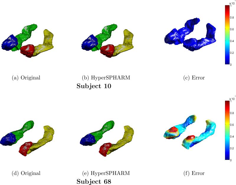
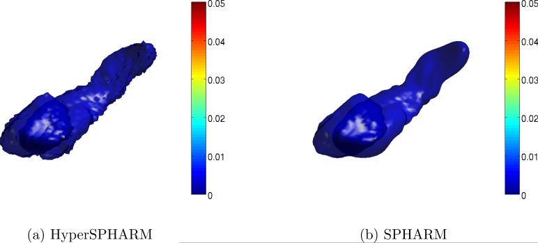
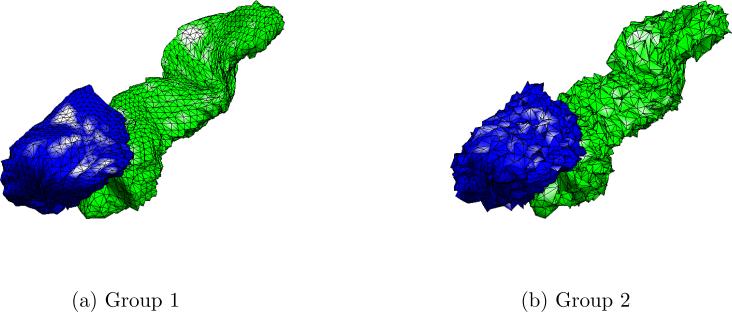
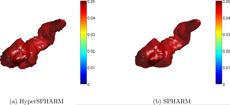
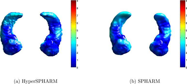
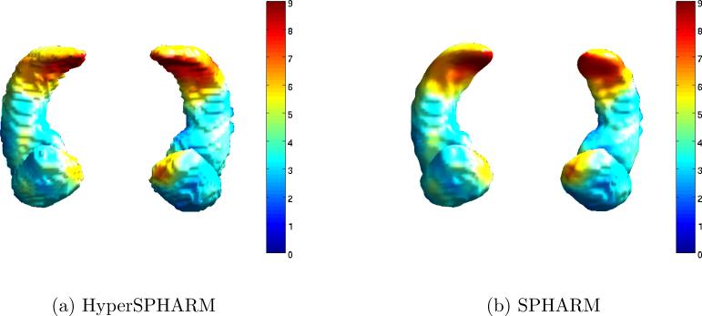
Image-based parcellation of the brain often leads to multiple disconnected anatomical structures, which pose significant challenges for analyses of morphological shapes. Existing shape models, such as the widely used spherical harmonic (SPHARM) representation, assume topological invariance, so are unable to simultaneously parameterize multiple disjoint structures. In such a situation, SPHARM has to be applied separately to each individual structure. We present a novel surface parameterization technique using 4D hyperspherical harmonics in representing multiple disjoint objects as a single analytic function, terming it HyperSPHARM. The underlying idea behind HyperSPHARM is to stereographically project an entire collection of disjoint 3D objects onto the 4D hypersphere and subsequently simultaneously parameterize them with the 4D hyperspherical harmonics. Hence, HyperSPHARM allows for a holistic treatment of multiple disjoint objects, unlike SPHARM. In an imaging dataset of healthy adult human brains, we apply HyperSPHARM to the hippocampi and amygdalae. The HyperSPHARM representations are employed as a data smoothing technique, while the HyperSPHARM coefficients are utilized in a support vector machine setting for object classification. HyperSPHARM yields nearly identical results as SPHARM, as will be shown in the paper. Its key advantage over SPHARM lies computationally; HyperSPHARM possess greater computational efficiency than SPHARM because it can parameterize multiple disjoint structures using much fewer basis functions and stereographic projection obviates SPHARM's burdensome surface flattening. In addition, HyperSPHARM can handle any type of topology, unlike SPHARM, whose analysis is confined to topologically invariant structures.
Keywords: Classification; Hippocampus & Amygdala; Hyperspherical harmonics; SPHARM; Shape analysis.
Copyright © 2015 Elsevier B.V. All rights reserved.
Figures
Similar articles
-
4D hyperspherical harmonic (HyperSPHARM) representation of multiple disconnected brain subcortical structures.Med Image Comput Comput Assist Interv. 2013;16(Pt 1):598-605. doi: 10.1007/978-3-642-40811-3_75. Med Image Comput Comput Assist Interv. 2013. PMID: 24505716 Free PMC article.
-
The 4D hyperspherical diffusion wavelet: A new method for the detection of localized anatomical variation.Med Image Comput Comput Assist Interv. 2014;17(Pt 3):65-72. doi: 10.1007/978-3-319-10443-0_9. Med Image Comput Comput Assist Interv. 2014. PMID: 25320783 Free PMC article.
-
A 4D hyperspherical interpretation of q-space.Med Image Anal. 2015 Apr;21(1):15-28. doi: 10.1016/j.media.2014.11.013. Epub 2015 Jan 3. Med Image Anal. 2015. PMID: 25624043 Free PMC article.
-
Parameterization-invariant shape comparisons of anatomical surfaces.IEEE Trans Med Imaging. 2011 Mar;30(3):849-58. doi: 10.1109/TMI.2010.2099130. Epub 2010 Dec 13. IEEE Trans Med Imaging. 2011. PMID: 21156390
-
Entropy-based particle correspondence for shape populations.Int J Comput Assist Radiol Surg. 2016 Jul;11(7):1221-32. doi: 10.1007/s11548-015-1319-6. Epub 2015 Dec 8. Int J Comput Assist Radiol Surg. 2016. PMID: 26646417 Free PMC article. Review.
Cited by
-
Automatic construction of statistical shape models using deformable simplex meshes with vector field convolution energy.Biomed Eng Online. 2017 Apr 24;16(1):49. doi: 10.1186/s12938-017-0340-0. Biomed Eng Online. 2017. PMID: 28438178 Free PMC article.
-
Spherical harmonics analysis reveals cell shape-fate relationships in zebrafish lateral line neuromasts.Development. 2024 Jan 15;151(2):dev202251. doi: 10.1242/dev.202251. Epub 2024 Jan 26. Development. 2024. PMID: 38276966 Free PMC article.
References
-
- Angenent S, Hacker S, Tannenbaum A, Kikinis R. On the laplace-beltrami operator and brain surface flattening. IEEE Transactions on Medical Imaging. 1999;18:700–711. - PubMed
-
- Aquilanti V, Cavalli S, Coletti C. The d-dimensional hydrogen atom: hyperspherical harmonics as momentum space orbitals and alternative Sturmian basis sets. Chemical Physics. 1997
-
- Benjamini Y, Hochberg Y. Controlling the false discovery rate: a practical and powerful approach to multiple testing. Journal of the Royal Statistical Society. 1995;57:289–300.
-
- Bonvallet B, Griffin N, Li J. 3D shape descriptors: 4D hyperspherical harmonics ’An exploration into the fourth dimension’. IASTED International Conference on Graphics and Visualization in Engineering. 2007:113–116.
Publication types
MeSH terms
Grants and funding
- P30 HD003352/HD/NICHD NIH HHS/United States
- UL1 TR000427/TR/NCATS NIH HHS/United States
- R01MH043454/MH/NIMH NIH HHS/United States
- P50 MH084051/MH/NIMH NIH HHS/United States
- P01 AG20166/AG/NIA NIH HHS/United States
- P50 MH84051/MH/NIMH NIH HHS/United States
- 5T15LM007359/LM/NLM NIH HHS/United States
- T15 LM007359/LM/NLM NIH HHS/United States
- P50 MH10031/MH/NIMH NIH HHS/United States
- UL1TR000427/TR/NCATS NIH HHS/United States
- R01 MH043454/MH/NIMH NIH HHS/United States
- P50 MH100031/MH/NIMH NIH HHS/United States
- P01 AG020166/AG/NIA NIH HHS/United States
- P50 AG033514/AG/NIA NIH HHS/United States
LinkOut - more resources
Full Text Sources
Other Literature Sources
