S values for 131I based on the ICRP adult voxel phantoms
- PMID: 25829162
- PMCID: PMC4729327
- DOI: 10.1093/rpd/ncv016
S values for 131I based on the ICRP adult voxel phantoms
Abstract
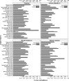
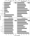
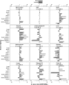
To improve the estimates of organ doses from nuclear medicine procedures using (131)I, the authors calculated a comprehensive set of (131)I S values, defined as absorbed doses in target tissues per unit of nuclear transition in source regions, for different source and target combinations. The authors used the latest reference adult male and female voxel phantoms published by the International Commission on Radiological Protection (ICRP Publication 110) and the (131)I photon and electron spectra from the ICRP Publication 107 to perform Monte Carlo radiation transport calculations using MCNPX2.7 to compute the S values. For each phantom, the authors simulated 55 source regions with an assumed uniform distribution of (131)I. They computed the S values for 42 target tissues directly, without calculating specific absorbed fractions. From these calculations, the authors derived a comprehensive set of S values for (131)I for 55 source regions and 42 target tissues in the ICRP male and female voxel phantoms. Compared with the stylised phantoms from Oak Ridge National Laboratory (ORNL) that consist of 22 source regions and 24 target regions, the new data set includes 1662 additional S values corresponding to additional combinations of source-target tissues that are not available in the stylised phantoms. In a comparison of S values derived from the ICRP and ORNL phantoms, the authors found that the S values to the radiosensitive tissues in the ICRP phantoms were 1.1 (median, female) and 1.3 (median, male) times greater than the values based on the ORNL phantoms. However, for several source-target pairs, the difference was up to 10-fold. The new set of S values can be applied prospectively or retrospectively to the calculation of radiation doses in adults internally exposed to (131)I, including nuclear medicine patients treated for thyroid cancer or hyperthyroidism.
Published by Oxford University Press 2015. This work is written by (a) US Government employee(s) and is in the public domain in the US.
Figures


References
-
- Beierwaltes W. H., Rabbani R., Dmuchowski C., Lloyd R. V., Eyre P., Mallette S. An analysis of “ablation of thyroid remnants” with I-131 in 511 patients from 1947–1984: experience at University of Michigan. J. Nucl. Med. 25, 1287–1293 (1984). - PubMed
-
- Meier D. A., Brill D. R., Becker D. V., Clarke S. E. M., Silberstein E. B., Royal H. D., Balon H. R. Procedure guideline for therapy of thyroid disease with (131)iodine. J. Nucl. Med. 43, 856–861 (2002). - PubMed
-
- Ron E., et al. Cancer mortality following treatment for adult hyperthyroidism. J. Am. Med. Assoc. 280, 347–355 (1998). - PubMed
-
- Willegaignon J., Stabin M. G., Guimaraes M. I., Malvestiti L. F., Sapienza M. T., Maroni M., Sordi G. M. Evaluation of the potential absorbed doses from patients based on whole-body 131I clearance in thyroid cancer therapy. Health Phys. 91, 123–127 (2006). - PubMed
-
- Smith T., Edmonds C. J. Radiation dosimetry in the treatment of thyroid carcinoma by I-131. Radiat. Prot. Dosim. 5, 141–149 (1983).
Publication types
MeSH terms
Substances
Grants and funding
LinkOut - more resources
Full Text Sources
Other Literature Sources

