. 2015 Jun;276(6):657-80.
doi: 10.1002/jmor.20370. Epub 2015 Mar 30.

Histology of the heterostracan dermal skeleton: Insight into the origin of the vertebrate mineralised skeleton

Affiliations

Histology of the heterostracan dermal skeleton: Insight into the origin of the vertebrate mineralised skeleton

Joseph N Keating et al. J Morphol. 2015 Jun.

Abstract

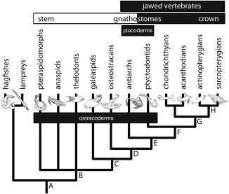
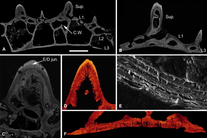
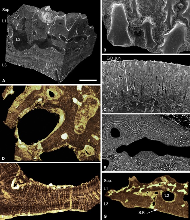
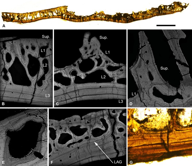
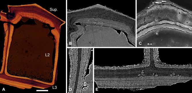
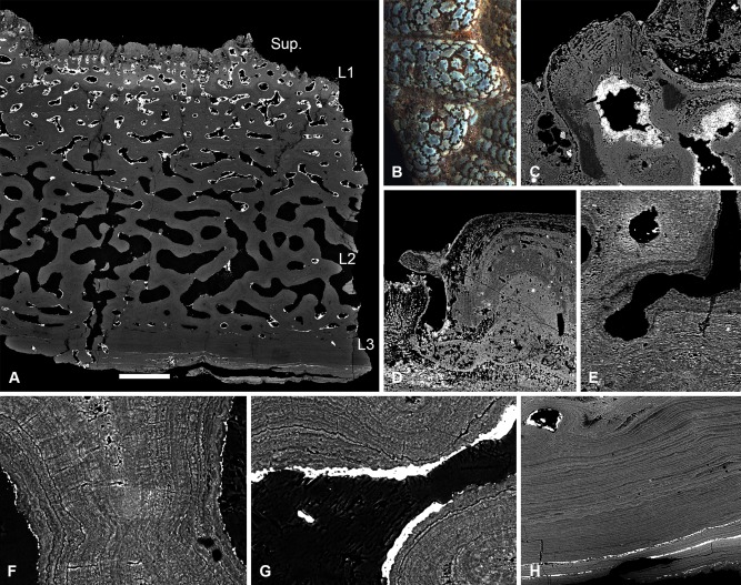
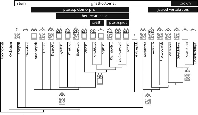
Living vertebrates are divided into those that possess a fully formed and fully mineralised skeleton (gnathostomes) versus those that possess only unmineralised cartilaginous rudiments (cyclostomes). As such, extinct phylogenetic intermediates of these living lineages afford unique insights into the evolutionary assembly of the vertebrate mineralised skeleton and its canonical tissue types. Extinct jawless and jawed fishes assigned to the gnathostome stem evidence the piecemeal assembly of skeletal systems, revealing that the dermal skeleton is the earliest manifestation of a homologous mineralised skeleton. Yet the nature of the primitive dermal skeleton, itself, is poorly understood. This is principally because previous histological studies of early vertebrates lacked a phylogenetic framework required to derive evolutionary hypotheses. Nowhere is this more apparent than within Heterostraci, a diverse clade of primitive jawless vertebrates. To this end, we surveyed the dermal skeletal histology of heterostracans, inferred the plesiomorphic heterostracan skeleton and, through histological comparison to other skeletonising vertebrate clades, deduced the ancestral nature of the vertebrate dermal skeleton. Heterostracans primitively possess a four-layered skeleton, comprising a superficial layer of odontodes composed of dentine and enameloid; a compact layer of acellular parallel-fibred bone containing a network of vascular canals that supply the pulp canals (L1); a trabecular layer consisting of intersecting radial walls composed of acellular parallel-fibred bone, showing osteon-like development (L2); and a basal layer of isopedin (L3). A three layered skeleton, equivalent to the superficial layer L2 and L3 and composed of enameloid, dentine and acellular bone, is possessed by the ancestor of heterostracans + jawed vertebrates. We conclude that an osteogenic component is plesiomorphic with respect to the vertebrate dermal skeleton. Consequently, we interpret the dermal skeleton of denticles in chondrichthyans and jawless thelodonts as independently and secondarily simplified. J. Morphol. 276:657-680, 2015. © 2015 The Authors Journal of Morphology Published by Wiley Periodicals, Inc.

Keywords: bone; dentine; dermoskeleton; enameloid; gnathostome; jawless; microstructure.


Figures

Figure 1
Figure 1
Phylogenetic relationships of the principal vertebrate groups from Donoghue and Keating (2014). Labelled internal branches refer to acquisition of key skeletal apomorphies. Origin of the vertebrate skeleton consisting of a splanchnocranium, neurocranium, fin rays and arcualia (A); origin of a mineralised dermal skeleton and of the canonical vertebrate mineralised tissues: bone, dentine and enameloid (B); origin of a mineralised neurocranium (C); cellular dermal and perichondral bone, neurocranium includes elements equivalent to the scapula and coracoid (D); mineralised splanchnocranium, evolution of jaws and pelvic girdle (E); mineralised axial skeleton (F), mineralised fin radials, teeth associated with spanchnocranium (G); and endochondral bone (H).
Figure 2
Figure 2
Histology of Loricopteraspis serrata. NRM‐PAL C.5939, SEM BSE section through the cephalothoracic shield (A); NRM‐PAL C.5940, SRXTM section through a scale unit (B); NRM‐PAL C.5941, SEM BSE section through a tubercle, showing the enemeloid capping layer (C); NRM‐PAL C.5940, SRXTM volume rendered virtual thin section of a tubercle, showing the arrangement of dentine canaliculi radiating from a pulp canal (D); NRM‐PAL C.5942, etched SEM section through a wall of L2, showing homogenous core and lamellar margins pervaded by an orthogonal fabric of thread‐like spaces (E); NRM‐PAL C.5940, SRXTM volume rendered virtual thin section of L3, showing Sharpey's fibres trending in two principal orientations (F). Sup., superficial layer; L1, layer 1; L2, layer 2; L3, layer 3; C.W., incomplete cross wall; E/D jun., Enameloid/dentine junction. Scale bar equals 606 μm in (A), 227 μm in (B), 179 μm in (C), 55 μm in (D), 30 μm in (E) and 56 μm in (F).
Figure 3
Figure 3
Histology of Tesseraspis tesselata. NHM P.73617, SRXTM sections through an isosurface model of a tessera (A); SEM of the external surface morphology of a tessera, showing two distinct tubercle generations, specimen lost (B); etched SEM section through the enameloid capping layer of the superficial layer, specimen lost (C); NHM P.73617, SRXTM volume rendered transverse section through L2, showing the architecture of the intersecting radial walls (D); NHM P.73618, SEM BSE section through L2 showing truncated centripetal lamellae interpreted as resorption (E); volume rendered virtual thin sections of NHM P.73617 (F, G); transverse section through a radial wall of L2, showing the arrangement of thread‐like spaces (F); section through the dermal skeleton of a tessera, showing the arrangement of Sharpey's fibres in L3 (G). S.F., Sharpey's fibres. Scale bar equals 193 μm in (A), 628 μm in (B), 47 μm in (C), 124 μm in (D), 68 μm in (E), 64 μm in (F) and 230 μm in (G).
Figure 4
Figure 4
Scanning electron microscopy BSE (A) and etched SEM sections (B–I) through the cephalothoracic dermal skeleton of Corvaspis kingi. NHM P.73613, Sectioned fragment from the centre of the headshield (A); NHM P.73614, superficial tubercle, showing centripetal dentine lamellae (B); NHM P.73614, enameloid capping layer, showing fine calibre ramifying canaliculi (C); NHM P.73615, section through the margin of the shield, showing the transition from cancellar to trabecular L2 (D); sections through radial walls of L2 (E–G), showing thread‐like spaces radiating from a homogenous core, NHM P.73614 (E), thread‐like spaces pervading the entire thickness of the wall, NHM P.73615 (F), and a region of linearly arranged thread‐like spaces, NHM P.73616 (G); NHM P.73614, L2 osteon, showing an intrinsic parallel fibred matrix (H); NHM P.73614, L3 showing an orthogonal fabric of Sharpey's fibres (I). LAG, line of arrested growth; I.F., intrinsic fibres. Scale bar equals 385 μm in (A), 44 μm in (B), 35 μm in (C), 569 μm in (D), 40 μm in (E), 55 μm in (F), 42 μm in (G), 16 μm in (H) and 62 μm in (I).
Figure 5
Figure 5
Light microscopy thin section (A, G) and SEM BSE polished sections (B–F) through the cephalothoracic shield of Phialaspis symondsi, NHM P.73619. Section from the centre to the margin of the shield, showing the transition from the central ‘plate’ to the peripheral tuberculated region (A); histological structure of the central ‘plate’ (B) and peripheral region (C); detail of the superficial layer/L1 of the peripheral region. L1 centripetal lamellae are truncated, suggesting the vascular canals were remodelled via resorption (D); osteon lamellae of L2 truncated by vascular space, indicating resorption (E); section through the peripheral region, showing line of arrested growth demarking the previous margin of the shield (F), L3, showing alternating light and dark bands (G). Scale bar equals 3.3 mm in (A), 567 μm in (B), 523 μm in (C), 263 μm in (D) 113 μm in (E), 403 μm in (F) and 279 μm in (G).
Figure 6
Figure 6
Scanning electron microscopy BSE sections of Amphiaspis sp., GIT 313‐32. Architecture of the cephalothoracic shield (A); section through a tubercle, showing centripetal dentine and compact vascular spaces of L1 (B); detail of the same tubercle, showing canaliculi perforating the surface (C); detail of centripetal lamellae which circumscribe the vasculature of L1, arrows points to truncated fibre spaces, interpreted as evidence of resorption (D); radial walls of L2, showing a homogenous core (E); detail of L3 (F). Scale bar equals 0.9 mm in (A), 393 μm in (B), 75 μm in (C), 115 μm in (D), 351 μm in (E) and 182 μm in (F).
Figure 7
Figure 7
Synchrotron radiation X‐ray tomographic microscopy sections, NHM P.73620 (A, B, D, E) and etched SEM sections, NHM P.73621 (C) of the cephalothoracic shield of Anglaspis macculloughi. Volume rendered virtual thin section, showing the histological structure of the shield (A); section through the superficial layer, showing centripetal dentine capped with a layer of SCE. Canaliculi pervade the dentine and ramify at the enameloid/dentine junction (B); detail of the enameloid capping layer (C); section through a radial wall of L2, showing centripetal lamellae developed about a compact lamellar core. The lamellae are warped in association with an orthogonal fabric of thread‐like spaces (D); section through L3 and the base of L2 (E). Scale bar equals 82 μm (A), 60 μm (B), 12 μm (C) and 41 μm (D, E).
Figure 8
Figure 8
Scanning electron microscopy BSE (A, B, G) and SRXTM (C–F) histological sections of Pteraspis sp. NRM‐PAL C.5943, structure of the cephalothoracic shield of Pteraspis (A); detail of the superficial layer and L1 of the same specimen (B); NRM‐PAL C.5944, section through the pineal plate (C); volume rendered virtual thin section of the tubercles, showing the canaliculi radiating from central pulp canals and ramifying at the enameloid/dentine junction, specimen lost during sampling (D); NRM‐PAL C.5945, volume rendered virtual thin section showing histological structure of the body scales (E); transverse volume rendered virtual thin section through L2, showing thread‐like spaces pervading the entire thickness of the radial walls, specimen lost during sampling (F); NRM‐PAL C.5943, section through L3 and the base of L2 (G). Pin., pineal window. Scale bar equals 373 μm in (A), 177 μm in (B), 339 μm in (C), 61 μm in (D), 149 μm in (E), 86 μm in (F) and 123 μm in (G).
Figure 9
Figure 9
Scanning electron microscopy BSE (NHM P.73622, A) and etched SEM (NHM P.73623, B–E) sections through the cephalothoracic shield of Loricopteraspis dairydinglensis. Architecture of the cephalothoracic dermal skeleton (A); detail of the superficial tubercles, showing centripetal dentine developed about a central pulp canal with a capping layer of SCE (B); detail of L2, showing an intersecting network of radial walls composed of centripetal lamellae with a homogenous core (C); section through a radial wall of L2, showing the homogenous core is pervaded by a mesh of coarse unmineralised spaces. The outer lamellar part is perforated by an orthogonal fabric of fine, thread‐like spaces, which warp the lamellae (D); section through L3, showing an orthogonal fabric of Sharpey's fibres (E). Scale bar equals 319 μm in (A), 50 μm in (B), 233 μm in (C), 71 μm in (D) and 51 μm in (E).
Figure 10
Figure 10
Histology of Psammosteus megalopteryx, NHM P.73624. SEM BSE sections (A, C–H) and LM image (B). Structure of the cephalothoracic dermal skeleton (A), detail of surface ornament consisting of concentric tubercle ‘islands’ separated by grooves (B); section through a tubercle, showing centripetal dentine lamellae pervaded by polarised canaliculi (C); superposition of a second generation of tubercles. The tubercle on the right is completely in filled with secondary dentine (D); section through L1 showing truncation of lamellae by the vasculature, interpreted as evidence of resorption (E); section through a radial wall of L2, showing centripetal apposition of lamellae about a homogenous core. A fine fabric of orthogonal thread‐like spaces passes through and warps the lamellae (F); lamellae truncated by the vascular space, suggesting resorption of L2 (G); detail of L3 (H). Scale bar equals 785 μm in (A), 1.9 mm in (B); 99 μm in (C), 84 μm in (D); 121 μm in (E), 43 μm in (F); 41 μm in (G); 157 μm in (H).
Figure 11
Figure 11
Evolutionary distribution of tissue types among total‐group gnathostomes. Phylogenetic relationships of pteraspidomorphs based on Janvier (1996), relationships among stem‐gnathostomes follow Donoghue and Keating (2014). Jawed vertebrate relations follow Zhu et al. (2013). Symbols represent the four‐layered structure of the dermal skeleton. Grey triangles correspond to dentine tubercles; white caps correspond to a capping layer of enameloid; stacked triangles corresponds to stacked tubercle generations; dark grey reticular band corresponds to L1; square box corresponds to cancellar L2; light grey reticular box corresponds to trabecular L2; bottom‐most band corresponds to L3. Symbols adjacent to internal branches represent the reconstructed ancestral state.

References

    1. Agassiz L. 1835. Recherches sur les Poissons Fossiles. Neuchatel: Petitpierre.
    1. Agassiz L. 1845. Monographie de Poissons Fossiles des Vieux Grès Rouges ou Système Dévonien (Old Red Sandstone) des Îles Britanniques et de Russie. Neuchâtel: Imprimerie de Petitpierre et Prince.
    1. Ball HW, Dineley DL, White E. 1961. The Old Red Sandstone of Brown Clee Hill and the Adjacent Area. London: British Museum (Natural History).
    1. Berg LS. 1940. Classification of Fishes both Recent and Fossil. Trav Inst Zool Acad Sci USSR 5:87–517.
    1. Blieck A. 1984. Les hétérostracés Ptéraspidiformes, agnathes du Silurien‐Dévonien du continent Nord‐Atlantique et des blocs avoisinants: Révision systématique, phylogénie, biostratigraphie, biogéographie. Paris: Centre national de la Recherche Scientifique.

Publication types

LinkOut - more resources