

Optimal number of angle images for calculating anterior angle volume and iris volume measurements
- PMID: 25829412
- PMCID: PMC4419775
- DOI: 10.1167/iovs.14-15883
Optimal number of angle images for calculating anterior angle volume and iris volume measurements
Abstract
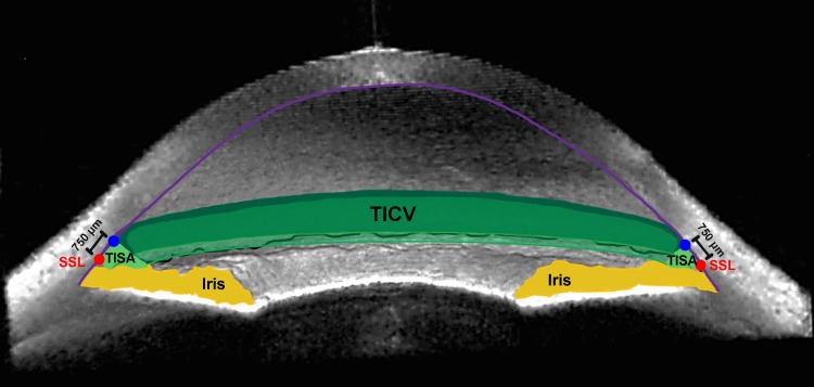
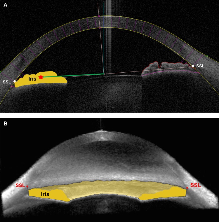
Purpose: We determined the optimal number of angle images required to obtain reliable measurements of trabecular-iris circumferential volume (TICV) and iris volume (IV) using swept-source Fourier domain anterior segment optical coherence tomography (SSFD-ASOCT) scans in narrow angle eyes.
Methods: Scleral spur landmarks (SSL) were manually identified on ASOCT angle images from 128 meridians from each of 24 eyes with chronic primary angle closure (PAC) spectrum of disease. The anterior and posterior corneal curves, and the anterior and posterior iris surfaces were identified automatically by the anterior chamber analysis and interpretation (ACAI) software, then manually examined and edited by the reader if required. Trabecular-iris circumferential volume at 750 μm from SSL (TICV750) and IV were subsequently calculated using varying numbers of angle images. Threshold error was determined to be less than the lower 95% confidence limit of mean absolute percent error (MAPE) of the change in TICV or IV resulting from laser peripheral iridotomy, which would be 17% for TICV and 5% for IV, based on previous studies. The optimal number of angle images was the smallest number of images where MAPE was less than this threshold for TICV and IV.
Results: A total of 32 equally-spaced angle images (16 meridians) was required to estimate TICV750 and 16 angle images (8 meridians) to estimate IV. Both were within 4.6% and 1.6% of MAPE, respectively.
Conclusions: It is possible to determine TICV and IV parameters reliably in narrow angles without evaluating all 128 meridians obtained with SSFD-ASOCT.
Figures
References
-
- Quigley HA,, Silver DM, Friedman DS,, et al. Iris cross-sectional area decreases with pupil dilation and its dynamic behavior is a risk factor in angle closure. J Glaucoma. 2009; 18: 173–179. - PubMed
-
- Quigley HA.Angle-closure glaucoma—simpler answers to complex mechanisms: LXVI Edward Jackson Memorial Lecture. Am J Ophthalmol. 2009; 148: 657–669. - PubMed
-
- Aptel F, Denis P.Optical coherence tomography quantitative analysis of iris volume changes after pharmacologic mydriasis. Ophthalmology. 2010; 117: 3–10. - PubMed
-
- Console JW, Sakata LM,, Aung T, Friedman DS,, He M.Quantitative analysis of anterior segment optical coherence tomography images: the Zhongshan Angle Assessment Program. Br J Ophthalmol. 2008; 92: 1612–1616. - PubMed
Publication types
MeSH terms
Grants and funding
LinkOut - more resources
Full Text Sources
Other Literature Sources
