. 2015 May 15;290(20):12779-92.
doi: 10.1074/jbc.M115.650325. Epub 2015 Mar 31.

Role of nucleotide binding and GTPase domain dimerization in dynamin-like myxovirus resistance protein A for GTPase activation and antiviral activity

Affiliations

Role of nucleotide binding and GTPase domain dimerization in dynamin-like myxovirus resistance protein A for GTPase activation and antiviral activity

Alexej Dick et al. J Biol Chem. .

Abstract

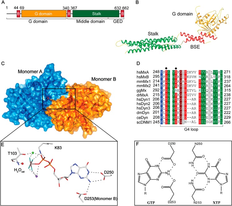
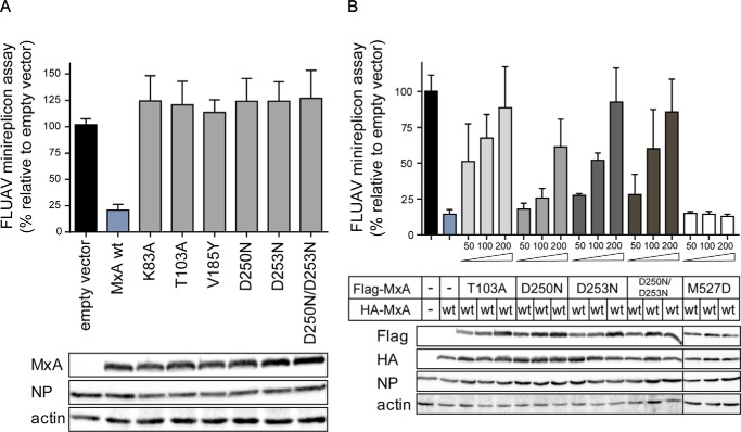
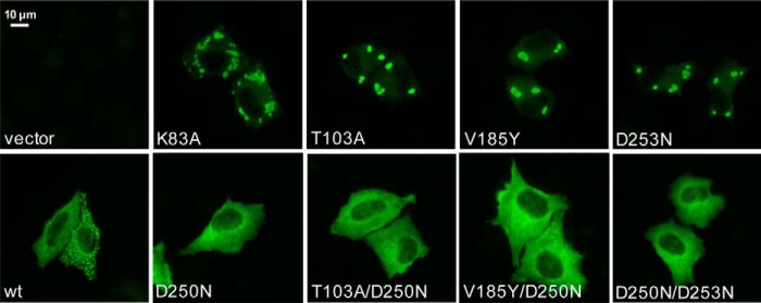
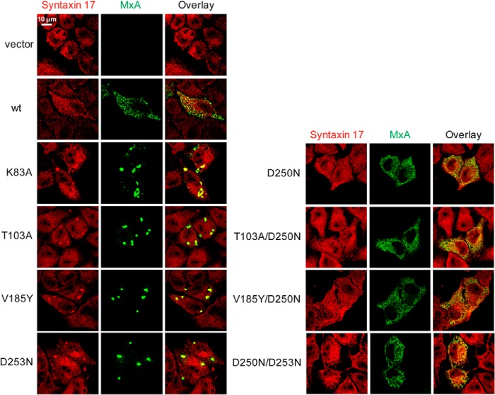
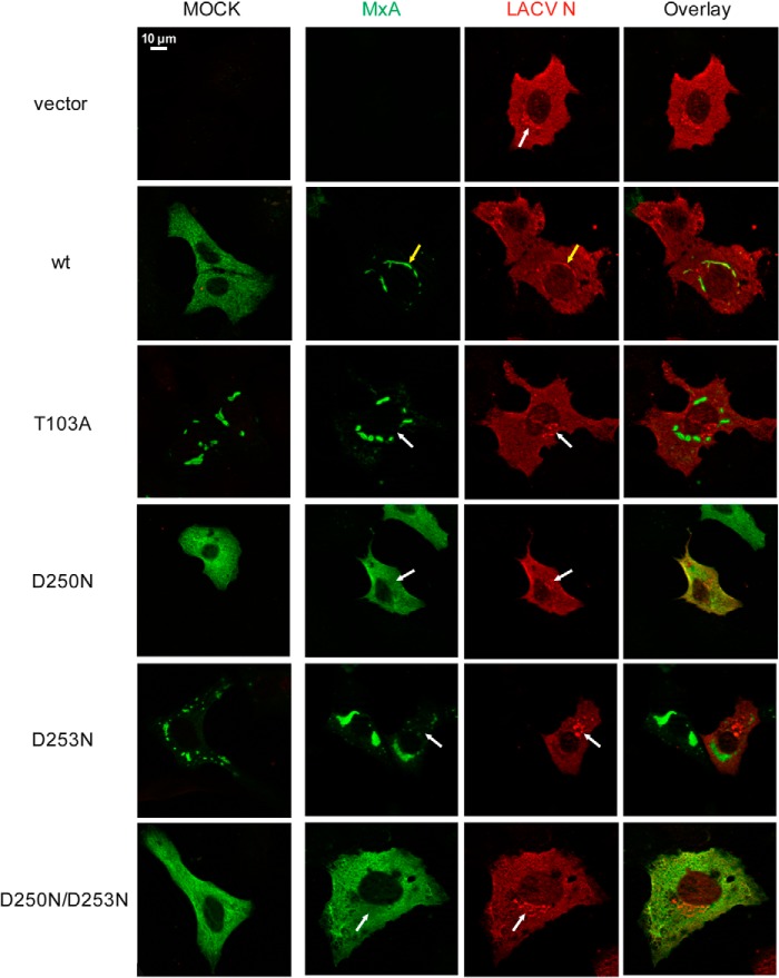
Myxovirus resistance (Mx) GTPases are induced by interferon and inhibit multiple viruses, including influenza and human immunodeficiency viruses. They have the characteristic domain architecture of dynamin-related proteins with an N-terminal GTPase (G) domain, a bundle signaling element, and a C-terminal stalk responsible for self-assembly and effector functions. Human MxA (also called MX1) is expressed in the cytoplasm and is partly associated with membranes of the smooth endoplasmic reticulum. It shows a protein concentration-dependent increase in GTPase activity, indicating regulation of GTP hydrolysis via G domain dimerization. Here, we characterized a panel of G domain mutants in MxA to clarify the role of GTP binding and the importance of the G domain interface for the catalytic and antiviral function of MxA. Residues in the catalytic center of MxA and the nucleotide itself were essential for G domain dimerization and catalytic activation. In pulldown experiments, MxA recognized Thogoto virus nucleocapsid proteins independently of nucleotide binding. However, both nucleotide binding and hydrolysis were required for the antiviral activity against Thogoto, influenza, and La Crosse viruses. We further demonstrate that GTP binding facilitates formation of stable MxA assemblies associated with endoplasmic reticulum membranes, whereas nucleotide hydrolysis promotes dynamic redistribution of MxA from cellular membranes to viral targets. Our study highlights the role of nucleotide binding and hydrolysis for the intracellular dynamics of MxA during its antiviral action.

Keywords: G protein; Mx proteins; antiviral response; catalytic mechanism; dynamin-like protein; innate immunity; interferon; membrane; viral replication.


Figures

FIGURE 1.
FIGURE 1.
Structure and GTPase domain dimerization of MxA. A, domain architecture; B, structure of human MxA (Protein Data Bank code 3SZR). C, homology model of the MxA GTPase domain dimer (residues 69–340) based on the crystal structure of the human dynamin1 GTPase domain-BSE construct in the GDP-AlF4-bound state (Protein Data Bank code 2X2E). D, sequence alignment of Mx and dynamin proteins in the G4 loop. Sequences of human MxA (Swiss-Prot accession P20591), human (hs) MxB (P20592), mouse (mm) Mx1 (P09922), mmMx2 (Q9WVP9), chicken (gg) Mx (Q90597), zebrafish (dr) MxA (Q8JH68), human dynamin1 (Q05193), human dynamin2 (P50570), human dynamin3 (Q9UQ16), Drosophila melanogaster (dm) dynamin (P27619), Caenorhabditis elegans (ce) dynamin (Q9U9I9), and Saccharomyces cerevisiae (sc) dynamin-related protein DNM1 (P54861) were aligned and manually adjusted. Residues with a conservation of greater than 70% are color-coded (D and E in red; R, K, and H in blue; N, Q, S, and T in gray; A, L, I, V, F, Y, W, M, and C in green, and P and G in brown). Asp-250 and Asp-253 are marked with a dot. E, details of the catalytic site. The red ball represents the catalytic water, the green ball a Mg2+ ion, and the purple ball a Na+ ion. Asp-250 stabilizes the purine base in cis, and Asp-253 of the neighboring monomer binds to it in trans. F, left, scheme showing the proposed binding mode of the guanine base by Asp-250 in cis and Asp-253 of the opposing molecule in trans. Right, xanthosine base-binding by Asn-250 and envisaged binding mode of Asn-253. Hydrogen bonds are depicted as dashed lines.
FIGURE 2.
FIGURE 2.
Nucleotide binding analysis. 1 mm solutions of the indicated nucleotide were titrated stepwise into 50 μm solutions of the indicated MxA mutants at 8 °C in an ITC device. Resulting heat changes were integrated, and the obtained values were fitted to a quadratic binding equation. The following KD values were derived from the fittings. A, M527D for GTPγS: KD = 15 ± 1 μm, n = 0.92 ± 0.02. B, M527D/K83A for GTPγS: KD = 39 ± 6 μm, n = 0.42 ± 0.03. C, M527D/T103A for GTPγS: KD = 28 ± 2 μm, n = 0.81 ± 0.02. D, M527D/D250N for XTPγS: KD = 7.8 ± 0.5 μm, n = 0.82 ± 0.01. E, M527D/D253N for GTPγS: KD = 9 ± 1 μm, n = 0.73 ± 0.01 and F, M527D/D250N/D253N for XTPγS: KD = 5.6 ± 0.4 μm, n = 0.87 ± 0.01. GTPγS (□) and XTPγS (○). Because of the reduced heat signal upon nucleotide binding, higher protein and ligand concentrations were used for the T103A and K83A mutants. For K83A, this resulted in increased protein precipitation that may explain the lowered binding number.
FIGURE 3.
FIGURE 3.
Analytical gel filtration analysis. Upon 15 min of incubation with 2 mm of the indicated nucleotide solutions, 50 μl of the indicated MxA mutants at a concentration of 2 mg/ml were applied to an S200 gel filtration column. A, M527D with GTPγS, GDP-AlF4, GDP, AlF4 alone or in the absence of nucleotides. B, M527D/K83A. C, M527D/T103A. D, M527D/D250N. E, M527D/D253N. F, M527D/D250N/D253N in the absence and presence of the indicated nucleotides.
FIGURE 4.
FIGURE 4.
Analysis of GTPase activities. A, protein concentration-dependent GTPase/XTPase activities of M527D and representative mutants. All reactions were carried out in the presence of 1 mm nucleotide at 37 °C, and GTP/XTP hydrolysis was monitored by HPLC analysis. The mean kobs was calculated from two independent experiments for each concentration, with the error bar showing the range of the two data points. M527D (GTP) (▿), M527D/K83A (GTP) (♢), M527D/T103A (GTP) (formula image
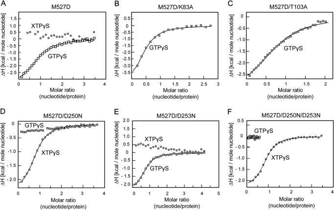
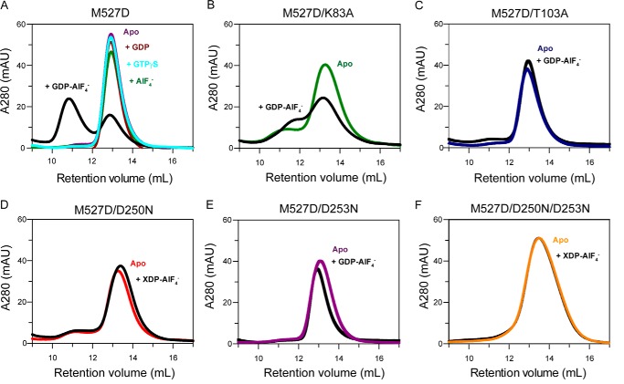
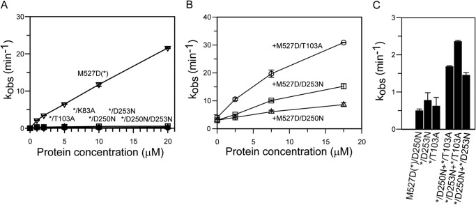
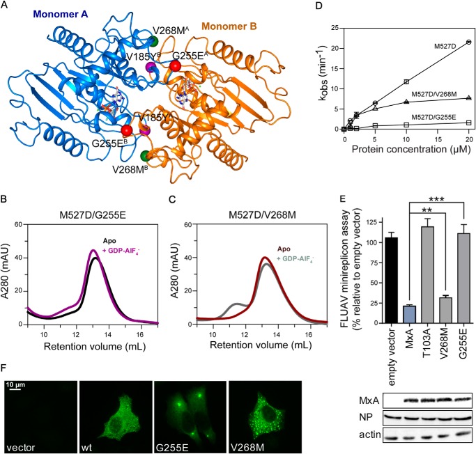
), M527D/D250N (XTP) (Δ), M527D/D253N (GTP) (□), and M527D/D250N/D253N (XTP) (○). B, GTPase activity of M527D can be stimulated by monomeric G domain mutants of MxA. 2.5 μ
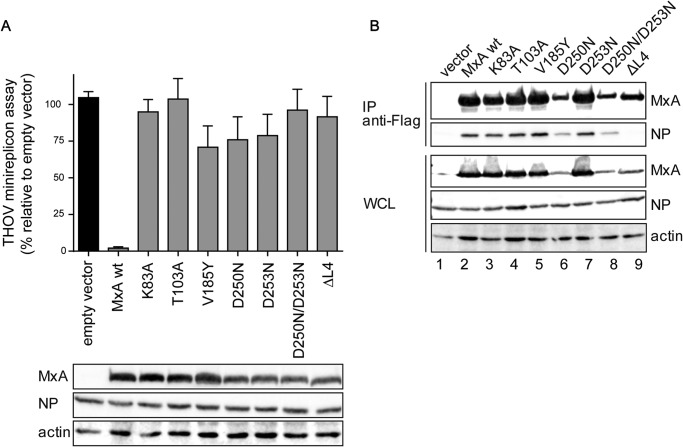
m M527D was incubated with the indicated concentrations of the corresponding mutant (see x axis). The mean kobs calculated from two independent experiments is indicated, with the error bar showing the range of the two data points. M527D+M527D/T103A (○), M527D+M527D/D250N (Δ), M527D+M527D/D253N (□). C, mixed GTPase assays, similar as in B, using 37.5 μm of each of the indicated M527D mutants. The mean kobs was calculated from two independent experiments, with the error bar showing the range of the two data points. When two monomeric G domain mutants were incubated together, their GTPase reactions were mostly additive.
FIGURE 5.
FIGURE 5.
G domain mutants interact with the viral NP. A, 293T cells were co-transfected with expression plasmids for the THOV minireplicon system, consisting of the viral polymerase subunits (10 ng each), NP (50 ng), and the pPolI-FF-Luc minigenome encoding firefly luciferase (50 ng) as well as expression plasmids for MxA or MxA mutants (100 ng) for 24 h. Firefly luciferase activity determined in the cell lysates was normalized to the activity of Renilla luciferase encoded by the co-transfected pRL-SV40 plasmid (10 ng). The activity in the absence of MxA, empty vector control, was set to 100%. Results are presented as means of technical duplicates of three independent experiments. Protein expression of FLAG-tagged Mx, viral NP, and β-actin was determined by Western blot analysis. B, co-precipitation of viral NP with MxA. 293T cells were transfected with FLAG-tagged MxA constructs and infected with THOV (10 multiplicities of infection). At 24 h post-infection, the cell lysates were subjected to FLAG-specific immunoprecipitation (IP). FLAG-MxA and co-precipitated THOV-NP as well as whole cell lysates (WCL) were analyzed by Western blot. Results of one experiment that is representative for three individual experiments are shown.
FIGURE 6.
FIGURE 6.
Functional GTPase is crucial for antiviral activity. A, 293T cells were co-transfected with expression plasmids for the FLUAV minireplicon system of VN/04, consisting of the viral polymerase subunits (10 ng each), NP (100 ng), and the pPolI-FF-Luc minigenome encoding firefly luciferase under the control of the viral promoter (50 ng) as well as expression plasmids for MxA or MxA mutants (300 ng). After 24 h, firefly luciferase activity was determined in the cell lysates and normalized to the activity of Renilla luciferase encoded by the co-transfected pRL-SV40 plasmid (10 ng). The activity in the absence of MxA, empty vector control, was set to 100%. Protein expression of FLAG-tagged MxA, viral NP, and β-actin were determined by Western blot analysis. B, dominant-negative effect of MxA mutants on WT MxA activity. HA-tagged WT MxA (300 ng) was co-transfected with the components of the VN/04 minireplicon system as described for A and increasing amounts (50, 100, and 200 ng) of the indicated FLAG-tagged MxA mutants. Results are presented as means of technical duplicates of three independent experiments.
FIGURE 7.
FIGURE 7.
Intracellular distribution of MxA G domain mutants. HeLa cells were transfected with expression plasmids for WT MxA or MxA mutants (50 ng). At 24 h post-transfection, cells were fixed and stained with a specific antibody against MxA. Results are representative for three individual experiments.
FIGURE 8.
FIGURE 8.
Co-localization study of MxA G domain mutants with syntaxin 17. HeLa cells were transfected with expression plasmids for WT MxA or MxA mutants (50 ng). At 24 h post-transfection, cells were fixed and stained with specific antibodies against MxA (green) and syntaxin 17 (red). Immunofluorescence analysis was performed using a confocal laser scanning microscope.
FIGURE 9.
FIGURE 9.
Complex formation of MxA with the LACV nucleoprotein. Vero cells were transfected with the indicated MxA expression constructs (50 ng) and infected with LACV. At 18 h post-infection, cells were fixed, and immunofluorescence analysis was performed using specific antibodies against MxA (green) and LACV-N (red). The mock panel shows overlay images of transfected and mock-infected cells. The white arrows indicate accumulation of N protein in the Golgi area that is devoid of an MxA signal. In WT MxA-expressing cells, yellow arrows indicate formation of ER resident MxA-N assemblies.
FIGURE 10.
FIGURE 10.
Effects of MxA polymorphisms in the G interface. A, homology model of the MxA GTPase domain dimer as in Fig. 1C. Interface residues are depicted as balls: Val-185 (purple), Gly-255 (red), and Val-268 (green) with the superscript letter indicating monomer A or B, respectively. B and C, analytical gel filtration analysis of M527D/G255E and M527D/V268M as in Fig. 3. D, protein concentration-dependent GTPase activities of M527D (○), M527D/V268M (□), and M527D/G255E (▵) as in Fig. 4A. E, FLUAV-minireplicon system of VN/04, as described in Fig. 6A. Significance was calculated with Student's t test (n = 3). **, p = 0.0058; ***, p = 0.0002. F, intracellular distribution of G255E and V268M in HeLa cells, as in Fig. 7.

References

    1. Verhelst J., Hulpiau P., Saelens X. (2013) Mx proteins: antiviral gatekeepers that restrain the uninvited. Microbiol. Mol. Biol. Rev. 77, 551–566 - PMC - PubMed
    1. Haller O., Staeheli P., Schwemmle M., Kochs G. (2015) Mx GTPases: dynamin-like antiviral machines of innate immunity. Trends Microbiol. 23, 154–163 - PubMed
    1. Haller O., Kochs G. (2011) Human MxA protein: an interferon-induced dynamin-like GTPase with broad antiviral activity. J. Interferon Cytokine Res. 31, 79–87 - PubMed
    1. Netherton C. L., Simpson J., Haller O., Wileman T. E., Takamatsu H. H., Monaghan P., Taylor G. (2009) Inhibition of a large double-stranded DNA virus by MxA protein. J. Virol. 83, 2310–2320 - PMC - PubMed
    1. Gordien E., Rosmorduc O., Peltekian C., Garreau F., Bréchot C., Kremsdorf D. (2001) Inhibition of hepatitis B virus replication by the interferon-inducible MxA protein. J. Virol. 75, 2684–2691 - PMC - PubMed

Publication types

MeSH terms

LinkOut - more resources