

Ultrasound vs. MRI in the assessment of rotator cuff structure prior to shoulder arthroplasty
- PMID: 25829757
- PMCID: PMC4354568
- DOI: 10.1016/j.jor.2015.01.003
Ultrasound vs. MRI in the assessment of rotator cuff structure prior to shoulder arthroplasty
Abstract
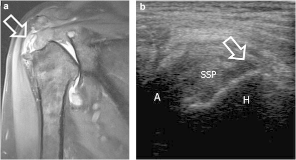
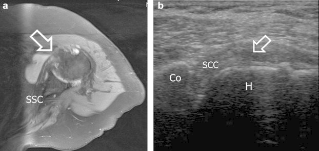
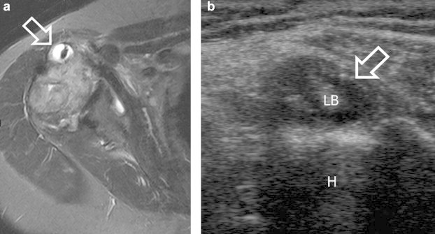
Background/aims: We compared the accuracy of US to 3 T Tesla MRI for the detection of rotator cuff and long biceps tendon pathologies before joint replacement.
Methods: 45 patients were prospectively included.
Results: For the supraspinatus tendon, the accuracy of US when using MRI as reference was 91.1%. For the infraspinatus tendon, the accuracy with MRI as reference was 84.4%. The subscapularis tendon was consistently assessed by US and MRI in 35/45 patients (accuracy 77.8%). For the long biceps tendon the accuracy was 86.7%.
Conclusion: US detection of rotator cuff and biceps tendon integrity is comparable to MRI and should be preferred in revision cases.
Keywords: MRI; Rotator cuff; Shoulder; Supraspinatus; Ultrasound.
Figures
References
-
- Omoumi P., Bafort A.C., Dubuc J.E., Malghem J., Vande Berg B.C., Lecouvet F.E. Evaluation of rotator cuff tendon tears: comparison of multidetector CT arthrography and 1.5-T MR arthrography. Radiology. 2012;264:812–822. - PubMed
-
- de Jesus J.O., Parker L., Frangos A.J., Nazarian L.N. Accuracy of MRI, MR arthrography, and ultrasound in the diagnosis of rotator cuff tears: a meta-analysis. AJR Am J Roentgenol. 2009;192:1701–1707. - PubMed
-
- Levine B.D., Motamedi K., Seeger L.L. Imaging of the shoulder: a comparison of MRI and ultrasound. Curr Sports Med Rep. 2012;11:239–243. - PubMed
-
- Zanetti M., Hodler J. Imaging of degenerative and posttraumatic disease in the shoulder joint with ultrasound. Eur J Radiol. 2000;35:119–125. - PubMed
-
- Lenza M., Buchbinder R., Takwoingi Y., Johnston R.V., Hanchard N.C., Faloppa F. Magnetic resonance imaging, magnetic resonance arthrography and ultrasonography for assessing rotator cuff tears in people with shoulder pain for whom surgery is being considered. Cochrane Database Syst Rev. 2013;9:CD009020. - PMC - PubMed
LinkOut - more resources
Full Text Sources
Other Literature Sources
