. 2015 Mar 28;7(1):36.
doi: 10.1186/s13148-015-0075-3. eCollection 2015.

Somatic cancer mutations in the MLL3-SET domain alter the catalytic properties of the enzyme

Affiliations

Somatic cancer mutations in the MLL3-SET domain alter the catalytic properties of the enzyme

Sara Weirich et al. Clin Epigenetics. .

Abstract

Background: Somatic mutations in epigenetic enzymes are frequently found in cancer tissues. The MLL3 H3K4-specific protein lysine monomethyltransferase is an important epigenetic enzyme, and it is among the most recurrently mutated enzymes in cancers. MLL3 mainly introduces H3K4me1 at enhancers.

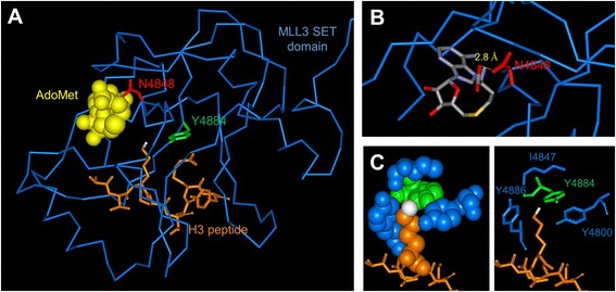
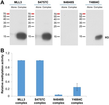
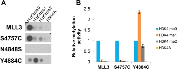
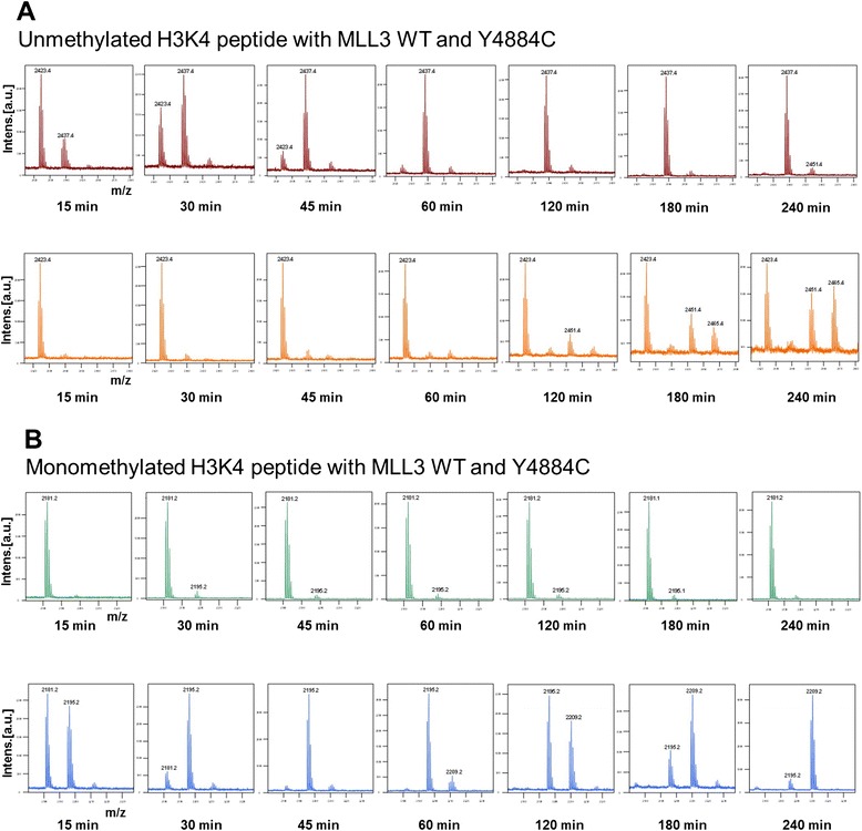
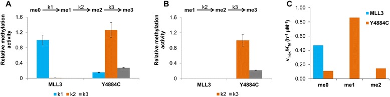
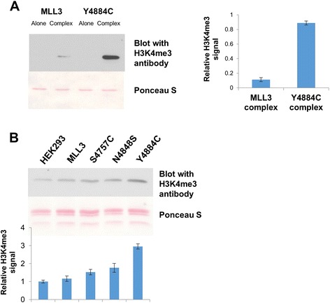
Results: We investigated the enzymatic properties of MLL3 variants that carry somatic cancer mutations. Asn4848 is located at the cofactor binding sites, and the N4848S exchange renders the enzyme inactive. Tyr4884 is part of an aromatic pocket at the active center of the enzyme, and Y4884C converts MLL3 from a monomethyltransferase with substrate preference for H3K4me0 to a trimethyltransferase with H3K4me1 as preferred substrate. Expression of Y4884C leads to aberrant H3K4me3 formation in cells.

Conclusions: Our data show that different somatic cancer mutations of MLL3 affect the enzyme activity in distinct and opposing manner highlighting the importance of experimentally studying the effects of somatic cancer mutations in key regulatory enzymes in order to develop and apply targeted tumor therapy.


Figures

Figure 1
Figure 1
Structure of the MLL1-SET domain bound to the H3 peptide and cofactor product S-adenosyl-L-homocysteine (AdoHcy) (pdb code 2W5Y). Note that an MLL3 structure currently is not available. (A) The protein is shown as blue ribbon, and the peptide is shown in orange in the stick model with the target nitrogen atom colored white. The residues corresponding to Asn4848 and Tyr4884 are displayed in red and green, respectively, the corresponding alignment of MLL1 and MLL3 is shown in Additional file 1: Figure S4. (B) Details of the MLL1-SET structure showing that N3906 (corresponding to N4848) is involved in an H-bond to AdoMet (shown in stick model with coloring by atom type). (C) Details of the MLL1-SET structures showing the hydrophobic and aromatic pocket of MLL1 surrounding the target nitrogen atom which consists of Y3942 (corresponding to Y4884, shown in green) and Y4800, I4847, and Y4885 (all designations refer to MLL3, residues shown in blue).
Figure 2
Figure 2
Activity of MLL3 protein variants. Recombinant histone H3 protein was methylated with radioactively labeled AdoMet by MLL3-SET wild-type and MLL3-SET mutant proteins either alone or in the presence of complex member proteins. (A) Example of an autoradiographic image of the SDS polyacrylamide gel. The methylation signal of H3 is indicated. (B) Quantitative analysis of the averages of duplicate experiments. The error bars indicate the standard error of the mean.
Figure 3
Figure 3
Substrate specificity of MLL3 protein variants. H3 (1 to 15) peptide arrays containing H3K4 at different methylation states and a K4A peptide were methylated with MLL3-SET wild-type and mutant proteins in the presence of complex members using radioactively labeled AdoMet. (A) Example of an autoradiographic image of the methylated peptide SPOT arrays, peptides with the corresponding lysine variants are indicated. (B) Quantitative analysis of the average methylation signals of two independent experiments. The methylation data were normalized to the H3K4me0 methylation signal obtained with the individual MLL3-SET variants. The error bars indicate the standard error of the mean.
Figure 4
Figure 4
Substrate specificity and product pattern of MLL3 protein variants. H3K4 unmethylated (H3; 1 to 19) and monomethylated H3 (1 to 17) peptides were methylated by MLL3-SET wild-type and MLL3-SET Y4884C mutant protein in the presence of complex members using unlabeled AdoMet. The samples were collected at different time points, and methylation was analyzed by mass spectrometry using the relative areas of the corresponding unmethylated and methylated peaks. (A) Matrix-assisted laser desorption/ionization (MALDI) spectra of the methylation of the H3K4me0 peptide with MLL3 wild-type (upper panel) and Y4884C (lower panel). The masses of the corresponding peptides are 2,423.4 Da (H3K4me0), 2,437.4 Da (H3K4me1), 2,451.4 Da (H3K4me2), and 2,465.4 Da (H3K4me3). (B) MALDI spectra of the methylation of the H3K4me1 peptide with MLL3 wild-type (upper panel) and Y4884C (lower panel). The masses of the corresponding peptides are 2,181.2 Da (H3K4me1), 2,195.2 Da (H3K4me2), and 2,209.2 Da (H3K4me3).
Figure 5
Figure 5
Summary of the analysis of the results of the peptide methylation experiments. Panels (A) and (B) show the average rate constants of MLL3-SET wild-type and MLL3-SET Y4884C variant for the methylation of H3K4me0 (A) and H3K4me1 (B) substrates determined at a peptide concentration of 10 μM. Error bars indicate the standard error of mean of two independent experiments. Data in panel (A) were normalized to k 1 of MLL3-SET, and data in panel (B) were normalized to k 2 of Y4884C. Panel (C) shows the v max/K M values for methylation of the un- (me0), mono- (me1), or dimethylated (me2) peptide substrates by MLL3-SET and Y4884C. These values were determined from methylation kinetics carried using 5, 10, 20, and 40 μM of peptide (Additional file 1: Figure S5).
Figure 6
Figure 6
Product specificity of MLL3-SET wild-type and Y4884C at protein level. (A) Methylation of recombinant histone H3 protein by MLL3-SET wild-type and Y4884C alone and in the presence of complex members using unlabeled AdoMet as cofactor. After methylation, the proteins were separated by SDS-PAGE, blotted and the methylation was detected using an H3K4 trimethylated antibody (upper panel). The lower panel shows the Ponceau S-stained image of the blot. The bar diagram shows the average methylation signal from two independent experiments. The error bars indicate the standard error of the mean. (B) Global histone H3K4me3 methylation analysis from HEK293 cells. The cells were transfected with different MLL3 variant plasmids, histones were isolated, and H3K4me3 methylation was probed by Western blot. The upper image shows the H3K4me3 signal and a Ponceau S stain as loading control. The bar diagram shows the quantification from three experiments. The error bars display the standard error of the mean. The signal obtained from the MLL3 was set to 1, and the other signals were normalized accordingly.

Similar articles

Cited by

References

    1. Shilatifard A. The COMPASS family of histone H3K4 methylases: mechanisms of regulation in development and disease pathogenesis. Annu Rev Biochem. 2012;81:65–95. doi: 10.1146/annurev-biochem-051710-134100. - DOI - PMC - PubMed
    1. Herz HM, Mohan M, Garruss AS, Liang K, Takahashi YH, Mickey K, et al. Enhancer-associated H3K4 monomethylation by Trithorax-related, the Drosophila homolog of mammalian Mll3/Mll4. Genes Dev. 2012;26(23):2604–20. doi: 10.1101/gad.201327.112. - DOI - PMC - PubMed
    1. Hu D, Gao X, Morgan MA, Herz HM, Smith ER, Shilatifard A. The MLL3/MLL4 branches of the COMPASS family function as major histone H3K4 monomethylases at enhancers. Mol Cell Biol. 2013;33(23):4745–54. doi: 10.1128/MCB.01181-13. - DOI - PMC - PubMed
    1. Dou Y, Milne TA, Ruthenburg AJ, Lee S, Lee JW, Verdine GL, et al. Regulation of MLL1 H3K4 methyltransferase activity by its core components. Nat Struct Mol Biol. 2006;13(8):713–9. doi: 10.1038/nsmb1128. - DOI - PubMed
    1. Steward MM, Lee JS, O’Donovan A, Wyatt M, Bernstein BE, Shilatifard A. Molecular regulation of H3K4 trimethylation by ASH2L, a shared subunit of MLL complexes. Nat Struct Mol Biol. 2006;13(9):852–4. doi: 10.1038/nsmb1131. - DOI - PubMed

LinkOut - more resources