. 2015 Aug;22(8):675-84.
doi: 10.1038/gt.2015.29. Epub 2015 Apr 1.

B7H6-specific chimeric antigen receptors lead to tumor elimination and host antitumor immunity

Affiliations

B7H6-specific chimeric antigen receptors lead to tumor elimination and host antitumor immunity

M-R Wu et al. Gene Ther. 2015 Aug.

Abstract

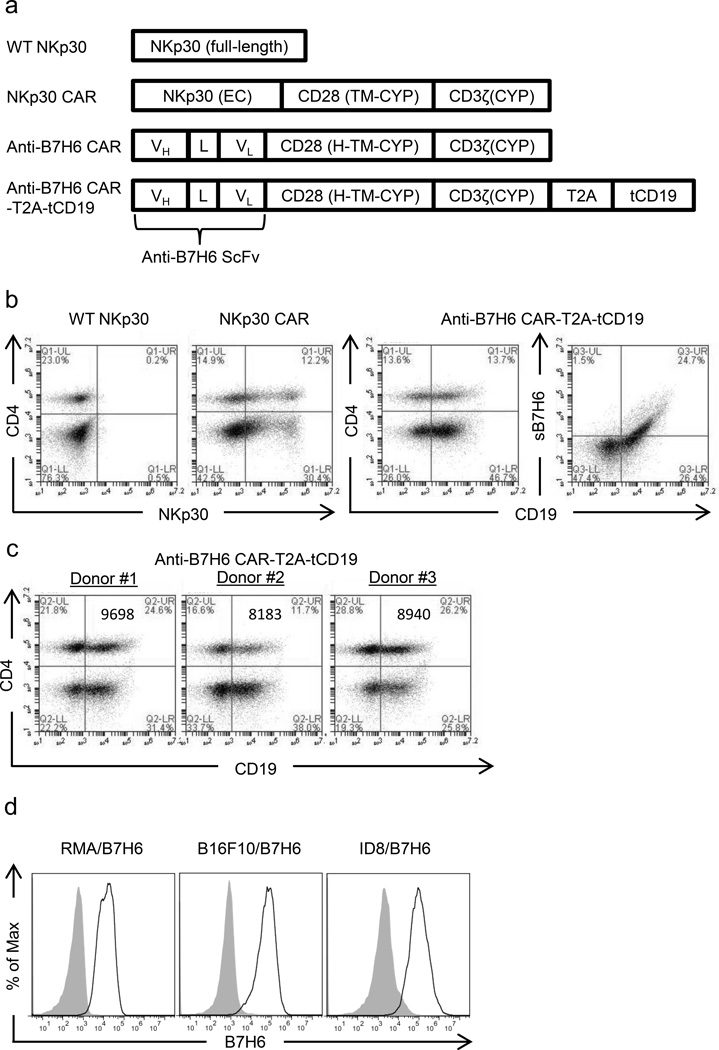
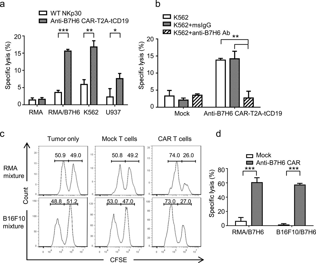
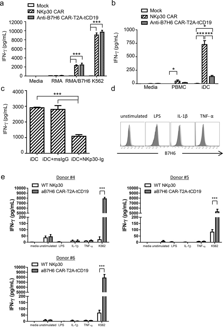
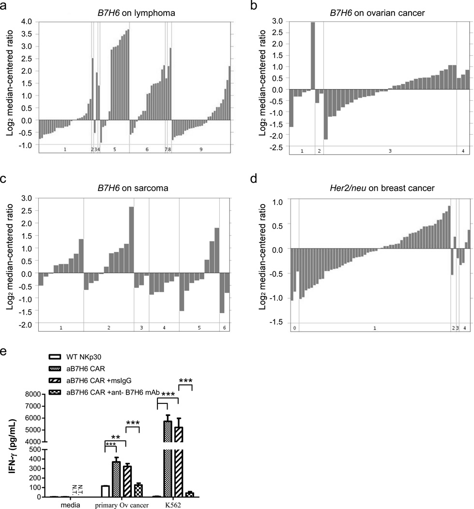
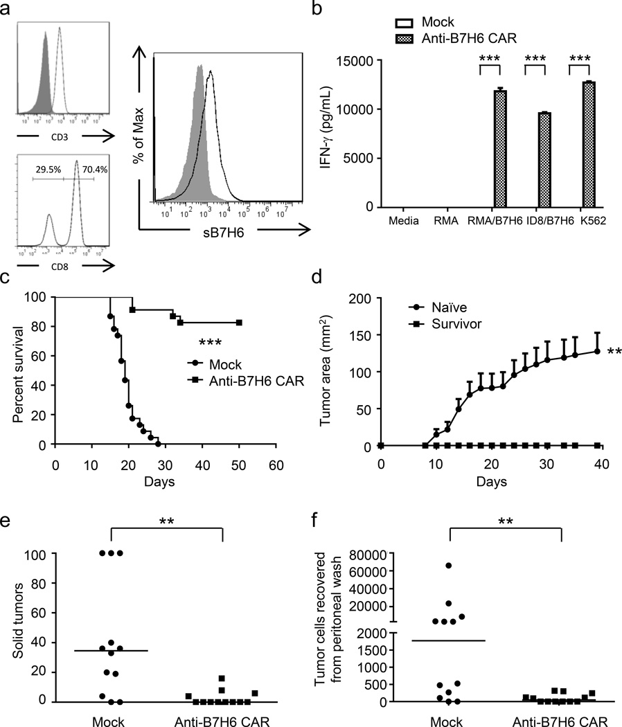
Chimeric antigen receptor (CAR) T-cell therapies have demonstrated durable and potentially curative therapeutic efficacy against B-cell leukemia in clinical trials. A CAR strategy can target any tumor surface antigens as long as an antigen-binding receptor can be generated. New CARs that target solid tumors and have the potential to target multiple tumor types are needed. In this study, B7H6, a ligand for the NK cell activating receptor NKp30, was targeted to create a CAR that targets multiple tumor types. B7H6 is expressed on various primary human tumors, including leukemia, lymphoma and gastrointestinal stromal tumors, but it is not constitutively expressed on normal tissues. B7H6-specific CAR T cells have robust cellular cytotoxicity and interferon-γ secretion when co-cultured with B7H6+ tumor cells, and they exhibit little self-reactivity to immature dendritic cells or pro-inflammatory monocytes. In vivo, B7H6-specific CAR T cells greatly enhanced the survival of RMA/B7H6 lymphoma-bearing mice. The long-term survivor mice were protected against a B7H6-deficient tumor re-challenge. This CAR therapy also decreased tumor burden in a murine ovarian cancer model. In conclusion, B7H6-specific CARs have the potential to treat B7H6+ hematologic and solid tumors.


Conflict of interest statement

CONFLICT OF INTEREST

Ming-Ru Wu and Leslie DeMars declare no potential conflict of interest.

Figures

Figure 1
Figure 1. Design and expression of NKp30-based CAR (NKp30 CAR) and B7H6-specific CARs (anti-B7H6 CARs)
(a) WT NKp30 is the full length wild-type NKp30 gene. A NKp30 CAR was created by fusing NKp30 extracellular (EC) domain with human CD28 transmembrane (TM), and cytoplasmic (CYP) domains, followed by a human CD3ζ CYP domain. A B7H6-specific CAR was created by fusing anti-B7H6 scFv DNA with the human CD28 hinge (H), transmembrane (TM), and cytoplasmic (CYP) domains, followed by a human CD3ζ CYP domain DNA. The anti-B7H6 CAR-T2A-tCD19 construct was created by combining the anti-B7H6 CAR DNA with a T2A sequence containing a furin cleavage site and a truncated (t) human CD19 DNA sequence. (b) Human PBMCs were transduced with WT NKp30, NKp30 CAR, or anti-B7H6 CAR-T2A-tCD19 constructs. Transduced T cells were stained with anti-CD4 mAbs, anti-NKp30 mAbs, soluble B7H6 (sB7H6), and/or anti-CD19 mAbs. CD4- T cells are CD8+ T cells. The data are representative of data from 3 different human donors. (c) Anti-B7H6 CAR expression on T cells from different PBMC donors were analyzed. The values in the graph represent the mean fluorescent intensities of CD19 expression for each sample. (d) RMA/B7H6, B16F10/B7H6, and ID8/B7H6 were stained with anti-B7H6 mAbs followed by goat anti-msIgG Abs (open histograms) or with goat anti-msIgG Abs only (filled histograms). Data shown are representative of 3 independent experiments.
Figure 2
Figure 2. In vitro cytotoxicity of B7H6-specific CAR T cells on tumor cell lines
(a) Human PBMCs transduced with WT NKp30 or anti-B7H6 CAR-T2A-tCD19 were co-cultured with B7H6- (RMA) or B7H6+ (RMA/B7H6, K562, U937) tumor cells at an E:T ratio 5:1 in a 6h LDH-release assay. The data are mean +SD of triplicates, and representative of 2 independent experiments. (b) K562 cells were pre-incubated with msIgG or anti-B7H6 mAbs. Mock (untransduced) or anti-B7H6 CAR-T2A-tCD19 human T cells were co-cultured with K562 at an E:T ratio 5:1 in a 6h LDH-release assay. (c) Mouse splenocytes untransduced (Mock) or transduced with the anti-B7H6 CAR were co-cultured with a 50% RMA + 50% RMA/B7H6 cell mixture or a 50% B16F10 + 50% B16F10/B7H6 cell mixture at an E:T ratio 5:1. RMA, B16F10 and RMA/B7H6, B16F10/B7H6 cells were labeled with 0.1uM (B7H6- cells) and 1uM CFSE (B7H6+ cells), respectively. After 8h, samples were harvested and analyzed by flow cytometry. Data shown were pre-gated on live cells and excluded T cells. (d) Specific lysis by Mock and anti-B7H6 CAR T cells against RMA/B7H6 and B16F10/B7H6 are shown as mean +SD. Data are representative of 2 independent experiments. An * indicates p < 0.05; ** indicates p < 0.005; *** indicates p < 0.0001.
Figure 3
Figure 3. B7H6-specific CAR T cells produce IFN-γ in response to B7H6 expressing tumors but not immature DCs or LPS, IL-1β, or TNF-α stimulated PBMCs
(a) Mock, NKp30 CAR, or anti-B7H6 CAR T cells were co-cultured with tumor cells at an E:T ratio 1:1. After 24h, cell-free conditioned media was harvested and IFN-γ concentration was determined by ELISA. The results are shown as mean + SD and are representative of 2 different PBMC donors. (b) Mock, NKp30 CAR, or anti-B7H6 CAR T cells were co-cultured with autologous PBMCs or autologous immature DCs at an E:T ratio 5:1 (105:2×10⁴). Cell-free media was harvested 24h later and IFN-γ concentration was measured. The results are shown as mean + SD and are representative of 2 PBMC different donors. (c) Autologous iDCs were pre-incubated with msIgG or NKp30-Ig for 45min before they were co-cultured with NKp30 CAR T cells at an E:T ratio 5:1 (105:2×10⁴). After 24h, cell-free culture media was harvested and IFN-γ concentration was determined by ELISA. The results are shown as mean + SD and are representative of 2 different donors. (d) Human PBMCs were cultured and stimulated with LPS, IL-1β, or TNF-α for 48h. Cells were harvested and stained with anti-CD14 mAbs and anti-B7H6 mAbs. Histograms shown are pre-gated on CD14+ monocytes. Filled histograms represent isotype controls and open histograms represent B7H6 expression. Data shown are representative of 3 different PBMC donors. (e) WT NKp30 or anti-B7H6 CAR T cells were co-cultured with unstimulated, LPS, IL-1β, or TNF-α stimulated autologous PBMCs for 24h. Cell free supernatant was harvested and IFN-γ concentration was determined by ELISA. The results of CAR T cells derived from three different donors are shown as mean + SD. An * indicates p < 0.05; ** indicates p < 0.005; *** indicates p < 0.0001.
Figure 4
Figure 4. B7H6 mRNA expression is upregulated in multiple types of primary human tumor samples, and B7H6-specific CAR T cells recognize human primary ovarian cancer
B7H6 mRNA expression from various primary human tumor samples was analyzed from the Oncomine database (https://www.oncomine.org). (a) B7H6 mRNA expression in B cell lymphoma patient samples. Cancer subsets are as indicated: 1. Activated B-cell-Like Diffuse Large B-Cell Lymphoma. 2. B-Cell Neoplasm (Not Specified). 3. B-cell Non-Hodgkin's Lymphoma. 4. Burkitt's Lymphoma. 5. Diffuse Large B-Cell Lymphoma. 6. Germinal Center B-Cell-Like Diffuse Large B-Cell Lymphoma. 7. Grade 3 Follicular Lymphoma. 8. Immunoblastic Lymphoma. 9. Type 3 Diffuse Large B-Cell Lymphoma. (b) B7H6 mRNA expression in ovarian cancer patient samples. Cancer subsets are as indicated: 1. Ovarian Clear Cell Adenocarcinoma. 2. Ovarian Endometrioid Adenocarcinoma. 3. Ovarian Serous Surface Papillary Carcinoma. 4. Undifferentiated Ovarian Carcinoma. (c) B7H6 mRNA expression in various types of sarcoma samples. Cancer subsets are as indicated: 1. Gastrointestinal Stromal Tumor. 2. Leiomyosarcoma. 3. Liposarcoma. 4. Malignant Fibrous Histiocytoma. 5. Monophasic Synovial Sarcoma. 6. Schwannoma. (d) Her2/neu mRNA expression in breast cancer samples. Disease subsets are as indicated: 0. Not specified. 1. Ductal Breast Carcinoma. 2. Ductal Breast Carcinoma in Situ. 3. Fibroadenoma. 4. Lobular Breast Carcinoma. Note the scale difference in each figure. (e) WT NKp30 or B7H6-specific CAR T cells were co-cultured with a primary human ovarian cancer sample at an E:T ratio 1:1. For blocking experiments, cancer samples were pre-incubated with 5µg of msIgG or anti-B7H6 mAbs before co-culturing with T cells. Cell-free conditioned media was harvested after 24h and IFN-γ concentration was determined by ELISA. The results shown are mean + SD and are representative of T cells made from 2 different PBMC donors. An * indicates p < 0.05; ** indicates p < 0.005; *** indicates p < 0.0001. N.T. indicates not tested.
Figure 5
Figure 5. B7H6-specific CAR T cells promote survival of RMA/B7H6 lymphoma-bearing mice and decrease tumor burden in ID8/B7H6 ovarian cancer-bearing mice
(a) Cell surface molecules on murine B7H6-specific CAR T cells. Expression of CD3, CD8, and the B7H6-specific CAR are shown. Filled histograms represent isotype controls and open histograms represent specific expression. More than 99% of cells in the cell preparation are CD3+ T cells. CD8- cells are CD4+ T cells. (b) Murine Mock T cells or anti-B7H6 CAR T cells were co-cultured with tumor cells at an E:T ratio 1:1 (RMA, RMA/B7H6, K562) or 4:1 (ID8/B7H6). After 24h, cell-free culture media was harvested and the IFN-γ concentration was measured by ELISA. The results are shown as mean + SD and are representative of 2 independent experiments. (c) B6 mice were injected with RMA/B7H6 (105 cells, i.v.) on day 0. Murine Mock T cells or anti-B7H6 CAR T cells were administered (5 × 10⁶ cells, i.v.) on days 5, 7, and 9. Data are presented in Kaplan-Meier survival curves and are pooled from 3 independent experiments (n=23 for each group). (d) Naïve and survived mice (from Figure 5c) were challenged with 104 RMA tumor cells subcutaneously, and tumor area was measured every other day. Data shown are mean + SEM and are pooled from 2 independent experiments (n=7 for each group). (e & f) ID8/B7H6 (GFP+) ovarian cancer cells (5 × 10⁶ cells, i.p.) were injected into B6 mice on day 0. Murine Mock T cells or anti-B7H6 CAR T cells were administered on day 7 (5 × 10⁶ cells, i.p.). The total number of solid tumors (stopped counting at 100) for each mouse on the peritoneal wall was counted (e) and the number of GFP+ tumor cells in the peritoneal wash (f) were measured on day 42. Each dot represents data from an individual mouse. Horizontal bars represent median values. These data are combined from two independent experiments. An * indicates p < 0.05; ** indicates p < 0.005; *** indicates p < 0.0001.

References

    1. Porter DL, Levine BL, Kalos M, Bagg A, June CH. Chimeric antigen receptor-modified T cells in chronic lymphoid leukemia. N Engl J Med. 2011;365(8):725–733. - PMC - PubMed
    1. Kalos M, Levine BL, Porter DL, Katz S, Grupp SA, Bagg A, et al. T cells with chimeric antigen receptors have potent antitumor effects and can establish memory in patients with advanced leukemia. Sci Transl Med. 2011;3(95):95ra73. - PMC - PubMed
    1. Grupp SA, Kalos M, Barrett D, Aplenc R, Porter DL, Rheingold SR, et al. Chimeric antigen receptor-modified T cells for acute lymphoid leukemia. N Engl J Med. 2013;368(16):1509–1518. - PMC - PubMed
    1. Brentjens RJ, Davila ML, Riviere I, Park J, Wang X, Cowell LG, et al. CD19-targeted T cells rapidly induce molecular remissions in adults with chemotherapy-refractory acute lymphoblastic leukemia. Sci Transl Med. 2013;5(177):177ra38. - PMC - PubMed
    1. Sadelain M, Brentjens R, Riviere I. The basic principles of chimeric antigen receptor design. Cancer Discov. 2013;3(4):388–398. - PMC - PubMed

Publication types

Substances