. 2015 Apr 1;10(4):e0123258.
doi: 10.1371/journal.pone.0123258. eCollection 2015.

The SKINT1-like gene is inactivated in hominoids but not in all primate species: implications for the origin of dendritic epidermal T cells

Affiliations

The SKINT1-like gene is inactivated in hominoids but not in all primate species: implications for the origin of dendritic epidermal T cells

Rania Hassan Mohamed et al. PLoS One. .

Abstract

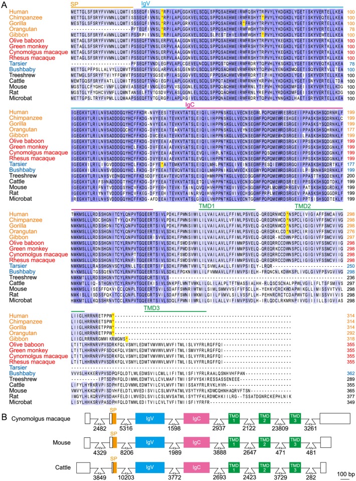
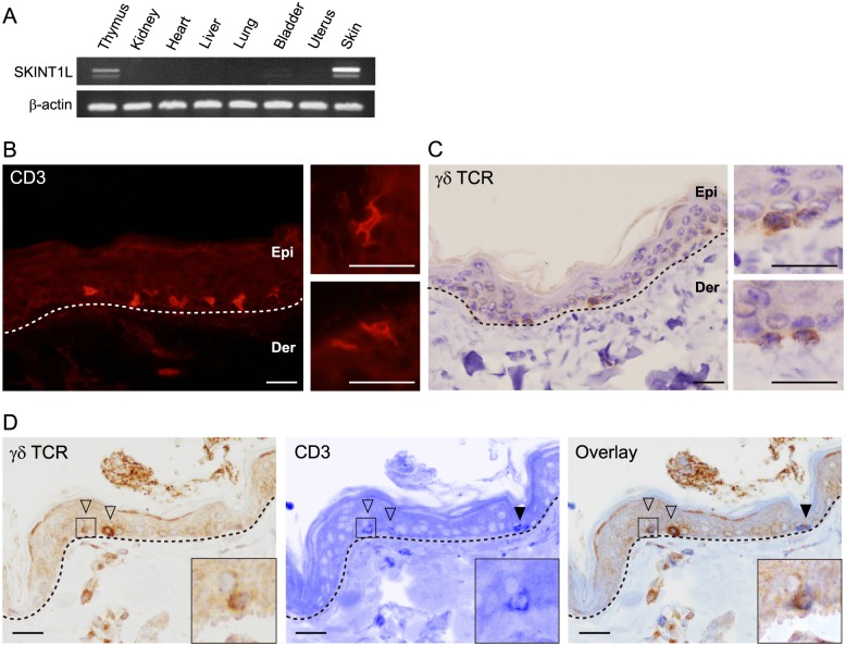
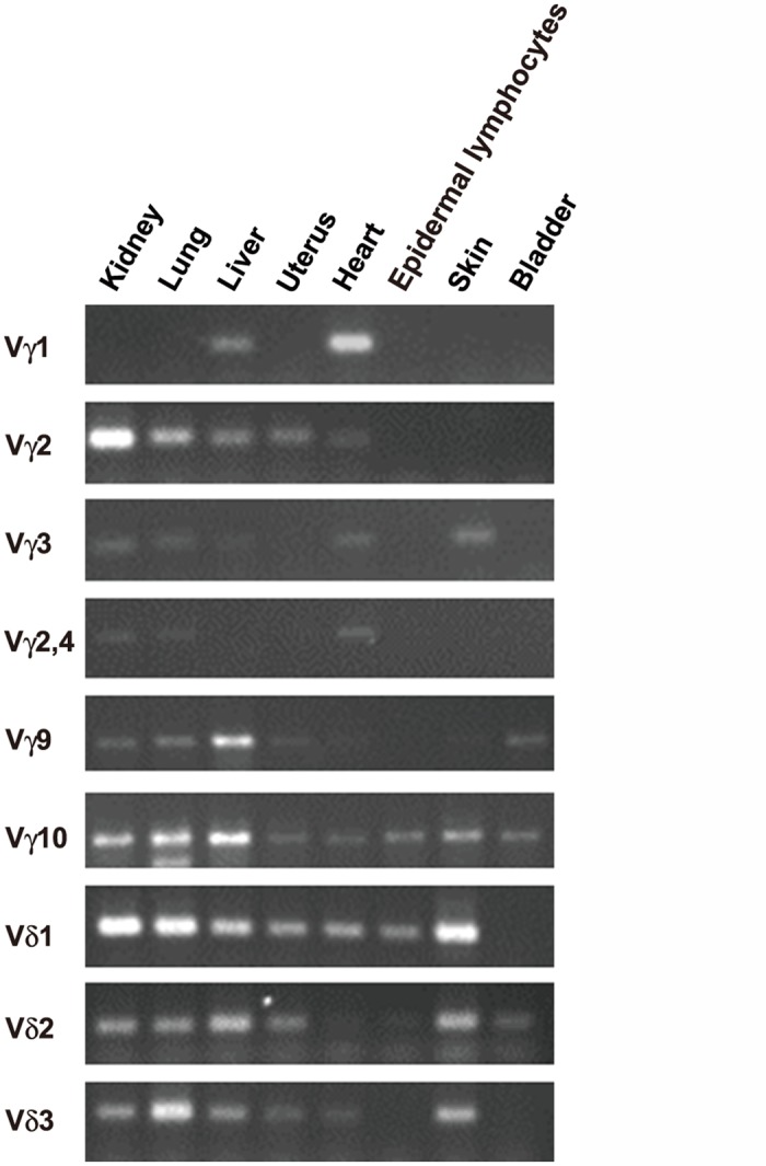
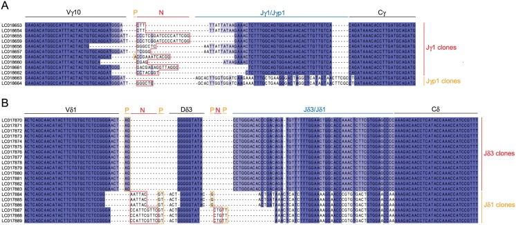
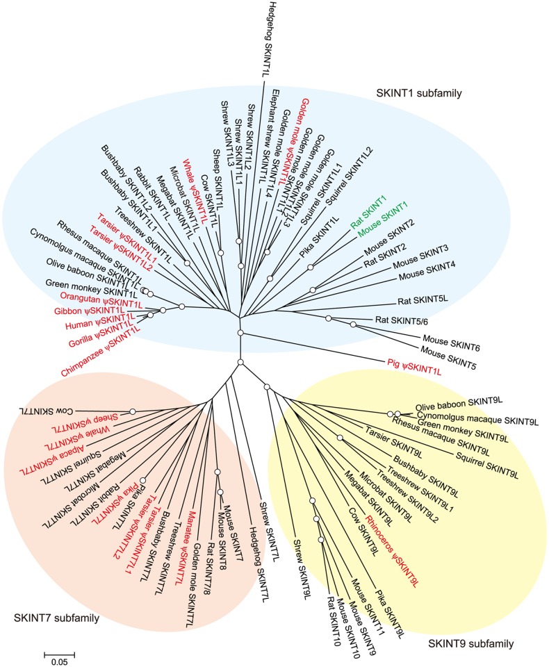
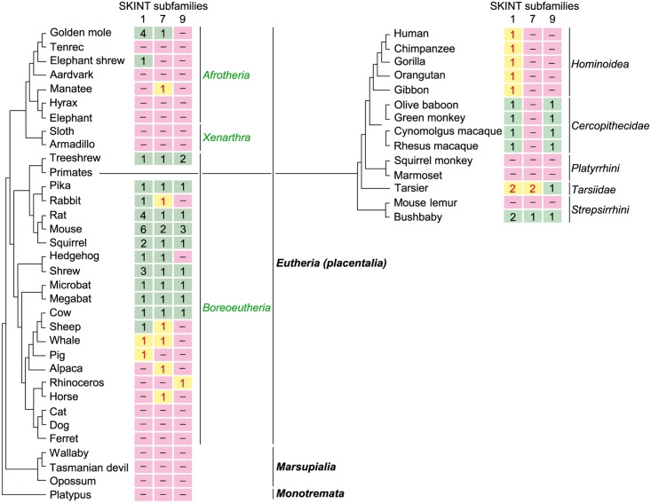
Dendritic epidermal T cells, which express an invariant Vγ5Vδ1 T-cell receptor and account for 95% of all resident T cells in the mouse epidermis, play a critical role in skin immune surveillance. These γδ T cells are generated by positive selection in the fetal thymus, after which they migrate to the skin. The development of dendritic epidermal T cells is critically dependent on the Skint1 gene expressed specifically in keratinocytes and thymic epithelial cells, suggesting an indispensable role for Skint1 in the selection machinery for specific intraepithelial lymphocytes. Phylogenetically, rodents have functional SKINT1 molecules, but humans and chimpanzees have a SKINT1-like (SKINT1L) gene with multiple inactivating mutations. In the present study, we analyzed SKINT1L sequences in representative primate species and found that all hominoid species have a common inactivating mutation, but that Old World monkeys such as olive baboons, green monkeys, cynomolgus macaques and rhesus macaques have apparently functional SKINT1L sequences, indicating that SKINT1L was inactivated in a common ancestor of hominoids. Interestingly, the epidermis of cynomolgus macaques contained a population of dendritic-shaped γδ T cells expressing a semi-invariant Vγ10/Vδ1 T-cell receptor. However, this population of macaque T cells differed from rodent dendritic epidermal T cells in that their Vγ10/Vδ1 T-cell receptors displayed junctional diversity and expression of Vγ10 was not epidermis-specific. Therefore, macaques do not appear to have rodent-type dendritic epidermal T cells despite having apparently functional SKINT1L. Comprehensive bioinformatics analysis indicates that SKINT1L emerged in an ancestor of placental mammals but was inactivated or lost multiple times in mammalian evolution and that Skint1 arose by gene duplication in a rodent lineage, suggesting that authentic dendritic epidermal T cells are presumably unique to rodents.


Conflict of interest statement

Competing Interests: The authors have declared that no competing interests exist.

Figures

Fig 1
Fig 1. Amino acid sequence alignment of SKINT1 and SKINT1L molecules.
A. Deduced SKINT1 and SKINT1L protein sequences of indicated species were aligned using the Clustal Omega program. Strictly and highly conserved residues are indicated in dark blue and light blue, respectively. Hominoid sequences contain multiple premature stop codons (indicated by red asterisks). The location of predicted domains is indicated on top of the sequences. Species names indicated in orange, red and blue represent hominoids, OWMs and prosimians, respectively. SP stands for signal peptides. Accession numbers are as follows: cattle, XP_005204826; microbat, XP_006107913.1; mouse, NP_001096132; rat, NP_001129388; and treeshrew, XP_006147332.1. Accession numbers and relevant information for primate sequences are given in S1 Table. B. The exon-intron organization of cynomolgus macaque SKINT1L was compared to that of mouse Skint1 and bovine SKINT1L. Exon-intron boundaries were predicted based on the consensus splice junction sequences and similarity of the deduced amino acid sequences to the mouse SKINT1 protein sequence. Numbers indicate the length of introns in base pairs. Open boxes indicate 5'- and 3'-untranslated regions. bp, base pairs.
Fig 2
Fig 2. Tissue distribution of SKINT1L transcripts and immunohistochemical identification of DETC-like cells in the macaque skin.
A. SKINT1L expression in cynomolgus macaque tissues was examined by reverse transcription PCR. β-actin was used as a control. Thymic tissues were obtained from a macaque fetus at embryonic day 100; the remaining tissues were procured from adult animals. A faint, lower band in the thymus and skin represents alternatively spliced transcripts encoding SKINT1L molecules with two TMDs. B-D: The basal and suprabasal layers of the epidermis of the cynomolgus macaque contain dendritic-shaped CD3+, γδ TCR+ cells. Paraffin sections of adult macaque skin were stained with cross-reacting antibody for human CD3 (B), cross-reacting antibody for human γδ TCR (C) or with both antibodies (D). Insets show higher magnification images of CD3+ dendritic-shaped cells (B), γδ TCR+ cells (C) and CD3/γδ TCR double-positive cells (D). Open and filled arrowheads indicate CD3+ γδ TCR+ cells and CD3+ γδ TCR- cells, respectively. Scale bar, 10 μm. Epi, epidermis; Der, dermis.
Fig 3
Fig 3. Cynomolgus macaque skin T cells predominantly express Vγ10Vδ1 TCR.
A. Genomic organization of cynomolgus macaque TCR Vγ and Vδ loci. Green and yellow boxes indicate functional and non-functional gene segments, respectively. Numbers given in roman numerals over the blankets indicate the subgroups of Vγ gene segments defined by Huck et al. [27]. GenBank accession numbers for the macaque genomic sequences subjected to analysis were NC_022274 for the Vγ locus and NC_022278 for the Vδ locus. kbp, kilobase pairs. Arrows indicate orientation of transcription. B. Expression of cynomolgus macaque TCR Vγ and Vδ gene segments in representative tissues. Reverse transcription PCR was conducted using a set of primer pairs specific for each TCR Vγ or Vδ gene segment shown in Table 1.
Fig 4
Fig 4. Junctional sequences of macaque TCR Vγ10- and Vδ1-chains.
Panels A and B show the junctional sequences of TCR Vγ10- and Vδ1-chains, respectively. Strictly and highly conserved residues are indicated in dark blue and light blue, respectively. P- and N-nucleotides are boxed in orange and red, respectively. The sequences were deposited in GenBank under accession numbers LC018653-LC018664 (Vγ10-chains) and LC017870-LC017889 (Vδ1-chains).
Fig 5
Fig 5. Phylogenetic tree of the SKINT family.
The neighbor-joining tree was constructed based on deduced amino acid sequences as described in Materials and Methods. Nodes with bootstrap confidence values over 80 for 1,000 replications are indicated by circles. Genes judged to be non-functional are indicated in red. Genes proven to be functional are in green. Genes with no obvious inactivating mutations are in black. Accession numbers of sequences and other relevant information are given in S2 Table.
Fig 6
Fig 6. Distribution of SKINT subfamily proteins across mammalian orders.
The phylogenetic relationship of mammals shown here is based on Murphy et al. [32] and Song et al. [33]. The copy number of SKINT genes belonging to each subfamily is shown for 47 mammalian species representing 22 orders. The copy number of functional and non-functional genes is indicated in black and red, respectively. The distribution of SKINT subfamily proteins in 14 primate species is shown separately on the right. Accession numbers of sequences and other relevant information are given in S2 Table.

Similar articles

Cited by

References

    1. Allison JP, Havran WL. The immunobiology of T cells with invariant γδ antigen receptors. Annu Rev Immunol. 1991; 9: 679–705. - PubMed
    1. Hayday AC. γδ cells: a right time and a right place for a conserved third way of protection. Annu Rev Immunol. 2000; 18: 975–1026. - PubMed
    1. Witherden DA, Havran WL. Molecular aspects of epithelial γδ T cell regulation. Trends Immunol. 2011; 32: 265–271. 10.1016/j.it.2011.03.005 - DOI - PMC - PubMed
    1. Yoshida S, Mohamed RH, Kajikawa M, Koizumi J, Tanaka M, Fugo K, et al. Involvement of an NKG2D ligand H60c in epidermal dendritic T cell-mediated wound repair. J Immunol. 2012; 188: 3972–3979. 10.4049/jimmunol.1102886 - DOI - PubMed
    1. Itohara S, Farr AG, Lafaille JJ, Bonneville M, Takagaki Y, Haas W, et al. Homing of a γδ thymocyte subset with homogeneous T-cell receptors to mucosal epithelia. Nature. 1990; 343: 754–757. - PubMed

Publication types

Associated data

LinkOut - more resources