

Diffusion tensor magnetic resonance imaging of trigeminal nerves in relapsing herpetic keratouveitis
- PMID: 25830672
- PMCID: PMC4382307
- DOI: 10.1371/journal.pone.0122186
Diffusion tensor magnetic resonance imaging of trigeminal nerves in relapsing herpetic keratouveitis
Abstract
Background: Corneal hypoesthesia is the landmark of HSV and VZV keratitis and can lead to neurotrophic keratitis. Diffusion tensor imaging (DTI) is a new magnetic resonance imaging (MRI) derived technique, which offers possibilities to study axonal architecture. We aimed at assessing the potential impact of recurrent HSV or VZV-related keratitis on the axonal architecture of trigeminal nerves using DTI.
Design: Prospective non-interventional study.
Participants: Twelve patients and 24 controls.
Methods: DTI using MRI of the trigeminal fibers and corneal esthesiometry using the Cochet-Bonnet esthesiometer were acquired for patients affected by unilateral and recurrent HSV or VZV-related keratitis (3 months after the last corneal inflammatory event), and control subjects with no history of ocular or neuronal disease affecting the trigeminal pathways.
Main outcome measures: Fractional anisotropy (FA) and apparent diffusion coefficient (ADC) were compared between the 2 eyes of both patients and controls, and correlated with corneal esthesiometry.
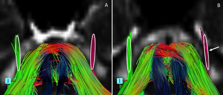
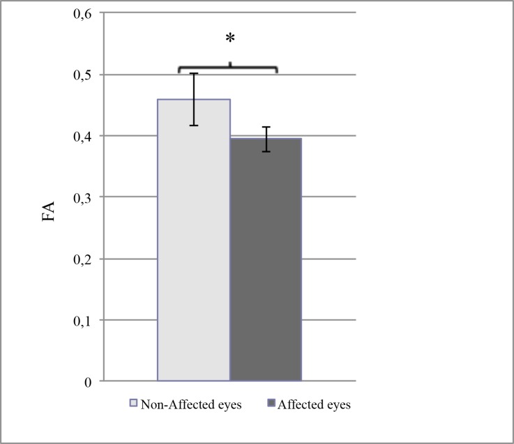
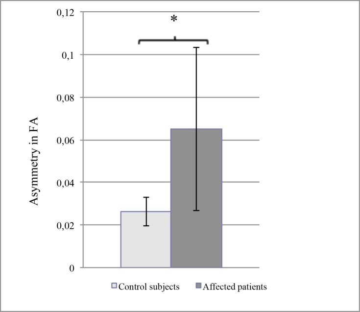
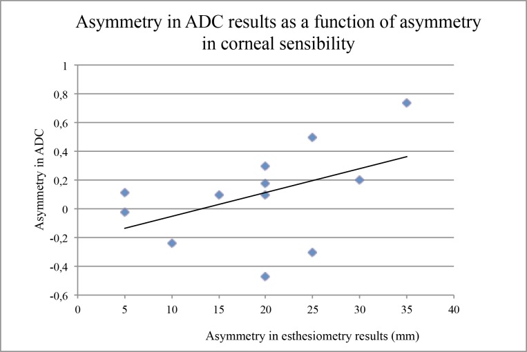
Results: FA was lower in the trigeminal fibers ipsilateral to the affected eye compared to the non-affected side (0.39±0.02 versus 0.46±0.04, P=0.03). This difference was more important than the intra-individual variability observed in controls. Concomitantly, the asymmetry in ADC results was significantly correlated with the loss of corneal sensitivity in the affected eye.
Conclusions: Corneal hypoesthesia related to HSV and VZV keratitis is associated with persistent modifications in the architecture and functionality of the trigeminal fibers. These results add further explanation to the pathogenesis of HSV and VZV-induced neurotrophic keratitis, which may occur despite an apparent quiescence of the disease.
Conflict of interest statement
Figures
Similar articles
-
Impact of herpetic stromal immune keratitis in corneal biomechanics and innervation.Graefes Arch Clin Exp Ophthalmol. 2018 Jan;256(1):155-161. doi: 10.1007/s00417-017-3826-3. Epub 2017 Oct 29. Graefes Arch Clin Exp Ophthalmol. 2018. PMID: 29082447
-
Impairment of lacrimal secretion in the unaffected fellow eye of patients with recurrent unilateral herpetic keratitis.Ophthalmology. 2013 Oct;120(10):1959-67. doi: 10.1016/j.ophtha.2013.02.037. Epub 2013 May 9. Ophthalmology. 2013. PMID: 23664465
-
Corneal Sub-Basal Nerve Changes in Patients with Herpetic Keratitis During Acute Phase and after 6 Months.Medicina (Kaunas). 2019 May 27;55(5):214. doi: 10.3390/medicina55050214. Medicina (Kaunas). 2019. PMID: 31137905 Free PMC article.
-
[Battle with herpes for 37 years].Nippon Ganka Gakkai Zasshi. 2015 Mar;119(3):145-66; discussion 167. Nippon Ganka Gakkai Zasshi. 2015. PMID: 25854108 Review. Japanese.
-
Herpes simplex keratitis.Prog Retin Eye Res. 2006 Jul;25(4):355-80. doi: 10.1016/j.preteyeres.2006.05.001. Epub 2006 Jun 27. Prog Retin Eye Res. 2006. PMID: 16807055 Review.
Cited by
-
Mapping fine-scale anatomy of gray matter, white matter, and trigeminal-root region applying spherical deconvolution to high-resolution 7-T diffusion MRI.MAGMA. 2018 Dec;31(6):701-713. doi: 10.1007/s10334-018-0705-9. Epub 2018 Sep 17. MAGMA. 2018. PMID: 30225801
-
[Pathogenesis and epidemiology of neurotrophic keratopathy].Ophthalmologe. 2019 Feb;116(2):109-119. doi: 10.1007/s00347-018-0823-9. Ophthalmologe. 2019. PMID: 30478498 Review. German.
-
Is There a Magnetic Resonance Imaging-Discernible Cause for Trigeminal Neuralgia? A Structured Review.World Neurosurg. 2017 Feb;98:89-97. doi: 10.1016/j.wneu.2016.10.104. Epub 2016 Oct 27. World Neurosurg. 2017. PMID: 27989975 Free PMC article. Review.
-
Clinical Applications for Diffusion MRI and Tractography of Cranial Nerves Within the Posterior Fossa: A Systematic Review.Front Neurosci. 2019 Feb 7;13:23. doi: 10.3389/fnins.2019.00023. eCollection 2019. Front Neurosci. 2019. PMID: 30809109 Free PMC article.
-
Structural and functional changes of binocular corneal innervation and ocular surface function after unilateral SMILE and tPRK.Br J Ophthalmol. 2024 Oct 22;108(11):1492-1499. doi: 10.1136/bjo-2023-324358. Br J Ophthalmol. 2024. PMID: 38527771 Free PMC article.
References
-
- de Melker H, Berbers G, Hahne S, Rumke H, van den Hof S, de Wit A, et al. The epidemiology of varicella and herpes zoster in The Netherlands: implications for varicella zoster virus vaccination. Vaccine. 2006;24: 3946–52. - PubMed
MeSH terms
LinkOut - more resources
Full Text Sources
Other Literature Sources
