

Abnormalities in hemispheric specialization of caudate nucleus connectivity in schizophrenia
- PMID: 25830688
- PMCID: PMC4630217
- DOI: 10.1001/jamapsychiatry.2014.3176
Abnormalities in hemispheric specialization of caudate nucleus connectivity in schizophrenia
Abstract
Importance: Hemispheric specialization of the human brain is a marker of successful neurodevelopment. Altered brain asymmetry that has been repeatedly reported in schizophrenia may represent consequences of disrupted neurodevelopment in the disorder. However, a complete picture of functional specialization in the schizophrenic brain and its connectional substrates is yet to be unveiled.
Objectives: To quantify intrinsic hemispheric specialization at cortical and subcortical levels and to reveal potential disease effects in schizophrenia.
Design, setting, and participants: Resting-state functional connectivity magnetic resonance imaging has been previously used to quantitatively measure hemispheric specialization in healthy individuals in a reliable manner. We quantified the intrinsic hemispheric specialization at the whole brain level in 31 patients with schizophrenia and 37 demographically matched healthy controls from November 28, 2007, through June 29, 2010, using resting-state functional magnetic resonance imaging.
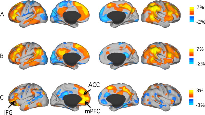
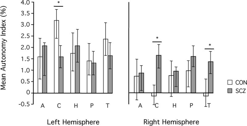
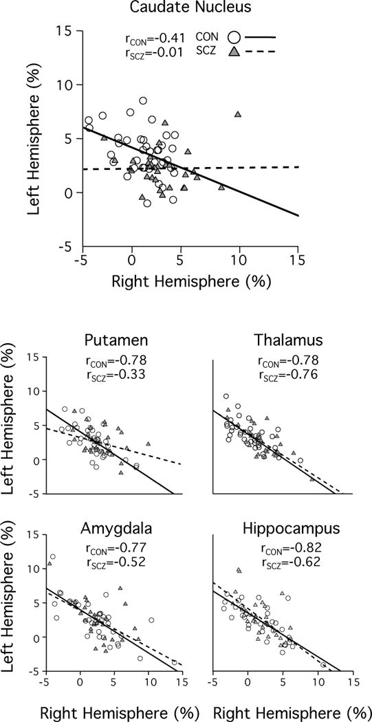
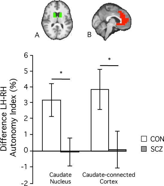
Results: The caudate nucleus and cortical regions with connections to the caudate nucleus had markedly abnormal hemispheric specialization in schizophrenia. Compared with healthy controls, patients exhibited weaker specialization in the left, but the opposite pattern in the right, caudate nucleus (P < .001). Patients with schizophrenia also had a disruption of the interhemispheric coordination among the cortical regions with connections to the caudate nucleus. A linear classifier based on the specialization of the caudate nucleus distinguished patients from controls with a classification accuracy of 74% (with a sensitivity of 68% and a specificity of 78%).
Conclusions and relevance: These data suggest that hemispheric specialization could serve as a potential imaging biomarker of schizophrenia that, compared with task-based functional magnetic resonance imaging measures, is less prone to the confounding effects of variation in task compliance, cognitive ability, and command of language.
Conflict of interest statement
The Authors declare no conflict of interests.
Figures
Similar articles
-
Interhemispheric connectivity and hemispheric specialization in schizophrenia patients and their unaffected siblings.Neuroimage Clin. 2019;21:101656. doi: 10.1016/j.nicl.2019.101656. Epub 2019 Jan 7. Neuroimage Clin. 2019. PMID: 30660663 Free PMC article.
-
Distinct hemispheric specialization of functional connectivity in schizophrenia with and without auditory verbal hallucinations.Neuroreport. 2019 Dec 18;30(18):1294-1298. doi: 10.1097/WNR.0000000000001364. Neuroreport. 2019. PMID: 31688422
-
Dopamine-Related Disruption of Functional Topography of Striatal Connections in Unmedicated Patients With Schizophrenia.JAMA Psychiatry. 2016 Aug 1;73(8):862-70. doi: 10.1001/jamapsychiatry.2016.0178. JAMA Psychiatry. 2016. PMID: 27145361 Free PMC article.
-
Intra- and Inter-hemispheric Connectivity Supporting Hemispheric Specialization.2016 Mar 11. In: Kennedy H, Van Essen DC, Christen Y, editors. Micro-, Meso- and Macro-Connectomics of the Brain [Internet]. Cham (CH): Springer; 2016. 2016 Mar 11. In: Kennedy H, Van Essen DC, Christen Y, editors. Micro-, Meso- and Macro-Connectomics of the Brain [Internet]. Cham (CH): Springer; 2016. PMID: 28590670 Free Books & Documents. Review.
-
Schizophrenia and the corpus callosum: developmental, structural and functional relationships.Behav Brain Res. 1994 Oct 20;64(1-2):203-11. doi: 10.1016/0166-4328(94)90132-5. Behav Brain Res. 1994. PMID: 7840887 Review.
Cited by
-
Brain functional specialization and cooperation in Parkinson's disease.Brain Imaging Behav. 2022 Apr;16(2):565-573. doi: 10.1007/s11682-021-00526-4. Epub 2021 Aug 24. Brain Imaging Behav. 2022. PMID: 34427879
-
Disrupted brain functional asymmetry at rest in patients with major depressive disorder associated with sleep disturbances.Brain Imaging Behav. 2024 Dec;18(6):1366-1375. doi: 10.1007/s11682-024-00924-4. Epub 2024 Sep 14. Brain Imaging Behav. 2024. PMID: 39276300
-
Resting-State Functional Connectivity Profile of Insular Subregions.Brain Sci. 2024 Jul 25;14(8):742. doi: 10.3390/brainsci14080742. Brain Sci. 2024. PMID: 39199437 Free PMC article.
-
Dynamic changes in brain lateralization correlate with human cognitive performance.PLoS Biol. 2022 Mar 17;20(3):e3001560. doi: 10.1371/journal.pbio.3001560. eCollection 2022 Mar. PLoS Biol. 2022. PMID: 35298460 Free PMC article.
-
Hemispheric lateralization abnormalities of the white matter microstructure in patients with schizophrenia and bipolar disorder.J Psychiatry Neurosci. 2017 Jun;42(4):242-251. doi: 10.1503/jpn.160090. J Psychiatry Neurosci. 2017. PMID: 28234211 Free PMC article.
References
-
- Toga AW, Thompson PM. Mapping brain asymmetry. Nat Rev Neurosci. 2003 Jan;4(1):37–48. - PubMed
-
- Gazzaniga MS. Cerebral specialization and interhemispheric communication. Brain. 2000 Jul 1;123(7):1293–1326. 2000. - PubMed
-
- Chi JG, Dooling EC, Gilles FH. Left-right asymmetries of the temporal speech areas of the human fetus. Arch Neurol. 1977 Jun;34(6):346–348. - PubMed
Publication types
MeSH terms
Grants and funding
LinkOut - more resources
Full Text Sources
Other Literature Sources
Medical
