Case Reports
. 2015;10(4):329-41.
doi: 10.1080/15592294.2015.1027853.

A novel Werner Syndrome mutation: pharmacological treatment by read-through of nonsense mutations and epigenetic therapies

Affiliations
Case Reports

A novel Werner Syndrome mutation: pharmacological treatment by read-through of nonsense mutations and epigenetic therapies

Ruben Agrelo et al. Epigenetics. 2015.

Abstract

Werner Syndrome (WS) is a rare inherited disease characterized by premature aging and increased propensity for cancer. Mutations in the WRN gene can be of several types, including nonsense mutations, leading to a truncated protein form. WRN is a RecQ family member with both helicase and exonuclease activities, and it participates in several cell metabolic pathways, including DNA replication, DNA repair, and telomere maintenance. Here, we reported a novel homozygous WS mutation (c.3767 C > G) in 2 Argentinian brothers, which resulted in a stop codon and a truncated protein (p.S1256X). We also observed increased WRN promoter methylation in the cells of patients and decreased messenger WRN RNA (WRN mRNA) expression. Finally, we showed that the read-through of nonsense mutation pharmacologic treatment with both aminoglycosides (AGs) and ataluren (PTC-124) in these cells restores full-length protein expression and WRN functionality.

Keywords: Epigenetics; PTC read-through therapy; Werner Syndrome; methylation; mutation.


Figures

Figure 1.
Figure 1.
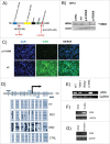
Novel WS mutation (A) The representation of the mutation localization shows the DNA sequence with codons in alternate colors as well as the protein sequence. (B) Sequencing results detecting the homozygous substitution c.3767 C > G in WRN in both patients (WS1 and WS2) and the same heterozygous mutation in the mother (HT). The two peaks in the chromatogram show the normal and mutated allele. No mutation was found in the wild type control (CTRL). (C) Photographs of the 2 patients showing the typical WS premature appearance. Their mother, who is heterozygous for the mutation, did not show the WS phenotype.
Figure 2 (See previous page).
WRN is not expressed in WS cells harboring the p.S1256X mutation: Methylation analysis of the WRN promoter in patients and controls. (A) The illustration shows the WRN protein domains, including the nuclear localization signal (NLS) and the positions of the p.S1256X and p.R369X (the most common mutation in Caucasians) mutations. The N-terminal epitope recognized by the 8H3 antibody and the C-terminal epitope recognized by the 4H12 antibody are shown. The 4H12 antibody only detects full-length WRN, whereas the 8H3 antibody recognizes both the full-length and truncated protein forms. (B) Western analysis of the WRN protein using the 4H12 antibody in the control (CTRL-WRN+/+), heterozygous (HT-WRN+/−), p.S1256X(WRN−/−), and p.R369X (WRN−/−) cell lines. Actin was used as a loading control. (C) Immunofluorescence using the 4H12 antibody (which only detects full-length WRN) in cell lines showing WRN expression and nuclear localization in HT (WRN+/+) and p.S1256X (WRN−/−) showing no WRN expression. Blue indicates DAPI staining (first panel), and green indicates WRN (second panel). The third panel includes the merged images. (D) Methylation analysis of the WRN promoter. A schematic description of the WRN CpG islands around the transcription start site is shown (long black arrow). CpG dinucleotides are represented as short vertical lines. Locations of bisulphite genomic sequencing PCR primers and methylation-specific PCR primers are indicated as white and gray arrows, respectively. The bisulphite genomic sequencing results are shown for 5 individual clones in B lymphocytes (HT, WS1 and WS2 for p.S1256X mutation and CTRL). (E) WRN mRNA expression levels for (HT, WS1 and WS2 for p.S1256X mutation and CTRL) in naïve B lymphocytes assessed by RT-PCR.GAPDH was used as a loading control. (F) Treatment with a demethylating agent (5-aza) reactivated WRN gene expression in the WS2 lymphoblastoid cell line. (G) Treatment with emetine increased WRN mRNA transcript levels in the WS2 lymphoblastoid cell line by inhibiting NMD.
Figure 3.
Figure 3.
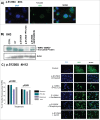
PTC read-through treatment in WS cells harboring the p.S1256X mutation restores the full-length WRN protein. (A) Immunofluorescence of WRN in cells harboring the p.S1256X mutation shows cytoplasmic localization of the truncated protein detected with the 8H3 antibody indicating that the protein is stable in the cytosol. (B) Western analysis of WRN protein with the 8H3 antibody, which recognizes the truncated forms of WRN, as follows: CTRL (WRN+/+), HT (WRN+/−), p.S1256X (WRN−/−), p.S1256X (WRN−/−) + PTC-124, and p.S1256X(WRN−/−) + gentamicin. In lane one (CTRL), only full-length WRN was detected. In the HT, both full-length WRN and the truncated protein forms were detected. In p.S1256X (WRN−/−), only the truncated form was detected. Upon treatment with PTC-124 or gentamicin, restoration of full-length WRN (upper band) was observed together with a reduction of truncated WRN (lower band). (C) Immunofluorescence using the 4H12 antibody in p.S1256X (WRN−/−) untreated cells (in which no WRN expression was detected) or p.S1256X (WRN−/−) cells treated with PTC-124, gentamicin, streptomycin, or G418 showing restored nuclear WRN expression. Plots of the percentage (%) of cells harboring the p.S1256X and p.R369X mutations and showing nuclear WRN expression after being treated with the 3 different AGs and PTC-124 by read-through therapy of PTC mutations are shown.
Figure 4 (See previous page).
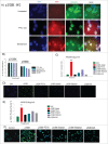
Restoration of nuclear localization and functionality of WRN as measured by induced apoptosis andγ-H2AX foci upon read-through treatment in WS cells harboring the pS1256X mutation. (A) After PTC-124 or AGs treatment of WS cells harboring the p.S1256X or p.R369X mutation, WRN was localized in the nucleolus, which is its main localization. Immunofluorescence shows the colocalization of WRN (red) and nucleolin (green) in p.S1256X(WRN−/−) cells treated with PTC-124 or gentamicin. (B) Plot representing the percentage (%) of WRN and nucleolin colocalization against treatment with PTC-124 and 3 different AGs in cells harboring the p.S1256X or p.R369Xmutation. (C) WS cells are sensitive to the 4NQO DNA-damaging agent. Decreased apoptosis was measured by the TUNEL assay in WS cells harboring the p.S1256X mutation and treated with PTC-124 or AGs. Upon treatment, the apoptosis levels decreased to l for some drugs, which was close to the levels in control cells (WRN+/+). Untreated WRN(−/−) was compared to WRN(+/+) and AGs or PTC-124 treated (WRN−/−). Error bars represent the standard deviation (n = 9); **P < 0.01. P-values were determined by Student's t-test. (D) The apoptotic cells are shown in green for the different conditions. (E) Statistical representation of γ-H2AX foci in WS cells harboring the p.S1256X mutation left untreated or treated with PTC-124 or AGs and control cells (WRN+/+ cells) upon 4NQO treatment. Error bars represent the standard deviation (n = 9); *P < 0.05. P-values were determined by Student's t-test. (F) Representative images showing γ-H2AX foci in WS cells left untreated or treated with PTC-124 or AGs.
Figure 5 (See previous page).
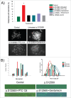
Chromosomal damage and impaired DNA replication are reduced by PTC read-through therapy in WS cells harboring the p.S1256X mutation. (A) Chromosomal breakage measured by cytogenetic analysis of metaphase chromosomes. Untreated WS cells harboring the p.S1256X mutation had increased fragility when compared to control (WRN +/+) cells. Fragility was decreased to the control cell level in mutant cells after cell treatment with PTC-124 or AGs. The metaphase spreads of control WS cells harboring the p.S1256X mutation left untreated or treated with PTC-124 and gentamicin are shown. Error bars represent the s.standard deviation (n = 9); *P < 0.05. P-values were determined by Student's t-test. (B) Analysis of DNA replication by fiber assays of control (WRN +/+) and WS cells harboring the p.S1256X mutation left untreated or treated with PTC-124 or gentamicin. The average fiber length for the control was 6–9 μm, and the average fiber length was 3–6 μm for the WS cells harboring the p.S1256X mutation. Upon WS cell treatment with PTC-124 or gentamicin, the average fiber was 6–9 μM, which was similar to the fiber length in the control. Two plots of one representative experiment out of 3 biological replicas are shown (n = 200 for each condition). The DNA fiber assays were performed with the control cells and WS cells harboring the p.S1256X mutation left untreated or treated with PTC-124 or gentamicin. BrdU-labeled fibers are stained in green.

References

    1. Epstein CJ, Martin GM, Schultz AL, Motulsky AG. Werner's syndrome a review of its symptomatology, natural history, pathologic features, genetics and relationship to the natural aging process. Medicine (Baltimore) 1966; 45:177-21; PMID:5327241 - PubMed
    1. Salk D. Werner's syndrome: a review of recent research with an analysis of connective tissue metabolism, growth control of cultured cells, and chromosomal aberrations. Hum Genet 1982; 62:1-5; PMID:6759366; http://dx.doi.org/10.1007/BF00295598 - DOI - PubMed
    1. Goto M. Hierarchical deterioration of body systems in Werner's syndrome: implications for normal ageing. Mech Ageing Dev 1997; 98:239-54; PMID:9352493 - PubMed
    1. Lauper JM, Krause A, Vaughan TL, Monnat RJ, Jr. Spectrum and risk of neoplasia in Werner syndrome: a systematic review. PLoS One 2013; 8(4):e59709; PMID:23573208; http://dx.doi.org/10.1371/journal.pone.0059709 - DOI - PMC - PubMed
    1. Goto M, Miller RW, Ishikawa Y, Sugano H. Excess of rare cancers in Werner syndrome (adult progeria).Cancer Epidemiol Biomarkers Prev 1996; 5:239-46; PMID:8722214 - PubMed

Publication types

LinkOut - more resources