. 2015 Apr 1;10(4):e0120798.
doi: 10.1371/journal.pone.0120798. eCollection 2015.

Characterization of phospho-(tyrosine)-mimetic calmodulin mutants

Affiliations

Characterization of phospho-(tyrosine)-mimetic calmodulin mutants

Silviya R Stateva et al. PLoS One. .

Abstract

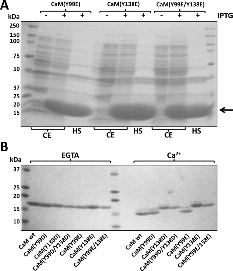
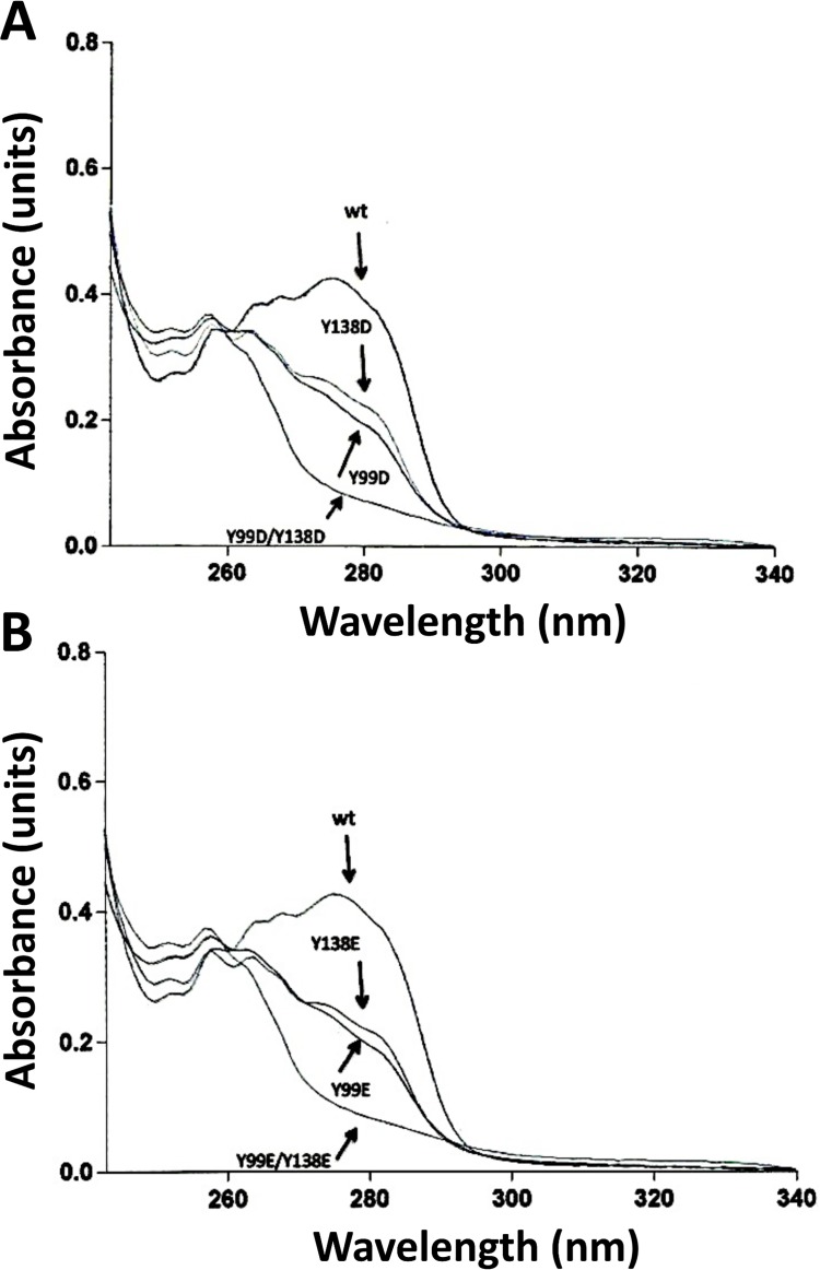
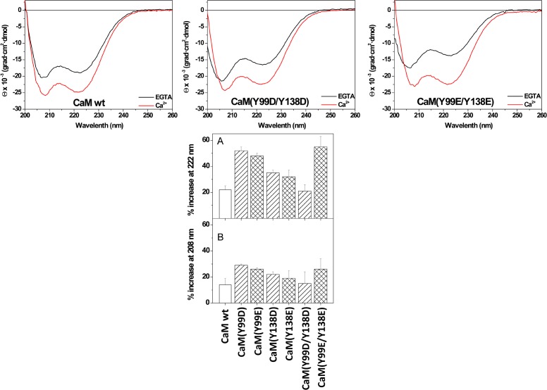
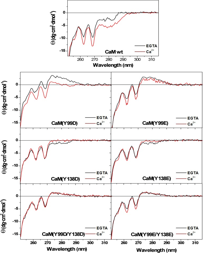
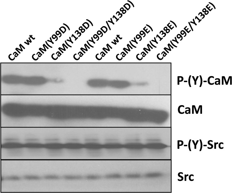
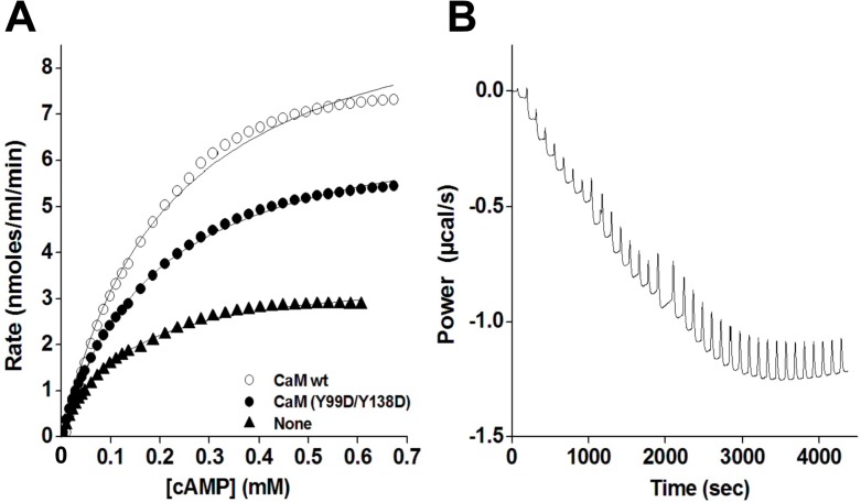
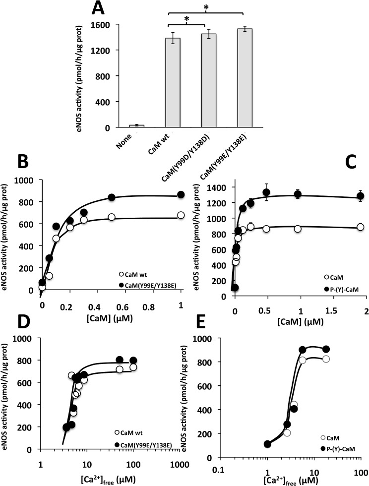
Calmodulin (CaM) phosphorylated at different serine/threonine and tyrosine residues is known to exert differential regulatory effects on a variety of CaM-binding enzymes as compared to non-phosphorylated CaM. In this report we describe the preparation and characterization of a series of phospho-(Y)-mimetic CaM mutants in which either one or the two tyrosine residues present in CaM (Y99 and Y138) were substituted to aspartic acid or glutamic acid. It was expected that the negative charge of the respective carboxyl group of these amino acids mimics the negative charge of phosphate and reproduce the effects that distinct phospho-(Y)-CaM species may have on target proteins. We describe some physicochemical properties of these CaM mutants as compared to wild type CaM, after their expression in Escherichia coli and purification to homogeneity, including: i) changes in their electrophoretic mobility in the absence and presence of Ca2+; ii) ultraviolet (UV) light absorption spectra, far- and near-UV circular dichroism data; iii) thermal stability in the absence and presence of Ca2+; and iv) Tb3+-emitted fluorescence upon tyrosine excitation. We also describe some biochemical properties of these CaM mutants, such as their differential phosphorylation by the tyrosine kinase c-Src, and their action as compared to wild type CaM, on the activity of two CaM-dependent enzymes: cyclic nucleotide phosphodiesterase 1 (PDE1) and endothelial nitric oxide synthase (eNOS) assayed in vitro.


Conflict of interest statement

Competing Interests: The authors have declared that no competing interests exist.

Figures

Fig 1
Fig 1. Expression and purification of the different CaM species.
(A) The figure shows the pattern of proteins of the bacterial cell extract (CE) and the corresponding heated supernatant (HS) from E. coli BL21(DE3)pLysS transformed with plasmids encoding CaM(Y99E), CaM(Y138E) and CaM(Y99E/Y138E) in the absence (-) and presence (+) of 0.5 mM IPTG for 4 h as described in Materials and Methods. The arrow points to the band of CaM induced by IPTG. (B) The different recombinant CaM species (≈ 1–2 μg) purified as described in Materials and Methods were separated by SDS-PAGE in the presence of 5 mM EGTA or 1 mM CaCl2 (Ca 2+) in the loading buffer, to observe the Ca2+-induced mobility shift.
Fig 2
Fig 2. Absorption spectra of the different CaM species.
The plots show UV-light absorption spectra of recombinant wild type (wt) and the indicated Y/D (panel A) and Y/E (panel B) CaM mutants (2 mg/ml) purified as described in Materials and Methods and dialyzed against 20 mM Tris-HCl (pH 7.5).
Fig 3
Fig 3. Ca2+-induced changes in the far-UV circular dichroism spectra of the different CaM species.
(Top panels) Far-UV CD spectra for wild type (wt) and the double Y/D and Y/E CaM mutants (118 μM) were recorded at 20°C in 20 mM Tris-HCl (pH 7.5), 0.1 M KCl, containing 1 mM EGTA (black lines) or 1 mM CaCl2 (red lines). (Bottom panels) The mean ± SEM (n = 3) increase in ellipticity in the far-UV circular dichroism signals at 222 nm (A) and 208 nm (B) of the indicated CaM species is represented as percentage taking as 100% the ellipticity of each CaM species in the presence of EGTA. Measurements were carried out at 20°C in 20 mM Tris-HCl (pH 7.5), 0.1 M KCl, containing 1 mM EGTA or 1 mM CaCl2, respectively. The concentration of CaM was 12 μM (A) and 118 μM (B).
Fig 4
Fig 4. Thermal stability of the different CaM species in the absence and presence of Ca2+.
The plots present the decrease in the CD signal at 222 nm, expressed as percentage of the ellipticity at 20°C, for wild type (wt) and the single and double Y/D and Y/E CaM mutants (12 μM) in 20 mM Tris-HCl (pH 7.5), 0.1 M KCl, in the presence of 1 mM EGTA (solid lines) and 1 mM CaCl2 (dashed lines) as described in Materials and Methods.
Fig 5
Fig 5. Ca2+-induced changes in the near-UV circular dichroism spectra of the different CaM species.
Near-UV CD spectra for wild type (wt) and the single and double Y/D and Y/E CaM mutants (118 μM) were recorded at 20°C in 20 mM Tris-HCl (pH 7.5), 0.1 M KCl, containing 1 mM EGTA (black lines) or 1 mM CaCl2 (red lines). Ellipticity values were normalized per mole of residue.
Fig 6
Fig 6. Tb3+-induced fluorescence emission spectra of the different CaM species.
(A) The fluorescence emission spectra (520–580 nm) of wild type (wt) CaM and the indicated CaM mutants (10 μM) was recorded in a buffer containing 10 mM Pipes-HCl (pH 6.5) and 100 mM KCl in the absence and presence of increasing concentrations of TbCl3 (10–200 μM) as described in Materials and Methods. (B, C) The plots present the emission fluorescence at 543 nm of wild type (wt) CaM and the indicated CaM mutants (10 μM) at increasing concentrations of TbCl3 in the conditions described in A.
Fig 7
Fig 7. Phosphorylation of different CaM species by c-Src.
The different CaM species (2 μg) were assayed for phosphorylation by recombinant c-Src as described in Materials and Methods. The samples were probed with an anti-phospho-tyrosine antibody to detect the tyrosine-phosphorylated CaM species (P-(Y)-CaM) and auto-phosphorylated c-Src (P-(Y)-Src). The membranes were striped and reprobed with anti-CaM and anti-Src antibodies as loading controls.
Fig 8
Fig 8. Effect of different CaM species on the activity of PDE1.
(A) The cyclic nucleotide PDE1 activity was assayed in the absence (None) and presence of the indicated CaM species (1.9 μM) in the presence of 100 μM free Ca2+ as described in Materials and Methods. The plot presents the average ± SEM of triplicate samples from three separate experiments (** p = 0.02 using the Student’s t test). (B) The plots present the PDE1 activity assayed as in A but using increasing concentrations of the indicated CaM species. (C) The plot presents the average ± SEM activity of PDE1 of three independent experiments assayed at increasing concentrations of non-phosphorylated CaM (open symbols) or phospho-(Y)-CaM (filled symbols) prepared as described in Materials and Methods. (D) The plot presents the PDE1 activity assayed as in A in the presence of the indicated CaM species (1.9 μM) at increasing concentrations of free Ca2+ using an EGTA/Ca2+ buffer as described in Materials and Methods. (E) The plot presents the average ± range activity of PDE1 assayed in the presence of non-phosphorylated CaM (open symbols) and phospho-(Y)-CaM (filled symbols) (0.97 μM) of two independent experiments assayed at increasing concentrations of free Ca2+ using an EGTA/Ca2+ buffer as described in Materials and Methods.
Fig 9
Fig 9. Effect of different CaM species on the kinetics of PDE1 determined by isothermal titration calorimetry.
(A) The plot presents the PDE1 activity (normalized for 0.04 units of enzyme) at increasing concentrations of cAMP in the absence (filled triangles) and presence of wild type CaM (open circles) or CaM(Y99D/Y138D) (filled circles) (3.8 μM) in a buffer containing 50 mM imidazol-HCl (pH 7.5), 200 mM NaCl, 5 mM MgCl2, 0.4 mM EGTA, and 0.5 mM CaCl2 using ITC as described in Materials and Methods setting the micro-calorimeter chamber 37°C. (B) The trace corresponds to a typical experiment showing the rate of heat produced by PDE1 over time after injections of different pulses of cAMP from which the activity of the enzyme can be derived as described in Materials and Methods. This particular trace corresponds to an experiment performed in the absence of CaM using 0.04 units of PDE1. Similar traces were obtained in other conditions tested.
Fig 10
Fig 10. Effect of different CaM species on the activity of eNOS.
(A) The activity of eNOS was assayed in the absence (None) and presence of the indicated CaM species (4.7 μM) in the presence of 1 mM free Ca2+ as described in Materials and Methods. The plot presents the average ± SEM of triplicate samples from three separate experiments (* p ≤ 0.05 using the Student’s t test). (B) The plot presents the eNOS activity assayed as in A but using increasing concentrations of the indicated CaM species. (C) The plot presents the average ± range activity of eNOS of two independent experiments assayed at increasing concentrations of non-phosphorylated CaM (open symbols) or phospho-(Y)-CaM (filled symbols) prepared as described in Materials and Methods. (D) The plot presents the eNOS activity assayed as in A in the presence of the indicated CaM species (4.7 μM) at increasing concentrations of free Ca2+ using an EGTA/Ca2+ buffer as described in Materials and Methods. (E) The plot presents the activity of eNOS assayed as in A in the presence of non-phosphorylated CaM (open symbol) and phospho-(Y)-CaM (filled symbols) (0.6 μM) assayed at increasing concentrations of free Ca2+ using an EGTA/Ca2+ buffer as described in Materials and Methods.

Similar articles

Cited by

References

    1. Chin D, Means AR. Calmodulin: a prototypical calcium sensor. Trends Cell Biol. 2000;10: 322–328. - PubMed
    1. Berchtold MW, Villalobo A. The many faces of calmodulin in cell proliferation, programmed cell death, autophagy, and cancer. Biochim Biophys Acta. 2014;1843: 398–435. 10.1016/j.bbamcr.2013.10.021 - DOI - PubMed
    1. Crotti L, Johnson CN, Graf E, De Ferrari GM, Cuneo BF, Ovadia M, et al. Calmodulin mutations associated with recurrent cardiac arrest in infants. Circulation. 2013;127: 1009–1017. 10.1161/CIRCULATIONAHA.112.001216 - DOI - PMC - PubMed
    1. Yin G, Hassan F, Haroun AR, Murphy LL, Crotti L, Schwartz PJ, et al. Arrhythmogenic calmodulin mutations disrupt intracellular cardiomyocyte Ca2+ regulation by distinct mechanisms J Am Heart Assoc. 2014;3: e000996 10.1161/JAHA.114.000996 - DOI - PMC - PubMed
    1. Ten Broeke R, Blalock JE, Nijkamp FP, Folkerts G. Calcium sensors as new therapeutic targets for asthma and chronic obstructive pulmonary disease. Clin Exp Allergy. 2004;34: 170–176. - PubMed

Publication types

Substances