Review
. 2015 Apr-Jun;28(2):160-172.

Current endoscopic ultrasound-guided approach to incidental subepithelial lesions: optimal or optional?

Affiliations
Review

Current endoscopic ultrasound-guided approach to incidental subepithelial lesions: optimal or optional?

Alexander J Eckardt et al. Ann Gastroenterol. 2015 Apr-Jun.

Abstract

Subepithelial lesions (SEL) are identified during endoscopic procedures on a regular basis. They can occur anywhere in the gastrointestinal (GI) tract and are located beneath the normal epithelial layer, which explains why a tissue diagnosis is difficult to obtain with routine biopsies. Endoscopic ultrasound (EUS) is used to further characterize these lesions. EUS can distinguish intramural lesion from extramural compression. Furthermore, it allows allocation of intramural lesions to a specific layer of the GI wall and offers additional information as to whether a lesion could be benign or malignant. EUS also assists in choosing the optimal means of tissue acquisition. The choice of tissue acquisition is based on a number of factors, such as tumor size, EUS features, and location within the GI tract or within a specific layer of the GI wall. Furthermore, local expertise and patient factors should be considered when deciding whether tissue acquisition, surgical intervention or follow up is recommended. In this review we offer an EUS-guided approach to the evaluation of incidental SEL based on current evidence and point out areas of uncertainty, which explain why the proposed algorithmic approach may be optional rather than optimal.

Keywords: Endoscopic ultrasonography; fine needle aspiration; gastrointestinal stromal tumor; subepithelial lesions.


Conflict of interest statement

Conflict of Interest: None

Figures
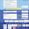
Figure 1
Figure 1
Stepwise algorithm for the work-up of an incidental upper gastrointestinal (GI) subepithelial lesion (SEL) The diagnostic algorithm is explained in the manuscript. It applies mainly for upper GI-SEL. Exceptions, such as rectal lesions, small bowel lesions, suspected leiomyoma in the esophagus and suspected neuroendocrine tumors must be considered. Broken lines depict alternative options
Figure 2
Figure 2
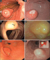
Endoscopic features of selected subepithelial lesions. (A) Gastric gastrointestinal stromal tumor with a central ulceration. (B) Inflammatory fibroid polyp with ulceration. (C) Lipoma in the transverse colon with a positive pillow sign after indentation with the closed biopsy forceps (arrow). (D) Lymphangioma with translucent appearance, the septated lesion appears lobulated on macroscopic inspection. (E) Pancreatic rest in the antrum with umbilication. (F) Granular cell tumor. A yellowish hue becomes particularly visible after superficial biopsy (small picture)
Figure 3
Figure 3
Endosonographic features of selected subepithelial lesions. (A) Hyperechoic gastric lipoma, arising from the 3rd endoscopic ultrasound layer (by courtesy of Prof. Dr. Uwe Will, Gera). (B) Small gastric leiomyoma with calcifications (atypical), arising from 4th hypoechoic layer. (C) Small gastric gastrointestinal stromal tumor (GIST), arising from 4th hypoechoic layer (arrowhead). (D) Large gastric GIST with distinct heterogeneity and small anechoic cavities (arrow heads), highly vascularized (contrast-enhanced power Doppler), arising from 4th hypoechoic layer. (E) Rare case of esophageal leiomyomatosis. The hypoechoic tumor nodules arise from the 4th hypoechoic layer. (F) Large esophageal leiomyoma arising from 4th hypoechoic layer
Figure 4
Figure 4
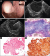
Endoscopic ultrasound (EUS)-guided fine-needle aspiration of a gastric subepithelial tumor. (A) Endoscopic image of an incidental medium-sized subepithelial lesion (SEL) arising from the gastric cardia. (B) EUS reveals a 2 cm, hypoechoic, homogeneous, lobulated SEL with distinct margins, arising from the 4th EUS layer (M. propria, arrow heads). Leiomyoma is suspected. (C) EUS fine needle aspiration using a 22G aspiration needle. (D) Smear cytology (May-Grünwald-Giemsa, x200) and (E) Histology of core particles (Hematoxylin-Eosin, x200) show fascicles of spindle-shaped tumor cells, suggestive of a mesenchymal tumor. (F) Immunohistochemistry (Desmin, x 200: strongly positive; CD117: negative, not shown here) confirms benign leiomyoma. Surgery is not necessary (Photomicrographs D-F by courtesy of Dr. Stephan Wagner, Königs Wusterhausen, Germany)

References

    1. Hedenbro JL, Ekelund M, Wetterberg P. Endoscopic diagnosis of submucosal gastric lesions. The results after routine endoscopy. Surg Endosc. 1991;5:20–23. - PubMed
    1. Jenssen C, Dietrich CF. Endoscopic ultrasound of gastrointestinal subepithelial lesions. Ultraschall Med. 2008;29:236–256. - PubMed
    1. Polkowski M. Endoscopic ultrasound and endoscopic ultrasound-guided fine-needle biopsy for the diagnosis of malignant submucosal tumors. Endoscopy. 2005;37:635–645. - PubMed
    1. Menon L, Buscaglia JM. Endoscopic approach to subepithelial lesions. Therap Adv Gastroenterol. 2014;7:123–130. - PMC - PubMed
    1. Eckardt AJ, Wassef W. Diagnosis of subepithelial tumors in the GI tract. Endoscopy, EUS, and histology: bronze, silver, and gold standard? Gastrointest Endosc. 2005;62:209–212. - PubMed

LinkOut - more resources