. 2015 Apr 7;112(14):E1744-53.
doi: 10.1073/pnas.1406551112. Epub 2015 Mar 23.

T-cell receptor α enhancer is inactivated in αβ T lymphocytes

Affiliations

T-cell receptor α enhancer is inactivated in αβ T lymphocytes

Beatriz del Blanco et al. Proc Natl Acad Sci U S A. .

Abstract

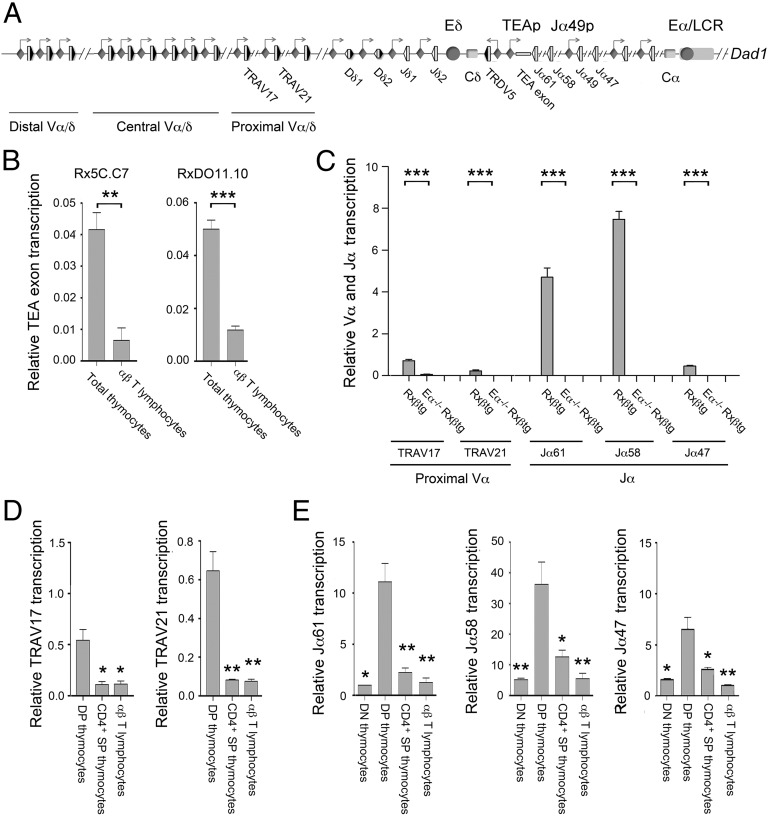
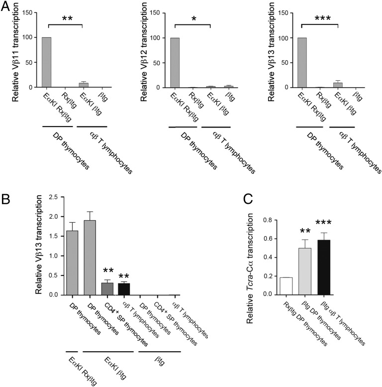
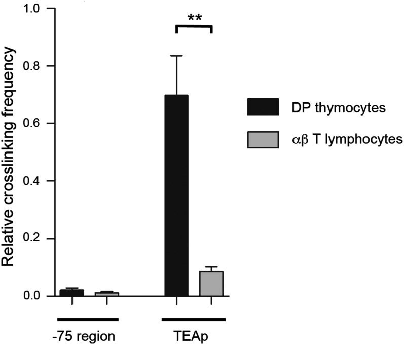
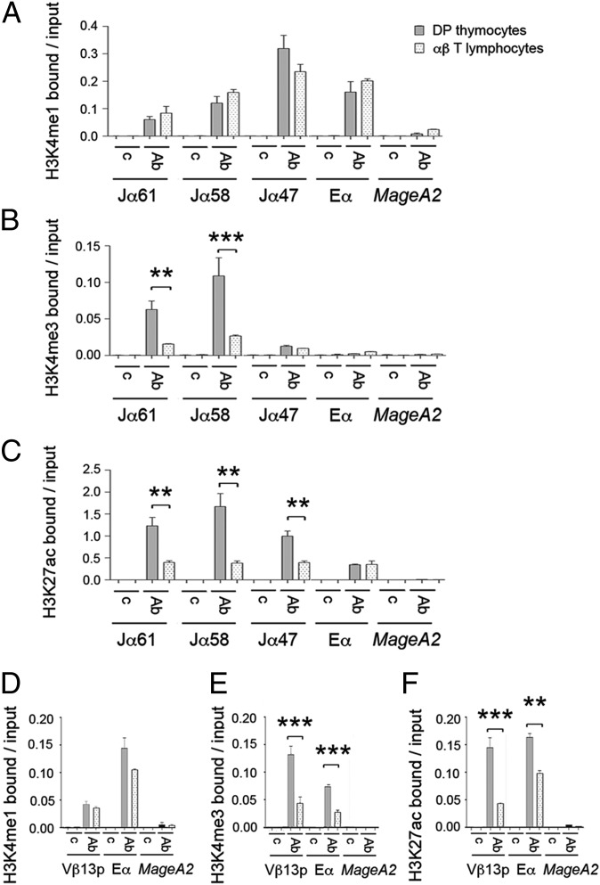
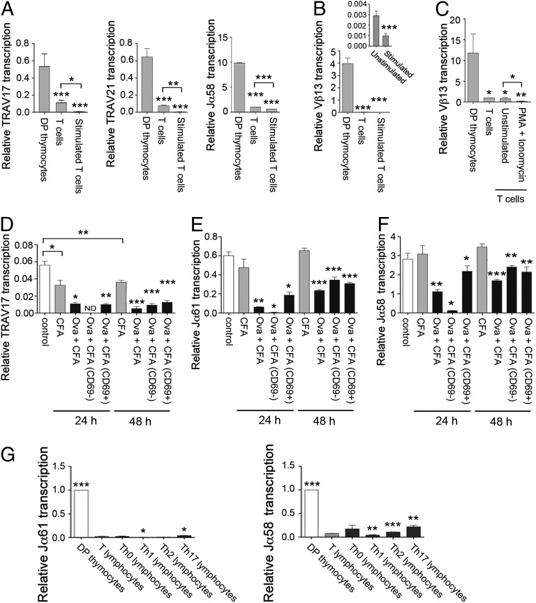
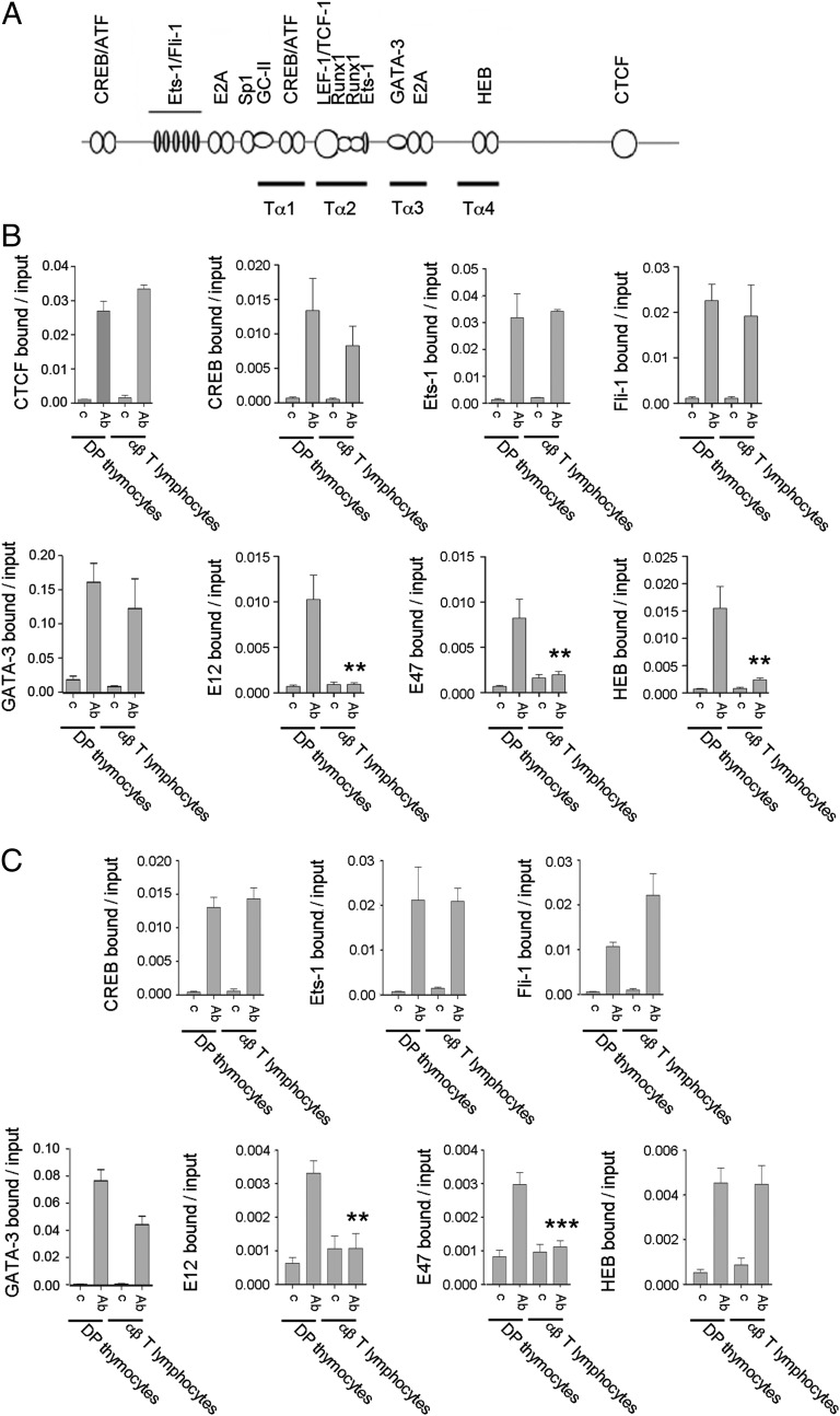
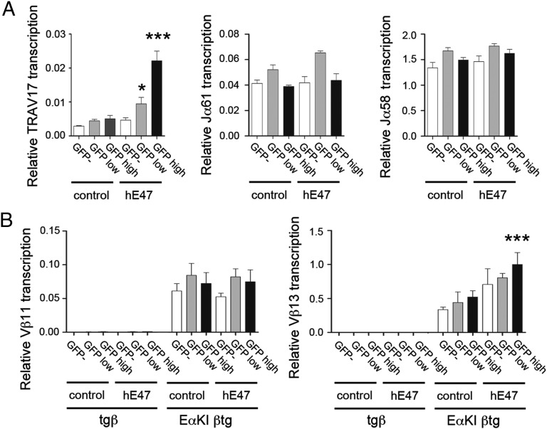
The Tcra enhancer (Eα) is essential for Tcra locus germ-line transcription and primary Vα-to-Jα recombination during thymocyte development. We found that Eα is inhibited late during thymocyte differentiation and in αβ T lymphocytes, indicating that it is not required to drive transcription of rearranged Tcra genes. Eα inactivation resulted in the disruption of functional long-range enhancer-promoter interactions and was associated with loss of Eα-dependent histone modifications at promoter and enhancer regions, and reduced expression and recruitment of E2A to the Eα enhanceosome in T cells. Enhancer activity could not be recovered by T-cell activation, by forced expression of E2A or by the up-regulation of this and other transcription factors in the context of T helper differentiation. Our results argue that the major function of Eα is to coordinate the formation of a chromatin hub that drives Vα and Jα germ-line transcription and primary rearrangements in thymocytes and imply the existence of an Eα-independent mechanism to activate transcription of the rearranged Tcra locus in αβ T cells.

Keywords: T-cell development; T-cell receptor; enhancer; transcription.


Conflict of interest statement

The authors declare no conflict of interest.

Figures

Fig. 1.
Fig. 1.
Tcra Eα-dependent transcription is inactivated in SP thymocytes and αβ T lymphocytes. (A) Schematic representation of Tcra/Tcrd genomic structure. The V, D, and J gene segments are represented by gray vertical rectangles, and the recombination signal sequences are shown as black (containing a 23-bp spacer) or white (containing a 12-bp spacer) triangles. Positions of the TEA exon, the Vα/δ, Dδ, Jδ, and Jα gene segments, and the Cδ and Cα regions are indicated. Arrows represent active sites for germ-line transcription. Promoters and enhancers are represented as diamonds and circles, respectively. The Tcra LCR is represented as a long horizontal rectangle. (B) Analysis of TEA exon transcription in Rx5C.C7 and RxDO11.10 thymocytes and T lymphocytes by RT-qPCR. (C) Analysis of proximal Vα and Jα transcription in DP thymocytes from Rxßtg and Eα−/− Rxßtg mice by RT-qPCR. (D) Analysis of TRAV17 and TRAV21 germ-line transcripts in RxDO11.10 sorted thymocyte populations and T lymphocytes by RT-qPCR. (E) Analysis of Jα transcription in RxDO11.10 sorted thymocyte populations and T lymphocytes by RT-qPCR. The results were normalized to those for Actb and represent the mean ± SEM of duplicate RT-qPCR from three independent experiments. Two-tailed Student’s t tests were used to determine the statistical significance between the values of the RT-qPCRs from total thymocytes vs. T lymphocytes (B), Rxßtg vs. Eα−/− Rxßtg thymocytes (C), and DP thymocytes vs. T lymphocytes (D and E) (*P < 0.05, **P < 0.005, and *** P < 0.0005).
Fig. 2.
Fig. 2.
Eαi is inactivated in SP thymocytes and αβ T lymphocytes. (A) Analysis of Vβ11, Vβ12, and Vβ13 transcription in EαKI Rxßtg and Rxßtg thymocytes and EαKI βtg and βtg T lymphocytes by RT-qPCR. (B) Analysis of Vβ13 transcription from EαKI Rxßtg DP thymocytes, EαKI βtg and βtg sorted thymocyte populations, and EαKI βtg and βtg T lymphocytes by RT-qPCR. (C) Analysis of Tcra-Cα transcription in Rxßtg DP thymocytes, sorted βtg DP thymocytes, and sorted βtg T lymphocytes by RT-qPCR. The results were normalized to those for Actb and represent the mean ± SEM of duplicate RT-qPCR from three independent experiments. Two-tailed Student’s t tests were used to determine the statistical significance between the values of the RT-qPCRs from EαKI Rxßtg or EαKI βtg DP thymocytes vs. EαKI βtg CD4+ SP thymocytes and T lymphocytes (A and B) or Rxßtg DP thymocytes vs. βtg DP thymocytes or αβ T lymphocytes (C) (*P < 0.05, **P < 0.005, and ***P < 0.0005).
Fig. 3.
Fig. 3.
The Eα-TEAp long-range interactions present in DP thymocytes are lost in αβ T lymphocytes. 3C analysis of physical contacts between Eα and TEAp or a genomic fragment located at −75 kb from the enhancer in DP thymocytes (black bars) and αβ T lymphocytes (gray bars) from RxDO11.10. Data were normalized to the ligation frequency between Eα and a neighboring HindIII fragment (Fig. S2 and Table S1). HindIII digestion efficiency and the relative interactions between Eα and the control fragment in DP thymocytes and T lymphocytes are shown in Fig. S2. The results represent the mean ± SEM of duplicate qPCR from three independent experiments. Two-tailed Student’s t tests were used to determine the statistical significance between the values of the qPCRs obtained after ligation of HindIII fragments from DP thymocytes vs. T lymphocytes (**P < 0.005).
Fig. 4.
Fig. 4.
Comparative histone H3 modification analyses in DP thymocytes and αβ T lymphocytes. Analysis of Tcra Jα and Eα histone H3K4me1 (A), H3K4me3 (B), and H3K27ac (C) modifications in DP thymocytes and T lymphocytes from RxDO11.10 mice by qChIP. Analysis of Vβ13p and Eαi histone H3K4me1 (D), H3K4me3 (E), and H3K27ac (F) modifications in DP thymocytes from EαKI Rxßtg mice and T lymphocytes from EαKI βtg by qChIp. MageA2, negative control (49). Data represent the mean ± SEM of duplicate qPCR analysis of three independent experiments. Two-tailed Student’s t test were used to determine the statistical significance between the values of the qChIPs from DP thymocytes vs. T lymphocytes (**P < 0.005 and ***P < 0.0005).
Fig. 5.
Fig. 5.
Eα and Eαi are not activated by TCR-mediated cell stimulation and Th differentiation in T lymphocytes. (A) Analysis of TRAV17, TRAV21, and Jα58 germ-line transcripts in sorted DP thymocytes and unstimulated and stimulated T lymphocytes from RxDO11.10 mice by RT-qPCR. (B) Analysis of Vβ13 transcription in EαKI Rxßtg thymocytes and unstimulated and stimulated EαKI βtg T lymphocytes by RT-qPCR. (C) Analysis of Vβ13 transcription in EαKI Rxßtg DP thymocytes, EαKI βtg T lymphocytes, and EαKI tgβ T lymphocytes left in complete medium (unstimulated) or incubated with PMA and ionomycin (PMA + Ionomycin) by RT-qPCR. Analysis of TRAV17 (D), Jα61 (E), and Jα58 (F) transcripts present in unstimulated and in vivo stimulated RxDO11.10 T lymphocytes by RT-qPCR. Indicated transcripts were compared in T lymphocytes from noninjected mice (control), CFA-injected mice (CFA), or Ova 323–339 + CFA-injected mice (Ova + CFA). Indicated transcripts were compared in sorted CD69 and CD69+ T populations from Ova 323–339 + CFA-immunized mice. (G) Analysis of Gata3, Tcfe2a, Jα61, and Jα58 transcripts in sorted DP thymocytes, T lymphocytes, and Th0, Th1, Th2, and Th17 populations from RxDO11.10 mice by RT-qPCR. The results were normalized to those for Actb and represent the mean ± SEM of duplicate RT-qPCR from three independent experiments. Two-tailed Student’s t tests were used to determine the statistical significance between the values of the RT-qPCRs from DP thymocytes vs. T lymphocytes (A-C), from unstimulated vs. stimulated T lymphocytes (AF), and from T lymphocytes vs. DP thymocytes or Th populations (G) (*P < 0.05, **P < 0.005, and ***P < 0.0005).
Fig. 6.
Fig. 6.
E2A is not present at the Eα enhanceosome assembled in αβ T lymphocytes. (A) Diagram depicts the location of the Tα1-Tα4 regions as well as the described TF binding sites. (B and C) For CTCF q ChIPs, chromatin was prepared from sorted DP and T lymphocytes from RxDO11.10 mice. For all other TF ChIPs, DP thymocyte chromatin was prepared from EαKI Rxßtg thymocytes and T lymphocyte chromatin was prepared from EαKI βtg T lymphocytes. Immunoprecipitation was performed with the indicated control (c) or specific Abs (Ab). DNA purified from the input and Ab-bound fractions was used as a template for qPCR to evaluate the presence of Eα (B) and Eαi (C). Data represent the mean ± SEM of duplicate qPCR from three independent experiments. Two-tailed Student’s t tests were used to determine the statistical significance between the values of the qChIPs from DP thymocytes vs. T lymphocytes (*P < 0.05, **P < 0.005, and ***P < 0.0005).
Fig. 7.
Fig. 7.
E47 can partially rescue specific Eα-dependent transcription in αβ T lymphocytes. (A) Forced expression of hE47 by retroviral transduction of T lymphocytes from RxDO11.10 mice induces TRAV17 transcription but not Jα61 and Jα58 transcription. (B) Forced expression of hE47 by retroviral transduction of T lymphocytes from EαKI βtg mice induces Vβ13 but not Vβ11 gene segment transcription. GFP, GFP-low, and GFP-high–transduced populations with MigR (control) or MigR-hE47 (hE47) were sorted, and the indicated transcripts were quantified by RT-qPCR. The results were normalized to those for Actb and represent the mean ± SEM of duplicate RT-qPCR from three independent experiments. Two-tailed Student’s t tests were used to determine the statistical significance between the RT-qPCR values from MigR- and MigR-hE47-transduced T lymphocytes by comparing MigR-control GFP cells with MigR-hE47 GFP cells, MigR-control GFP-low cells with MigR-hE47 GFP-low cells, and MigR-control GFP-high cells with MigR-hE47 GFP-high cells (*P < 0.05 and ***P < 0.0005).

Similar articles

Cited by

References

    1. Cobb RM, Oestreich KJ, Osipovich OA, Oltz EM. Accessibility control of V(D)J recombination. Adv Immunol. 2006;91:45–109. - PubMed
    1. Rothenberg EV, Taghon T. Molecular genetics of T cell development. Annu Rev Immunol. 2005;23:601–649. - PubMed
    1. Taghon T, Yui MA, Pant R, Diamond RA, Rothenberg EV. Developmental and molecular characterization of emerging β- and γδ-selected pre-T cells in the adult mouse thymus. Immunity. 2006;24(1):53–64. - PubMed
    1. Ciofani M, et al. Obligatory role for cooperative signaling by pre-TCR and Notch during thymocyte differentiation. J Immunol. 2004;172(9):5230–5239. - PubMed
    1. Sleckman BP, Bardon CG, Ferrini R, Davidson L, Alt FW. Function of the TCR α enhancer in αβ and γδ T cells. Immunity. 1997;7(4):505–515. - PubMed

Publication types

MeSH terms

LinkOut - more resources