. 2015 Apr 7;112(14):E1792-9.
doi: 10.1073/pnas.1503941112. Epub 2015 Mar 23.

Structure of the paramyxovirus parainfluenza virus 5 nucleoprotein-RNA complex

Affiliations

Structure of the paramyxovirus parainfluenza virus 5 nucleoprotein-RNA complex

Maher Alayyoubi et al. Proc Natl Acad Sci U S A. .

Abstract

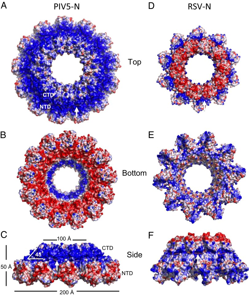
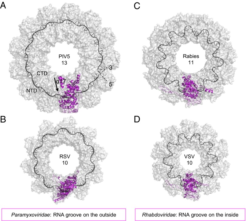
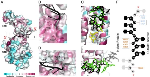
Parainfluenza virus 5 (PIV5) is a member of the Paramyxoviridae family of membrane-enveloped viruses with a negative-sense RNA genome that is packaged and protected by long filamentous nucleocapsid-helix structures (RNPs). These RNPs, consisting of ∼2,600 protomers of nucleocapsid (N) protein, form the template for viral transcription and replication. We have determined the 3D X-ray crystal structure of the nucleoprotein (N)-RNA complex from PIV5 to 3.11-Å resolution. The structure reveals a 13-mer nucleocapsid ring whose diameter, cavity, and pitch/height dimensions agree with EM data from early studies on the Paramyxovirinae subfamily of native RNPs, indicating that it closely represents one-turn in the building block of the RNP helices. The PIV5-N nucleocapsid ring encapsidates a nuclease resistant 78-nt RNA strand in its positively charged groove formed between the N-terminal (NTD) and C-terminal (CTD) domains of its successive N protomers. Six nucleotides precisely are associated with each N protomer, with alternating three-base-in three-base-out conformation. The binding of six nucleotides per protomer is consistent with the "rule of six" that governs the genome packaging of the Paramyxovirinae subfamily of viruses. PIV5-N protomer subdomains are very similar in structure to the previously solved Nipah-N structure, but with a difference in the angle between NTD/CTD at the RNA hinge region. Based on the Nipah-N structure we modeled a PIV5-N open conformation in which the CTD rotates away from the RNA strand into the inner spacious nucleocapsid-ring cavity. This rotation would expose the RNA for the viral polymerase activity without major disruption of the nucleocapsid structure.

Keywords: atomic structure; nucleocapsid ring; nucleoprotein; paramyxovirus; ribonucleoprotein.


Conflict of interest statement

The authors declare no conflict of interest.

Figures

Fig. 1.
Fig. 1.
(A) Western blot of purified PIV5-N after proteolysis. Lane 1, molecular weight ladder. Lane 2, 2 μg of PIV5-N. (B) Electron microscopy of the purified PIV5-N protein shown in A. Top left inset magnifies one of the observed ring structures. (Scale bar, 50 nm for big box and 10 nm for Inset).
Fig. 2.
Fig. 2.
(A) Cartoon representation of PIV5-N nucleocapsid ring 13-mer structure (top view) with chains colored differently and the RNA strand shown in black. (B) Side view (from outside of ring) of PIV5-N monomer with domains colored accordingly: Narm (3–31) in dark blue, N-terminal domain (NTD) (32–263) in light blue, C-terminal domain (CTD) (264–372) in salmon, and Carm (373–401) in red. The bound RNA is shown in black. (C) RSV-N monomer structure with RNA (black) bound in the RNA binding pocket. (D) Nipah-N monomer structure missing both Narm and Carm. (E) View of the protomer–protomer interface from inside of ring, showing the interaction of the Narm and Carm with neighboring protomers N − 1 and N + 1. The CTD α11–η2–α12 hydrophobic pocket is labeled in blue font.
Fig. 3.
Fig. 3.
Molecular surface representation of PIV5-N (AC) and RSV-N (DF) nucleocapsid rings colored by the electrostatic potential, ranging from −10 kT/e (red) to +10 kT/e (blue). RNA strand (salmon) is indicated by white arrow for clarity. PIV5-N charge distribution is more polarized than RSV-N with positive charges on the outside-top of the ring and negative charges on the inside-bottom of the ring.
Fig. 4.
Fig. 4.
Surface representation (gray) of the nucleocapsid ring structures from different negative-strand RNA viruses. Paramyxoviridae virus nucleocapsid like PIV5-N 13-mer (A) and RSV 10-mer (B) with RNA (black cartoon) bound from the outside of the ring. Rhabdoviridae viruses like rabies 11-mer (C) and VSV 10-mer (D) with the RNA bound on the inside of the ring. One N-protomer in each ring is displayed as cartoon (magneta). Arrow in A indicates direction of helix α17.
Fig. 5.
Fig. 5.
Conserved RNA binding pocket. (A) Consurf figure mapping amino acid conservation on PIV5-N structure based on alignment in Fig. S1 with conservation color scale shown at bottom. Each monomer binds to six nucleotides (black) and extends into the binding pocket of neighboring N molecules (gray). Two major binding pockets. (B) Close-up view of pocket 1: constitutes of conserved residues from NTD and binds the bases facing inward. (C) Close-up view of pocket 1: superposing PIV5-N RNA (black) and RSV-N RNA (green). Nucleotides from neighboring protomers are shown in gray (PIV5-N) and yellow (RSV-N). RNA is shown in stick representation. (D) Close-up view of pocket 2: constitutes of conserved residues from CTD and binds bases facing outward. (E) Same as C but for pocket 2. (F) Schematic representation figure showing the interaction of PIV5-N residues from pocket 1 (colored in light blue) and pocket 2 (colored in salmon) with RNA nucleotides (same color scheme as in A). The RNA is shown in 5′–3′ direction from bottom-to-top. Circles represent phosphates, pentagons represent the ribose sugars, hexagons represent the bases.
Fig. 6.
Fig. 6.
Open vs. closed conformation. (A) Superposition of PIV5-N (gray) CTD (Left) and NTD (Right) domains with the corresponding domains from Nipah-N (blue, Upper) and RSV-N (green, Lower). (B) Superposing full-length PIV5-N (gray) and Nipah-N (blue) at their NTD domains shows their respective CTDs at a 20° angle. The (arrow) indicates PIV5-N flexible hinge region between NTD and CTD. (C) Overlay of PIV5-N open conformation model (gray) on Nipah-N (blue). (D) Modeling of Nipah-P fragment (green) on PIV5-N structure overlaps with the Narm from N + 1 (box1) and Carm from N − 1 (box2). (E) Modeling the PIV5-N open conformation in the nucleocapsid ring structure shows CTD tilting toward the central-cavity of the ring away from the RNA (black cartoon). The N-protomer in open conformation is colored according to Fig. 2. (F) Close-up view of the exposed RNA groove in the PIV5-N open conformation.

References

    1. Lamb RA, Parks GD. Paramyxoviridae: The viruses and their replication. In: Knipe DM, Howley PM, editors. Fields Virology. 6th Ed. Wolters Kluwer/Lippincott, Williams and Wilkins; Philadelphia: 2013. pp. 957–995.
    1. Hull RN, Minner JR, Smith JW. New viral agents recovered from tissue cultures of monkey kidney cells. I. Origin and properties of cytopathogenic agents S.V.1, S.V.2, S.V.4, S.V.5, S.V.6, S.V.11, S.V.12 and S.V.15. Am J Hyg. 1956;63(2):204–215. - PubMed
    1. Goswami KKA, Lange LS, Mitchell DN, Cameron KR, Russell WC. Does simian virus 5 infect humans? J Gen Virol. 1984;65(Pt 8):1295–1303. - PubMed
    1. Arnheiter H, Davis NL, Wertz G, Schubert M, Lazzarini RA. Role of the nucleocapsid protein in regulating vesicular stomatitis virus RNA synthesis. Cell. 1985;41(1):259–267. - PubMed
    1. Tawar RG, et al. Crystal structure of a nucleocapsid-like nucleoprotein-RNA complex of respiratory syncytial virus. Science. 2009;326(5957):1279–1283. - PubMed

Publication types

Associated data

LinkOut - more resources