Stem cell conditioned culture media attenuated albumin-induced epithelial-mesenchymal transition in renal tubular cells
- PMID: 25832005
- PMCID: PMC4401473
- DOI: 10.1159/000373984
Stem cell conditioned culture media attenuated albumin-induced epithelial-mesenchymal transition in renal tubular cells
Abstract
Background: Proteinuria-induced epithelial-mesenchymal transition (EMT) plays an important role in progressive renal tubulointerstitial fibrosis in chronic renal disease. Stem cell therapy has been used for different diseases. Stem cell conditioned culture media (SCM) exhibits similar beneficial effects as stem cell therapy. The present study tested the hypothesis that SCM inhibits albumin-induced EMT in cultured renal tubular cells.
Methods: Rat renal tubular cells were treated with/without albumin (20 µmg/ml) plus SCM or control cell media (CCM). EMT markers and inflammatory factors were measured by Western blot and fluorescent images.
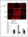
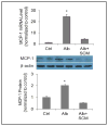
Results: Albumin induced EMT as shown by significant decreases in levels of epithelial marker E-cadherin, increases in mesenchymal markers fibroblast-specific protein 1 and α-smooth muscle actin, and elevations in collagen I. SCM inhibited all these changes. Meanwhile, albumin induced NF-κB translocation from cytosol into nucleus and that SCM blocked the nuclear translocation of NF-κB. Albumin also increased the levels of pro-inflammatory factor monocyte chemoattractant protein-1 (MCP)-1 by nearly 30 fold compared with control. SCM almost abolished albumin-induced increase of MCP-1.
Conclusion: These results suggest that SCM attenuated albumin-induced EMT in renal tubular cells via inhibiting activation of inflammatory factors, which may serve as a new therapeutic approach for chronic kidney diseases.
© 2015 S. Karger AG, Basel.
Figures


Similar articles
-
Hypoxia inducible factor-1α mediates the profibrotic effect of albumin in renal tubular cells.Sci Rep. 2017 Nov 20;7(1):15878. doi: 10.1038/s41598-017-15972-8. Sci Rep. 2017. PMID: 29158549 Free PMC article.
-
Mesenchymal stem cells modulate albumin-induced renal tubular inflammation and fibrosis.PLoS One. 2014 Mar 19;9(3):e90883. doi: 10.1371/journal.pone.0090883. eCollection 2014. PLoS One. 2014. PMID: 24646687 Free PMC article.
-
Arctigenin suppresses transforming growth factor-β1-induced expression of monocyte chemoattractant protein-1 and the subsequent epithelial-mesenchymal transition through reactive oxygen species-dependent ERK/NF-κB signaling pathway in renal tubular epithelial cells.Free Radic Res. 2015;49(9):1095-113. doi: 10.3109/10715762.2015.1038258. Epub 2015 May 13. Free Radic Res. 2015. PMID: 25968940
-
Albumin-induced epithelial mesenchymal transformation.Nephron Exp Nephrol. 2012;120(3):e91-102. doi: 10.1159/000336822. Epub 2012 May 16. Nephron Exp Nephrol. 2012. PMID: 22613868
-
Renal toxicity of albumin and other lipoproteins.Curr Opin Nephrol Hypertens. 1995 Jul;4(4):369-73. doi: 10.1097/00041552-199507000-00015. Curr Opin Nephrol Hypertens. 1995. PMID: 7552106 Review.
Cited by
-
FKBP51 promotes migration and invasion of papillary thyroid carcinoma through NF-κB-dependent epithelial-to-mesenchymal transition.Oncol Lett. 2018 Dec;16(6):7020-7028. doi: 10.3892/ol.2018.9517. Epub 2018 Sep 27. Oncol Lett. 2018. PMID: 30546435 Free PMC article.
-
Characterization of the iPSC-derived conditioned medium that promotes the growth of bovine corneal endothelial cells.PeerJ. 2019 Apr 16;7:e6734. doi: 10.7717/peerj.6734. eCollection 2019. PeerJ. 2019. PMID: 31024764 Free PMC article.
-
Hypoxia inducible factor-1α mediates the profibrotic effect of albumin in renal tubular cells.Sci Rep. 2017 Nov 20;7(1):15878. doi: 10.1038/s41598-017-15972-8. Sci Rep. 2017. PMID: 29158549 Free PMC article.
-
Establishment of renal proximal tubule cell lines derived from the kidney of p53 knockout mice.Cytotechnology. 2019 Feb;71(1):45-56. doi: 10.1007/s10616-018-0261-1. Epub 2019 Jan 2. Cytotechnology. 2019. PMID: 30603921 Free PMC article.
-
Renal Medullary Overexpression of Sphingosine-1-Phosphate Receptor 1 Transgene Attenuates Deoxycorticosterone Acetate (DOCA)-Salt Hypertension.Am J Hypertens. 2023 Aug 5;36(9):509-516. doi: 10.1093/ajh/hpad046. Am J Hypertens. 2023. PMID: 37171128 Free PMC article.
References
-
- Rodriguez-Iturbe B, Johnson RJ, Herrera-Acosta J. Tubulointerstitial damage and progression of renal failure. Kidney Int. 2005;68:S82–S86. - PubMed
-
- Hills CE, Squires PE. TGF-beta1-induced epithelial-to-mesenchymal transition and therapeutic intervention in diabetic nephropathy. Am J Nephrol. 2010;31:68–74. - PubMed
Publication types
MeSH terms
Substances
Grants and funding
LinkOut - more resources
Full Text Sources
Other Literature Sources
Research Materials
Miscellaneous
