. 2015 Apr;42(4):1895-903.
doi: 10.1118/1.4915495.

Spatially constrained incoherent motion method improves diffusion-weighted MRI signal decay analysis in the liver and spleen

Affiliations

Spatially constrained incoherent motion method improves diffusion-weighted MRI signal decay analysis in the liver and spleen

Vahid Taimouri et al. Med Phys. 2015 Apr.

Abstract

Purpose: To evaluate the effect of the spatially constrained incoherent motion (SCIM) method on improving the precision and robustness of fast and slow diffusion parameter estimates from diffusion-weighted MRI in liver and spleen in comparison to the independent voxel-wise intravoxel incoherent motion (IVIM) model.

Methods: We collected diffusion-weighted MRI (DW-MRI) data of 29 subjects (5 healthy subjects and 24 patients with Crohn's disease in the ileum). We evaluated parameters estimates' robustness against different combinations of b-values (i.e., 4 b-values and 7 b-values) by comparing the variance of the estimates obtained with the SCIM and the independent voxel-wise IVIM model. We also evaluated the improvement in the precision of parameter estimates by comparing the coefficient of variation (CV) of the SCIM parameter estimates to that of the IVIM.

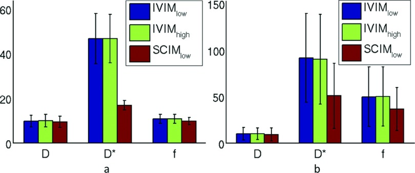
Results: The SCIM method was more robust compared to IVIM (up to 70% in liver and spleen) for different combinations of b-values. Also, the CV values of the parameter estimations using the SCIM method were significantly lower compared to repeated acquisition and signal averaging estimated using IVIM, especially for the fast diffusion parameter in liver (CVIV IM = 46.61 ± 11.22, CVSCIM = 16.85 ± 2.160, p < 0.001) and spleen (CVIV IM = 95.15 ± 19.82, CVSCIM = 52.55 ± 1.91, p < 0.001).

Conclusions: The SCIM method characterizes fast and slow diffusion more precisely compared to the independent voxel-wise IVIM model fitting in the liver and spleen.


Figures

FIG. 1.
FIG. 1.
Five ROIs selected in the liver and spleen were overlaid on the DW-MRI with b-values (a) and (d) 5 s/mm2, (b) and (e) 100 s/mm2, and (c) and (f) 600 s/mm2. (a)–(c) ROI 1 (lower right lobe) in purple and ROI 4 (lower spleen) in cyan and (d)–(f) ROI 2 (upper right lobe) in blue, ROI 3 (left lobe) in pink, and ROI 5 (upper spleen) in orange.
FIG. 2.
FIG. 2.
Representative example of the observed DW-MRI signal and the estimated models calculated by the IVIM and SCIM estimators from all 7 b-values (5, 50, 100, 200, 400, 600, and 800).
FIG. 3.
FIG. 3.
Bar plot of the CV of the incoherent motion parameters as estimated from group1 in (a) liver and (b) spleen.
FIG. 4.
FIG. 4.
(a) The DW-MR image illustrates the liver and spleen of a subject in group 2, (b)–(d) D, D*, and f parameter maps estimated using the IVIM model, (e)–(g) the corresponding IVIM parameter maps smoothed with a Gaussian filter (σ2 = 1 mm), and (h)–(j) D, D*, and f parameter maps estimated using the SCIM method.
FIG. 5.
FIG. 5.
Bar plot of the CV of the incoherent motion parameters as estimated from group 2 in (a) liver and (b) spleen using the IVIM method, the IVIM method while smoothing the parameter maps (IVIMσ), and the SCIM method.

Similar articles

Cited by

References

    1. Le Bihan D., Breton E., Lallemand D., Grenier P., Cabanis E., and Laval-Jeantet M., “MR imaging of intravoxel incoherent motions: Application to diffusion and perfusion in neurologic disorders,” Radiology 161, 401–407 (1986).10.1148/radiology.161.2.3763909 - DOI - PubMed
    1. Kiryu S., Dodanuki K., Takao H., Watanabe M., Inoue Y., Takazoe M., Sahara R., and Unuma K., “Free-breathing diffusion-weighted imaging for the assessment of inflammatory activity in Crohn’s disease,” J. Magn. Reson. Imaging 29, 880–886 (2009).10.1002/jmri.21725 - DOI - PubMed
    1. Oto A., Kayhan A., Williams J. T., Fan X., Yun L., Arkani S., and Rubin D. T., “Active Crohn’s disease in the small bowel: Evaluation by diffusion weighted imaging and quantitative dynamic contrast enhanced MR imaging,” J. Magn. Reson. Imaging 33, 615–624 (2011).10.1002/jmri.22435 - DOI - PubMed
    1. Oussalah A., Laurent V., Bruot O., Bressenot A., Bigard M. A., Regent D., and Peyrin-Biroulet L., “Diffusion-weighted magnetic resonance without bowel preparation for detecting colonic inflammation in inflammatory bowel disease,” Gut 59, 1056–1065 (2010).10.1136/gut.2009.197665 - DOI - PubMed
    1. Lemke A., Laun F. B., Simon D., Stieltjes B., and Schad L. R., “An in vivo verification of the intravoxel incoherent motion effect in diffusion-weighted imaging of the abdomen,” Magn. Reson. Med. 64, 1580–1585 (2010).10.1002/mrm.22565 - DOI - PubMed

Publication types