. 2015 Jun;16(2):91-9.
doi: 10.1007/s10969-015-9197-2. Epub 2015 Apr 2.

Crystal structures of Mycobacterial MeaB and MMAA-like GTPases

Affiliations

Crystal structures of Mycobacterial MeaB and MMAA-like GTPases

Thomas E Edwards et al. J Struct Funct Genomics. 2015 Jun.

Abstract

The methylmalonyl Co-A mutase-associated GTPase MeaB from Methylobacterium extorquens is involved in glyoxylate regulation and required for growth. In humans, mutations in the homolog methylmalonic aciduria associated protein (MMAA) cause methylmalonic aciduria, which is often fatal. The central role of MeaB from bacteria to humans suggests that MeaB is also important in other, pathogenic bacteria such as Mycobacterium tuberculosis. However, the identity of the mycobacterial MeaB homolog is presently unclear. Here, we identify the M. tuberculosis protein Rv1496 and its homologs in M. smegmatis and M. thermoresistibile as MeaB. The crystal structures of all three homologs are highly similar to MeaB and MMAA structures and reveal a characteristic three-domain homodimer with GDP bound in the G domain active site. A structure of Rv1496 obtained from a crystal grown in the presence of GTP exhibited electron density for GDP, suggesting GTPase activity. These structures identify the mycobacterial MeaB and provide a structural framework for therapeutic targeting of M. tuberculosis MeaB.


Figures

Figure 1
Figure 1
Mutliple sequence alignment of Mtb Rv1496 (MytuD.00200.a, PDB ID 3MD0) and its orthologs in M. smegmatis (MysmA.00200.b, PDB ID 3NXS), M. thermoresistibile (MythA.00200.a, PDB ID 3TK1), Methylobacterium extorquens (MeaB, PDB ID 2QM7 [5]), Homo sapiens (MMAA, PDB ID 2WWW [6]), and Escherichia coli (ArgK/YgfD, PDB ID 2P67, no primary citation).
Figure 2
Figure 2
Crystal structure of Mtb Rv1496 exhibits a homodimer with an N-terminal α-helical domain, a central G-domain bound to GDP and a C-terminal α-helical dimerization domain (a). The GDP-bound Mtb Rv1496 crystal structure overlaid with those of its orthologs (b) from M. smegmatis (Msm) shown in yellow ribbons and M. thermoresistible (Mth) shown in orange ribbons. The Mtb Rv1496 crystal structure overlaid with MeaB from M. extorquens (PDB ID 2QM7) shown in cyan ribbons (c). The biologically relevant dimer is shown for each structure. The Mtb Rv1496 crystal structure overlaid with MMAA from Homo sapiens (PDB ID 2WWW) shown in magenta ribbons (d); due to differences in the biological dimer detailed previously for human versus bacterial MMAA/MeaB proteins [6], we are only showing the monomer.
Figure 3
Figure 3
Recognition of GDP (a) or GTP (b) by M. tuberculosis Rv1496 and GDP by the Rv1496 orthologs from M. smegmatis (c) or M. thermoresistible (d). For Mtb Rv1496-GDP the |Fo|-|Fc| map is shown in green mesh contoured at 3.0 σ prior to modelling GDP. For Mtb Rv1496 crystals grown in the presence of GTP, the 2|Fo|-|Fc| map is shown in blue mesh contoured at 1.0 σ. Electron density was only present for the α and β phosphates of “GTP” and the model was otherwise nearly identical to the GDP bound structure, leading us to hypothesize that Rv1496 may have hydrolyzed the GDP to GTP. Only one amino acid differs between all three species, V245 in Mtb is V241 in Msm yet L239 in Mth (labelled in red).
Figure 4
Figure 4
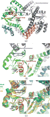
Examination of the conformational plasticity of the switch III loop in MeaB and its mycobacterial orthologs. (a) The Mth Rv1496 homodimer is shown as colored in Figure 2, but with the addition of the P-loop colored in red, the switch I loop colored in orange, the switch II loop colored in purple, and the switch III loop colored in blue. Close up (b) and overlay (c) of the P- and switch loops of MeaB, Mtb Rv1496 and its Msm and Mth orthologs, demonstrating structural rigidity of the GDP recognizing P-loop, but structural plasticity of the three switch loops.

References

    1. Korotkova N, Lidstrom ME. MeaB is a component of the methylmalonyl-CoA mutase complex required for protection of the enzyme from inactivation. J Biol Chem. 2004;279:13652–13658. - PubMed
    1. Korotkova N, Chistoserdova L, Kuksa V, Lidstrom ME. Glyoxylate regeneration pathway in the methylotroph Methylobacterium extorquens AM1. J Bacteriol. 2002;184:1750–1758. - PMC - PubMed
    1. Padovani D, Banerjee R. A G-protein editor gates coenzyme B12 loading and is corrupted in methylmalonic aciduria. Proc Natl Acad Sci U S A. 2009;106:21567–21572. - PMC - PubMed
    1. Dobson CM, Wai T, Leclerc D, Kadir H, Narang M, Lerner-Ellis JP, Hudson TJ, Rosenblatt DS, Gravel RA. Identification of the gene responsible for the cblB complementation group of vitamin B12-dependent methylmalonic aciduria. Hum Mol Genet. 2002;11:3361–3369. - PubMed
    1. Hubbard PA, Padovani D, Labunska T, Mahlstedt SA, Banerjee R, Drennan CL. Crystal structure and mutagenesis of the metallochaperone MeaB: insight into the causes of methylmalonic aciduria. J Biol Chem. 2007;282:31308–31316. - PubMed

Publication types

MeSH terms

LinkOut - more resources